


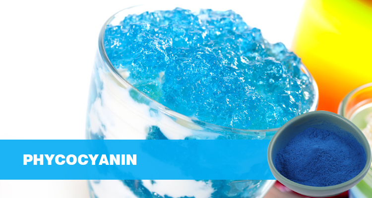
Phycocyanin (PC), a treasure crafted by nature, is primarily extracted from blue-green algae, an ancient and remarkable group of organisms, of which Spirulina and Arthrospira are prominent representatives. Its captivating blue color, a hue that seems to blend the depths of the ocean and the mystical starry sky, is unique, striking, and unforgettable. A molecular analysis reveals that phycocyanin has a molecular weight of approximately 110 kDa and is composed of a clever combination of α and β subunits. It also contains an open-ring tetrapyrrole structure. This structure not only endows phycocyanin with unique pigmentary properties, enabling it to play a key role in photosynthesis, efficiently capturing and transmitting light energy to sustain algae, but also imparts many of the properties of proteins, with a rich amino acid sequence that lays the foundation for its biological activity. This unique molecular structure, like a precisely engineered molecular machine, gives phycocyanin its potent antioxidant and anti-inflammatory properties, making it a core functional component that protects multiple organ health. As an antioxidant, it acts like a fierce guardian, rapidly identifying and eliminating free radicals generated in the body. These restless “troublemakers” wreak havoc on cellular structures and biomolecules. Phycocyanin effectively blocks these destructive actions, reducing oxidative stress damage to cells, thereby delaying cell aging and preventing various diseases caused by oxidative stress. Phycocyanin also excels in the anti-inflammatory field, precisely modulating inflammation-related signaling pathways, inhibiting the excessive release of inflammatory factors, and reducing the damage to tissues and organs caused by inflammatory responses, thereby safeguarding the body’s health and stability.
Bioavailability and Benefits
In the past, the application of phycocyanin was limited by its low bioavailability, like a bound sword, unable to fully unleash its powerful effects. However, with the rapid development of technology, the emergence of nano-delivery technology has brought new hope for the application of phycocyanin. Through nanotechnology techniques such as liposome encapsulation, phycocyanin is coated with a special “protective coat.” This coat not only effectively protects phycocyanin from degradation in the gastrointestinal tract but also facilitates its absorption, significantly increasing its intestinal absorption rate to 35%. This represents a significant improvement compared to traditional extracts, effectively opening the door for phycocyanin to function, allowing it to more smoothly enter the human circulatory system and reach various tissues and organs, exerting its health benefits.
Phycocyanin’s multi-target mechanism of action offers unique advantages in protecting organ health. It not only directly scavenges free radicals, reducing oxidative damage to cells and tissues, and preventing organ disease at its source, but also penetrates deep into cells to modulate organ-specific signaling pathways. For example, in the liver, phycocyanin can modulate the Nrf2 signaling pathway, activate the expression of a series of antioxidant enzymes, and enhance the liver’s antioxidant defenses. In the cardiovascular system, it can regulate the PI3K/Akt signaling pathway, inhibiting inflammation and apoptosis, protecting the integrity of vascular endothelial cells, and maintaining normal cardiovascular function. This level of protection from cells to organs enables phycocyanin to comprehensively protect the health of human organs, just like a comprehensive health guard, providing meticulous care for each organ of the body.
 Core Mechanism of Organ Protection: Driven by Dual Antioxidant and Anti-Inflammatory Pathways
 Core Mechanism of Organ Protection: Driven by Dual Antioxidant and Anti-Inflammatory Pathways
In the complex physiological processes that maintain human organ health, phycocyanin acts as a multifaceted guardian. Leveraging its potent antioxidant and anti-inflammatory properties, and through unique molecular mechanisms, it safeguards organ health at multiple levels, playing a crucial protective role at the cellular and molecular levels.
(I) Free Radical “Scavenger”: Blocking the Chain of Oxidative Damage
During normal physiological metabolism, the human body continuously produces free radicals, a natural part of life. However, when free radical production exceeds the body’s scavenging capacity, it’s like opening a Pandora’s box, and the shadow of oxidative stress quietly looms. Excessive free radicals, particularly superoxide anions (O₂⁻・) and hydroxyl radicals (・OH), are highly chemically active, acting like rampaging “molecular bombs,” indiscriminately attacking various biomolecules within cells. These free radicals react without hesitation with unsaturated fatty acids in cell membranes, triggering a chain reaction of lipid peroxidation. Once lipid peroxidation initiates, it’s like lighting a string of firecrackers. The structure and function of cell membranes are severely damaged, altering membrane fluidity and permeability, disrupting the balance of intracellular and extracellular material exchange, and significantly disrupting normal cellular function. Furthermore, free radicals can damage vital biomolecules within cells, such as DNA and proteins, leading to serious consequences like gene mutations and loss of protein function. These changes underlie the development and progression of many chronic diseases, such as cardiovascular disease, neurodegenerative diseases, and cancer.
The emergence of phycocyanin, like a ray of hope in the darkness, offers hope for resolving this dilemma. Its unique molecular structure exhibits remarkable free radical scavenging capabilities, making it a “killer” of intracellular free radicals. The aromatic amino acid residues in phycocyanin, such as tryptophan and histidine, act like carefully crafted “traps,” efficiently capturing superoxide anions (O₂⁻・) and hydroxyl radicals (・OH) through conjugation, stabilizing these dangerous free radicals and rendering them inactive, preventing them from attacking biomolecules within the cell. Phycocyanin also reacts specifically with lipid peroxides, acting like a skilled craftsman, converting them into stable lipid peroxide-phycocyanin complexes. This effectively inhibits the progression of lipid peroxidation and protects cells from further oxidative damage. Research data shows that in an oxidative stress model, malondialdehyde (MDA) levels were significantly reduced by over 40% after phycocyanin treatment. This significant decrease in MDA, a hallmark product of lipid peroxidation, directly demonstrates the powerful efficacy of phycocyanin in inhibiting lipid peroxidation. It also demonstrates that phycocyanin can effectively reduce free radical damage to cell membranes and other biological structures, maintaining normal cellular morphology and function.
In addition to directly scavenging free radicals, phycocyanin also has a more profound strategic significance: it activates the cell’s endogenous antioxidant defense system, awakening a group of dormant “guardians” and building a strong antioxidant defense line. Phycocyanin can activate the Nrf2 signaling pathway, which acts as a “command center” for intracellular antioxidant defense. Once activated by phycocyanin, it initiates a series of instructions, inducing the expression of antioxidant enzyme genes within the cell. Superoxide dismutase (SOD) and glutathione peroxidase (GSH-Px) are key members of this defense system. Superoxide dismutase (SOD) catalyzes the dismutation of superoxide anions (O₂⁻・), converting them into hydrogen peroxide (H₂O₂) and oxygen, effectively removing superoxide anions and reducing their cellular damage. Glutathione peroxidase (GSH-Px), using glutathione as a substrate, reduces hydrogen peroxide (H₂O₂) and lipid peroxides to water and the corresponding alcohols, further eliminating harmful substances produced by oxidative stress. When phycocyanin upregulates the activity of superoxide dismutase (SOD) and glutathione peroxidase (GSH-Px), the antioxidant capacity within cells is significantly enhanced, enabling them to better cope with free radical attacks and maintain intracellular redox balance. In animal experiments, feeding animals a phycocyanin-rich diet significantly increased the activity of superoxide dismutase (SOD) and glutathione peroxidase (GSH-Px) in their tissues, with increases ranging from 30% to 50% compared to the control group. This demonstrates the remarkable effectiveness of phycocyanin in activating the endogenous antioxidant defense system, providing more comprehensive and long-lasting protection for cells. This dual mechanism of action, combining direct free radical scavenging with activation of the endogenous antioxidant defense system, makes phycocyanin highly effective in interrupting the oxidative damage chain. It provides a solid foundation for cellular and organ health from multiple perspectives and levels, effectively preventing and mitigating organ damage and disease caused by oxidative stress.
(II) Inflammation “Regulatory Valve”: Inhibiting the Release of Proinflammatory Mediators
Inflammation is a normal defensive response of the body to external stimuli, acting like a warning signal. When the body is exposed to pathogen invasion, physical or chemical damage, or other stimuli, the inflammatory response rapidly activates. Various cells and molecules of the immune system rapidly flock to the damaged area, attempting to eliminate the pathogen and repair damaged tissue. However, when the inflammatory response becomes uncontrolled and overactivated, it becomes a double-edged sword, causing severe damage to the body’s own tissues and organs and becoming the root cause of many diseases. The nuclear factor-κB (NF-κB) pathway plays a key role in the inflammatory response. It acts as the “master switch” of the inflammatory response. Once activated, it triggers a complex chain reaction, leading to the massive release of multiple proinflammatory cytokines, such as tumor necrosis factor-α (TNF-α) and interleukin-6 (IL-6). These pro-inflammatory factors act like a swarm of uncontrolled “destroyers.” They stimulate the activation and aggregation of immune cells, leading to dilation and increased permeability of blood vessels at the site of inflammation, triggering inflammatory symptoms such as redness, swelling, and pain. Furthermore, they further activate other inflammatory signaling pathways, creating a vicious cycle that amplifies the inflammatory response and severely damages the normal structure and function of surrounding tissues and organs. Long-term chronic inflammation is also closely linked to the development and progression of numerous chronic diseases, including cardiovascular disease, diabetes, and neurodegenerative disorders, posing a serious threat to human health.
Phycocyanin plays a crucial role in regulating inflammatory responses. It precisely targets inflammatory signaling pathways, acting like a skilled “tuner,” modulating the intensity of the inflammatory response to maintain a moderate level and preventing excessive inflammation from causing damage to the body. Studies have found that phycocyanin can effectively block the activation of the NF-κB pathway. By inhibiting the phosphorylation of IκBα, it prevents the translocation of NF-κB from the cytoplasm to the nucleus, thereby disrupting NF-κB’s regulatory effects on proinflammatory genes and reducing the transcription and secretion of proinflammatory factors such as tumor necrosis factor-α (TNF-α) and interleukin-6 (IL-6). In a lipopolysaccharide (LPS)-induced inflammation model, a commonly used experimental model for studying inflammatory responses, phycocyanin intervention significantly suppressed the intensity of the inflammatory response, decreasing it by 55% compared to the untreated group. Levels of proinflammatory factors such as tumor necrosis factor-α (TNF-α) and interleukin-6 (IL-6) were also significantly reduced. This demonstrates the powerful ability of phycocyanin to inhibit the NF-κB pathway and reduce the secretion of proinflammatory factors, effectively alleviating the damage caused by inflammatory responses to tissues and organs. In addition to inhibiting the NF-κB pathway, phycocyanin can also alleviate inflammation through other pathways. It inhibits the release of inflammatory mediators such as histamine and 5-hydroxytryptamine. Histamine and 5-hydroxytryptamine are important mediators of inflammation, causing inflammatory symptoms such as vasodilation, increased vascular permeability, and tissue edema. By inhibiting the release of these inflammatory mediators, phycocyanin can effectively reduce localized inflammatory edema and tissue damage, alleviating the pain and discomfort caused by inflammation. In an allergic inflammation model, phycocyanin intervention significantly reduced the release of histamine and 5-hydroxytryptamine, and significantly improved edema and itching at the inflamed site, further confirming the positive role of phycocyanin in inhibiting the release of inflammatory mediators and alleviating inflammatory symptoms. Phycocyanin regulates inflammatory responses through multiple targets and pathways. It effectively inhibits the over-activation of inflammatory responses by blocking the NF-κB pathway, inhibiting the secretion of pro-inflammatory factors, and reducing the release of inflammatory mediators, providing powerful protection for organ health. It has great potential in preventing and treating inflammation-related diseases.
 Precise Organ-Targeted Protection
 Precise Organ-Targeted Protection
In the complex and delicate human body, each organ functions like a finely tuned component, working together to maintain the normal course of life. Phycocyanin, with its unique bioactivity, can penetrate deeply into various organs, targeting their physiological characteristics and common damage mechanisms, exerting precise and effective protective effects, thus building a solid defense for organ health.
(I) Liver: The Core Guardian of Metabolism and Detoxification
As the largest organ in the human body, the liver functions like a highly efficient “biochemical factory,” playing an irreplaceable and crucial role in numerous physiological processes, including metabolism, detoxification, and immune regulation. It undertakes the important task of synthesizing, decomposing, transforming, and storing various nutrients ingested by the body, while also facing the invasion of various harmful substances, such as chemical toxins, drugs, and alcohol. In modern life, due to factors such as environmental pollution, unhealthy diet, and excessive alcohol consumption, the liver faces unprecedented challenges, becoming highly susceptible to damage. This, in turn, can lead to a range of liver diseases, such as chemical liver damage and non-alcoholic fatty liver disease, which pose a serious threat to human health. Phycocyanin, with its potent antioxidant and anti-inflammatory properties, has become a loyal guardian of liver health, safeguarding normal liver function in multiple ways.
1. Protection against Chemical Liver Damage
Among the many factors that cause liver damage, chemical toxins are a significant threat. Carbon tetrachloride (CCl₄), a typical hepatotoxic chemical, is widely used in experimental studies to induce chemical liver damage. Upon entering the human body, CCl₄ is metabolized by the liver’s cytochrome P450 enzyme system to produce highly reactive trichloromethyl free radicals (・CCl₃) and trichloromethyl peroxide free radicals (・OO CCl₃). These free radicals act like frenzied “destroyers,” inducing lipid peroxidation within hepatocytes, severely damaging the structure and function of liver cell membranes and subsequently inducing hepatocyte apoptosis, significantly impacting the liver’s normal metabolic and detoxification functions.
Phycocyanin demonstrates remarkable protective capabilities against CCl₄-induced liver damage. Studies have found that phycocyanin can significantly reduce the rate of CCl₄-induced hepatocyte apoptosis by 38%. Its mechanism of action is primarily through activation of the Nrf2 pathway. The Nrf2 pathway is a key intracellular antioxidant stress response pathway. Upon activation by phycocyanin, Nrf2 enters the cell nucleus and binds to the antioxidant response element (ARE), initiating the transcription and expression of a series of antioxidant enzyme genes, such as heme oxygenase-1 (HO-1) and NAD(P)H quinone oxidoreductase 1 (NQO1). These antioxidant enzymes effectively scavenge intracellular free radicals, enhance hepatocyte membrane stability, and reduce damage to hepatocyte membranes caused by lipid peroxidation, thereby protecting hepatocytes from the threat of apoptosis. In related cell experiments, hepatocytes pretreated with phycocyanin showed a significant decrease in malondialdehyde (MDA) levels and a significant increase in the activities of antioxidant enzymes such as superoxide dismutase (SOD) and glutathione peroxidase (GSH-Px) after exposure to CCl₄. This strongly suggests that phycocyanin, by activating the Nrf2 pathway, enhances the antioxidant defenses of hepatocytes, effectively alleviating CCl₄-induced oxidative damage and reducing hepatocyte apoptosis.
In addition to chemical toxins, alcohol is also a common cause of liver damage. After entering the body, alcohol is primarily metabolized in the liver. Its metabolite, acetaldehyde, is highly toxic and can bind to biomacromolecules such as proteins and nucleic acids within hepatocytes, forming acetaldehyde-protein adducts. This alters the structure and function of these proteins and nucleic acids, thereby affecting the normal metabolism and physiological function of hepatocytes. Acetaldehyde also damages mitochondria within hepatocytes, leading to a decrease in mitochondrial membrane potential, impaired respiratory chain function, reduced ATP production, and disrupted cellular energy metabolism, further exacerbating hepatocyte damage. Long-term, heavy drinking can also lead to fatty liver degeneration, causing alcoholic fatty liver disease, which can progress to cirrhosis and liver cancer in severe cases. Phycocyanin has significant benefits in alcohol detoxification and liver protection. It accelerates the clearance of acetaldehyde, a metabolite of ethanol, effectively reducing mitochondrial damage, thereby enhancing the liver’s ability to detoxify. Several active ingredients in phycocyanin, such as tryptophan and histidine, specifically bind to acetaldehyde, forming stable complexes. This reduces acetaldehyde concentrations in the body and mitigates its toxic effects on liver cells. Phycocyanin also activates acetaldehyde dehydrogenase (ALDH) in the liver, accelerating the conversion of acetaldehyde to acetic acid and promoting its metabolism and excretion. In animal experiments, supplementing phycocyanin with alcohol-administered ethanol resulted in significantly reduced acetaldehyde levels in the blood and liver of mice. The morphology and function of liver mitochondria were significantly improved, mitochondrial membrane potential returned to normal, respiratory chain enzyme activity was enhanced, and ATP production significantly increased. This suggests that phycocyanin can effectively mitigate alcohol-induced damage to liver mitochondria, protect the energy metabolism function of hepatocytes, enhance the liver’s ability to detoxify, and reduce the risk of alcohol-induced liver injury.
2. Promotes Hepatocyte Regeneration
The regenerative capacity of hepatocytes is crucial for liver repair and functional recovery. Following liver injury, hepatocytes initiate a regenerative process, proliferating and differentiating to repair damaged liver tissue. However, in certain pathological conditions, such as severe liver injury and chronic liver disease, this regenerative capacity can be inhibited, hindering the liver’s repair process and impairing normal liver function. Phycocyanin plays a key role in promoting hepatocyte regeneration. It can upregulate HGF (hepatocyte growth factor) expression and promote the proliferation of hepatic progenitor cells, significantly increasing the rate of liver tissue repair by 25%. HGF is an important cell growth factor that binds to the c-Met receptor on the surface of hepatocytes, activating multiple downstream signaling pathways, such as the Ras-Raf-MEK-ERK pathway and the PI3K-Akt pathway. Activation of these signaling pathways promotes DNA synthesis, cell division, and proliferation in hepatocytes, while also inhibiting apoptosis and promoting hepatocyte survival and regeneration. Phycocyanin, by upregulating HGF expression, provides strong support for hepatocyte regeneration and accelerates the repair of damaged liver tissue. In a partial hepatectomy model, mice treated with phycocyanin showed significantly faster liver regeneration than controls, with a significant increase in the number of proliferating cell nuclear antigen (PCNA)-positive cells in liver tissue. This indicates that phycocyanin effectively promotes hepatocyte proliferation and accelerates liver regeneration and repair.
Non-alcoholic fatty liver disease (NAFLD) is a common liver disease characterized by excessive fat accumulation in the liver, leading to hepatocyte steatosis, inflammatory infiltration, and fibrosis. In severe cases, it can progress to cirrhosis and liver cancer. The development of non-alcoholic fatty liver disease (NAFLD) is associated with a variety of factors, including obesity, insulin resistance, hyperlipidemia, and metabolic syndrome. A key factor in the development of NAFLD is the disrupted lipid metabolism within hepatocytes. Excessive fatty acids accumulate within hepatocytes, leading to increased triglyceride synthesis and decreased fatty acid oxidation and transport, resulting in lipid deposition.
Phycocyanin can effectively improve lipid deposition in NAFLD, reducing hepatic triglyceride levels by 22%. Its mechanism of action primarily involves regulating signaling pathways and gene expression related to lipid metabolism. Phycocyanin can activate the AMPK signaling pathway in the liver. AMPK is a key regulator of intracellular energy metabolism. Activated AMPK inhibits the synthesis of fatty acids and triglycerides while promoting fatty acid oxidation and transport. Specifically, AMPK inhibits the activity of acetyl-CoA carboxylase (ACC), reducing the synthesis of malonyl-CoA and thereby inhibiting de novo fatty acid synthesis. AMPK also activates carnitine acyltransferase 1 (CPT1), promoting the entry of fatty acids into mitochondria for β-oxidation and accelerating fatty acid catabolism. Furthermore, phycocyanin regulates the expression of genes related to lipid metabolism in the liver, such as peroxisome proliferator-activated receptor α (PPARα) and fatty acid binding protein 1 (FABP1). Changes in the expression of these genes help maintain lipid homeostasis in the liver and reduce lipid deposition in hepatocytes. In an animal model of non-alcoholic fatty liver disease induced by a high-fat diet, phycocyanin administration significantly reduced liver steatosis, triglyceride and cholesterol levels, decreased inflammatory cell infiltration in the liver, and decreased the expression of inflammatory factors. This suggests that phycocyanin can effectively improve the lipid metabolism disorders and inflammatory state in non-alcoholic fatty liver disease, and has a promising preventive and therapeutic effect on non-alcoholic fatty liver disease.
(II) Cardiovascular System: Precision Maintenance of Circulatory Dynamics
 (II) Cardiovascular System: Precision Maintenance of Circulatory Dynamics
The cardiovascular system is like the human body’s “transportation network.” The heart, acting as the “power pump” of this network, continuously and rhythmically contracts and relaxes, propelling blood through the vessels, delivering oxygen and nutrients to tissues and organs throughout the body while carrying metabolic waste back to the liver and kidneys for processing. Blood vessels, like the crisscrossing “pipelines,” are responsible for the transport and distribution of blood. Endothelial cells form the barrier lining the vascular walls, not only maintaining the normal structure and function of the vessels but also participating in many important physiological processes, such as vasodilation, coagulation regulation, and inflammatory responses. However, under the influence of modern lifestyles, the cardiovascular system faces numerous challenges, including risk factors such as hypertension, hyperlipidemia, hyperglycemia, smoking, and obesity. These factors can lead to cardiovascular diseases such as endothelial dysfunction, atherosclerosis, and myocardial ischemia, seriously threatening human health. Phycocyanin, with its unique biological activity, can precisely maintain the cardiovascular system in multiple ways, reducing the risk of cardiovascular disease and providing strong support for heart and vascular health.
1. Strengthening the Vascular Endothelial Barrier
Endothelial cells serve as the first line of defense in the vascular lining. Their functional integrity is crucial for maintaining normal vascular function. When endothelial cells are stimulated by various risk factors, such as oxidative stress, inflammation, and hypertension, endothelial dysfunction can occur, manifesting as decreased nitric oxide (NO) synthesis, increased release of vasoconstrictor factors, inflammatory cell adhesion, and platelet aggregation. These changes further promote the development and progression of atherosclerosis, increasing the risk of cardiovascular disease. Nitric oxide (NO) is an important vasodilator, produced from L-arginine by nitric oxide synthase (NOS) in endothelial cells. NO diffuses into vascular smooth muscle cells, activates guanylate cyclase, and increases intracellular cyclic guanosine monophosphate (cGMP) levels, leading to vascular smooth muscle relaxation, vasodilation, reduced vascular resistance, and the maintenance of normal blood pressure and circulation. Phycocyanin plays a crucial role in strengthening the vascular endothelial barrier. It promotes nitric oxide (NO) synthesis, enhancing vasodilation by 30%, and thus effectively delaying the formation of atherosclerotic plaques. Through its potent antioxidant capacity, phycocyanin scavenges free radicals within endothelial cells, reducing oxidative damage to these cells, protecting nitric oxide synthase (NOS) activity, and promoting NO synthesis and release. Furthermore, phycocyanin modulates signaling pathways related to endothelial function, such as the PI3K-Akt pathway and the eNOS-NO pathway. Activating these pathways promotes eNOS phosphorylation, increasing its activity and further increasing NO production. In vitro cell experiments showed that treatment of vascular endothelial cells with phycocyanin significantly increased intracellular NO content and enhanced vasodilation. In animal studies, animals fed a phycocyanin-rich diet showed significant improvement in endothelial function, with a significant reduction in the area and thickness of atherosclerotic plaques. This suggests that phycocyanin can promote NO synthesis, enhance vasodilation, effectively protect the endothelial barrier, and slow the progression of atherosclerosis.
Abnormal proliferation of vascular smooth muscle cells is a key pathological basis for atherosclerosis and vascular remodeling. During the development of atherosclerosis, vascular smooth muscle cells, stimulated by inflammatory factors and growth factors, shift from a contractile to a synthetic phenotype. This enhanced proliferation leads to migration beneath the vascular intima, where they synthesize and secrete large amounts of extracellular matrix, resulting in intimal thickening and stenosis, increasing the risk of thrombosis. Phycocyanin inhibits vascular smooth muscle cell proliferation, reducing intimal thickness by 18%, thereby effectively reducing the risk of thrombosis. Phycocyanin inhibits cell proliferation by inhibiting signaling pathways related to cell proliferation, such as the MAPK and NF-κB pathways, reducing the expression of cell cycle proteins and growth factors, and preventing vascular smooth muscle cells from transitioning from the G1 phase to the S phase, thereby inhibiting cell proliferation. Phycocyanin can also induce vascular smooth muscle cell apoptosis, reducing cell number and further alleviating intimal thickening. In animal models of vascular injury, phycocyanin treatment significantly reduced intimal thickening and significantly inhibited the proliferation and migration of vascular smooth muscle cells. This indicates that phycocyanin can effectively inhibit abnormal vascular smooth muscle cell proliferation, maintain normal vascular structure and function, and reduce the risk of thrombosis.
2. Cardiomyocyte Energy Protection
Cardiomyocytes are the primary executors of cardiac contraction and relaxation, and their normal energy metabolism is crucial for maintaining the heart’s pumping function. Cardiomyocyte energy primarily derives from aerobic respiration in mitochondria, where nutrients are converted into ATP through oxidative phosphorylation, providing energy for cardiomyocyte contraction and relaxation. However, in pathological conditions such as myocardial ischemia-reperfusion injury, cardiomyocyte mitochondrial function can be severely affected, leading to impaired energy metabolism, decreased ATP production, intracellular calcium overload, and cardiomyocyte apoptosis and necrosis, severely impacting cardiac function. Mitochondrial complex I is a crucial component of the mitochondrial respiratory chain, participating in electron transport and proton transport, and playing a key role in ATP production. During myocardial ischemia-reperfusion injury, mitochondrial complex I activity is inhibited, resulting in blocked electron transport, an inability to establish a proton gradient, and reduced ATP production. Phycocyanin can enhance mitochondrial complex I activity, increasing ATP production by 28%, thereby effectively ameliorating myocardial ischemia-reperfusion injury. Through its antioxidant properties, phycocyanin scavenges free radicals within cardiomyocytes, reduces oxidative stress-induced mitochondrial damage, and protects the structure and function of mitochondrial complex I. Phycocyanin can also regulate signaling pathways related to mitochondrial biogenesis and function, such as the PGC-1α pathway and the Nrf1 pathway, promoting mitochondrial biogenesis and repair, and improving mitochondrial function and efficiency. In an animal model of myocardial ischemia-reperfusion, pretreatment with phycocyanin significantly increased mitochondrial complex I activity and ATP content in myocardial tissue, significantly reduced the rate of cardiomyocyte apoptosis, and significantly improved cardiac function. This suggests that phycocyanin can effectively protect cardiomyocyte energy metabolism and mitigate myocardial ischemia-reperfusion injury by enhancing mitochondrial complex I activity and increasing ATP production.
Oxidative stress is a key factor contributing to cardiomyocyte apoptosis. During myocardial ischemia-reperfusion, the massive production of oxygen free radicals disrupts redox homeostasis in cardiomyocytes, activating apoptotic signaling pathways such as the caspase-3 pathway and the mitochondrial apoptosis pathway, leading to cardiomyocyte apoptosis. The size of myocardial infarction is an important indicator of the severity of myocardial damage. Reducing myocardial infarction size is crucial for protecting cardiac function and reducing mortality. Phycocyanin can reduce oxidative stress-induced cardiomyocyte apoptosis and reduce the size of myocardial infarction by 20%. Phycocyanin inhibits myocardial apoptosis by scavenging free radicals in myocardial cells, inhibiting oxidative stress and reducing the expression of apoptosis-related proteins such as Caspase-3 and Bax, while increasing the expression of anti-apoptotic proteins such as Bcl-2. In an animal model of myocardial infarction, phycocyanin treatment significantly reduced the number of apoptotic cells in myocardial tissue, significantly reduced the size of myocardial infarction, and significantly improved cardiac systolic and diastolic function. This suggests that phycocyanin can effectively inhibit oxidative stress-induced cardiomyocyte apoptosis, reduce the size of myocardial infarction, and protect cardiac function.
(III) Gastrointestinal Tract: Expert in Mucosal Barrier Repair
The gastrointestinal tract, a vital site for digestion and absorption in the human body, is like a busy “production line,” responsible for digesting food, absorbing nutrients, and excreting waste. The gastrointestinal mucosa is the first line of defense against invading pathogens and harmful substances. Composed of epithelial cells, mucus, and immune cells, it provides multiple functions, including mechanical, chemical, and immune barriers, effectively protecting the gastrointestinal tract from damage. However, in daily life, the gastrointestinal tract is frequently irritated by various factors, such as gastric acid, Helicobacter pylori infection, nonsteroidal anti-inflammatory drugs, alcohol, and spicy foods. These factors can damage the gastrointestinal mucosa, leading to diseases such as gastritis, gastric ulcers, enteritis, and ulcerative colitis, impairing normal gastrointestinal function. Phycocyanin, with its unique biological activity, can precisely repair and regulate the gastrointestinal mucosal barrier, maintaining gastrointestinal health.
Repair of Gastric Mucosal Damage
The gastric mucosa is the innermost layer of the stomach wall. It comes into direct contact with digestive juices such as gastric acid and pepsin, and is also exposed to external pathogens and harmful substances, making it vulnerable to damage. Gastric acid, a key component of gastric juice, plays a vital role in food digestion. However, when gastric acid secretion is excessive or the gastric mucosal defenses are weakened, it can damage the gastric mucosa, leading to lesions such as gastric erosion and ulcers. Gastric ulcers are a common digestive system disease characterized by defects and ulcer formation in the gastric mucosa. Patients often experience symptoms such as upper abdominal pain, acid reflux, and belching, which seriously affect their quality of life.
Phycocyanin, an alkaline protein, effectively neutralizes gastric acid and regulates gastric pH, thereby accelerating the healing of gastric mucosal ulcers by 40%. Basic amino acid residues in phycocyanin, such as arginine and lysine, bind to hydrogen ions in gastric acid, reducing the concentration of gastric acid and alleviating irritation and damage to the gastric mucosa. Phycocyanin also promotes the proliferation and repair of gastric mucosal cells, upregulating the expression of genes associated with cell proliferation and repair, such as epidermal growth factor (EGF) and transforming growth factor-α (TGF-α). These growth factors stimulate the division and proliferation of gastric mucosal cells, promoting ulcer healing. In an animal model of gastric ulcers, treatment with phycocyanin significantly reduced the size of gastric ulcers, reduced their depth, and accelerated their healing rate. The expression levels of EGF and TGF-α in gastric mucosal tissue also increased significantly, indicating that phycocyanin can effectively promote the healing of gastric ulcers by neutralizing gastric acid and promoting the proliferation and repair of gastric mucosal cells.
The gastric mucosa is covered with a thick layer of mucus, composed of glycoproteins secreted by gastric epithelial cells. It lubricates and protects the gastric mucosa, protecting it from erosion by gastric acid and pepsin. Helicobacter pylori is a Gram-negative bacterium that colonizes the gastric mucosa. By secreting substances such as urease and cytotoxins, it damages the mucus layer and epithelial cells, leading to gastritis, gastric ulcers, and gastric cancer. Phycocyanin promotes the synthesis of glycoproteins in the mucus layer, strengthening the mucosal barrier and reducing H. pylori adhesion. Phycocyanin stimulates signaling pathways within gastric epithelial cells, such as the PI3K-Akt and MAPK pathways, promoting the expression of genes involved in glycoprotein synthesis and increasing glycoprotein content in the mucus layer, thereby strengthening the thickness and function of the mucosal barrier. Furthermore, phycocyanin, through its antimicrobial and anti-inflammatory properties, inhibits the growth and reproduction of H. pylori, reducing its adhesion and damage to the gastric mucosa. In in vitro experiments, after treating gastric mucosal epithelial cells with phycocyanin, the content of glycoprotein secreted by the cells increased significantly and the thickness of the mucus layer increased; in an animal model infected with Helicobacter pylori, the number of Helicobacter pylori in the gastric mucosal tissue decreased significantly after intervention with phycocyanin.
Application Transformation: From Mechanism Research to the Health Industry
With the deepening of research on the organ-protective mechanisms of phycocyanin, significant progress has been made in its application and transformation within the health industry. From functional foods to pharmaceuticals, phycocyanin is bringing new benefits to people’s health with its unique biological activities.
(I) Functional Foods and Dietary Supplements
In the functional foods and dietary supplements sector, phycocyanin is gradually emerging as a new option for those pursuing a healthier lifestyle. Researchers have carefully formulated phycocyanin into chewable tablets. This convenient dosage form offers consumers a simple and effective supplemental option. Clinical studies have demonstrated that daily intake of phycocyanin chewable tablets at the recommended dose of 20-50mg significantly increases liver GSH (glutathione) levels by 18%. As a key antioxidant in the liver, increased GSH levels enhance the liver’s antioxidant capacity, better protecting against free radical attack and reducing oxidative stress damage to liver cells, thereby effectively preventing and improving liver diseases such as chemical-induced liver damage and non-alcoholic fatty liver disease. To further expand the application of phycocyanin in functional foods, researchers have ingeniously combined it with spirulina polysaccharides to develop a stomach-protecting beverage. Spirulina polysaccharides inherently possess numerous biological activities, including immune regulation and antioxidant activity. When combined with phycocyanin, the two work synergistically, significantly enhancing the protection and repair of the gastrointestinal mucosa. Clinical trials have shown that this stomach-protecting beverage has a 63% remission rate for chronic gastritis. It works through multiple pathways. On the one hand, phycocyanin neutralizes gastric acid, reducing irritation and damage to the gastric mucosa and promoting the proliferation and repair of gastric mucosal cells. On the other hand, spirulina polysaccharides enhance the gastric mucosal barrier function, inhibit the adhesion of Helicobacter pylori, and reduce inflammatory responses. This combination of the two benefits chronic gastritis patients, effectively alleviating symptoms such as stomach pain, bloating, and acid reflux, and improving their quality of life.
(II) Targeted Formulations in the Pharmaceutical Field
In the pharmaceutical field, the application of phycocyanin is developing further towards targeted formulations, offering new hope for disease treatment. Researchers have used advanced liposome encapsulation technology to formulate phycocyanin into an injectable solution. This targeted formulation precisely targets the liver, providing strong support for protecting against chemotherapy-induced liver damage. In a Phase III clinical trial, patients treated with the liposome-encapsulated phycocyanin injection achieved a 41% improvement in liver enzyme levels. During chemotherapy, commonly used chemotherapy drugs such as cisplatin and doxorubicin, while killing cancer cells, can also severely damage liver cells, leading to elevated liver enzyme levels and impairing normal liver function. Liposome-encapsulated phycocyanin injection effectively mitigates chemotherapy-induced liver damage by inhibiting oxidative stress and inflammation, protecting liver cell structure and function, reducing liver enzyme levels, and promoting liver repair and regeneration, thereby improving chemotherapy tolerance and enhancing treatment efficacy.
For allergic rhinitis, a common respiratory disease, researchers have developed a nasal formulation of phycocyanin, offering patients a new treatment option. Clinical studies have shown that the intensity of histamine stimulation in patients treated with this formulation decreased by 50%. Allergic rhinitis is an inflammatory response to the nasal mucosa caused by allergens. Histamine is a key mediator in allergic reactions. Its release causes vasodilation and increased permeability in the nasal mucosa, leading to symptoms such as itching, sneezing, and runny nose. Phycocyanin nasal formulations can effectively repair damaged nasal mucosa in patients with allergic rhinitis, modulate the immune response, and inhibit the release of inflammatory mediators such as histamine, thereby alleviating nasal inflammation, alleviating symptoms, and improving patients’ quality of life.
(III) Safety and Dosage Consensus
Safety and dosage are of great concern in the application of phycocyanin. After rigorous research and evaluation, the European Food Safety Authority (EFSA) has determined that the safe upper limit of phycocyanin intake is 200 mg per day. Extensive experimental data demonstrates that within this dosage range, phycocyanin does not cause significant toxic effects on vital organs such as the liver and kidneys, providing safety assurance for its use in functional foods and pharmaceuticals. However, long-term or excessive intake may still pose potential risks, such as increased metabolic burden on the liver and kidneys. While there is currently no conclusive clinical evidence indicating serious health problems, caution is advised during use.
Regarding drug interactions, particular caution is required when using phycocyanin with medications such as warfarin. Warfarin is a commonly used anticoagulant that works by inhibiting the synthesis of vitamin K-dependent coagulation factors. Phycocyanin may compete with warfarin for plasma protein binding sites, thereby affecting warfarin’s blood concentration and anticoagulant effect. Therefore, to avoid the risk of drug interactions, a two-hour interval is recommended when using phycocyanin and warfarin simultaneously to ensure that both drugs can safely and effectively exert their respective effects and protect the patient’s medication safety.
Challenges and Future: Decoding the Deep Potential of Blue Guardian
 Challenges and Future: Decoding the Deep Potential of Blue Guardian
(I) Organ-Specific Target Analysis
Although phycocyanin has demonstrated significant efficacy in multi-organ protection, its specific targets and mechanisms of action in different organs remain under investigation. Clarifying the differential mechanisms of phycocyanin’s protection in organs such as the kidney and lung is key to further exploring its potential. For example, in the kidney, studies have found that energy metabolism in renal tubular epithelial cells is crucial for maintaining normal renal function. However, whether phycocyanin specifically improves energy metabolism in renal tubular epithelial cells through the AMPK pathway remains unclear. Further investigation of this mechanism will help reveal the unique mode of action of phycocyanin in kidney protection and provide more precise theoretical support for the prevention and treatment of kidney disease. Regarding the lung, phycocyanin’s protective effect on lung tissue may involve regulating multiple pathways, including inflammatory cell infiltration, oxidative stress, and extracellular matrix remodeling. However, the specific molecular targets and signaling pathways require further investigation. Comprehensively elucidating the targets and signaling networks of phycocyanin in different organs through advanced technologies such as single-cell sequencing and proteomics will lay a solid foundation for its precise application in clinical treatment.
(II) Delivery System Optimization
Although the bioavailability of phycocyanin has improved, significant room for improvement remains. Developing more efficient delivery systems is a key approach to enhancing the organ-protective effects of phycocyanin. For example, the development of intestinal pH-responsive microcapsules is of great significance. These microcapsules precisely release phycocyanin according to varying intestinal pH conditions, thereby preventing degradation in adverse environments such as gastric acid and ensuring efficient absorption and utilization within the intestine. By rationally designing the materials and structures of microcapsules, it is possible to increase the bioavailability of phycocyanin to over 50%, enabling more effective accumulation in target organs and enhancing their protective effects. Furthermore, the continuous advancement of nanotechnology offers more possibilities for phycocyanin delivery. For example, the use of novel nanocarriers such as nanoliposomes and nanomicelles not only improves the stability and solubility of phycocyanin but also enables targeted delivery to specific organs through surface modification, further enhancing its efficacy in organ protection.
Due to its natural pleiotropic properties, phycocyanin is evolving from a marine bioactive ingredient to a core functional factor in multi-organ protection. With the deepening of mechanism research and innovation in delivery technology, this blue guardian is expected to play a greater role in preventing organ degenerative diseases and improving organ function, becoming a bridge connecting nature’s gifts with precise health.
