Hydroxytyrosol: Decoding the dual codes of natural antioxidants and anti-aging

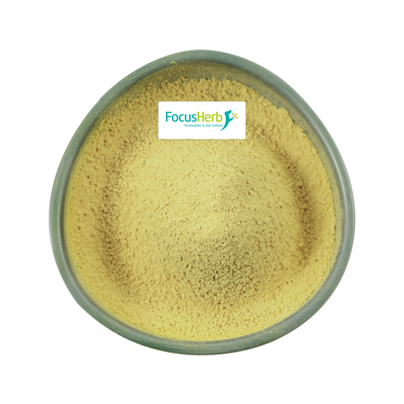
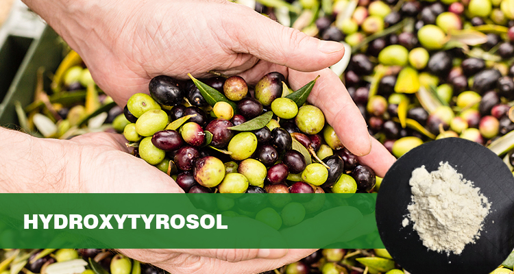
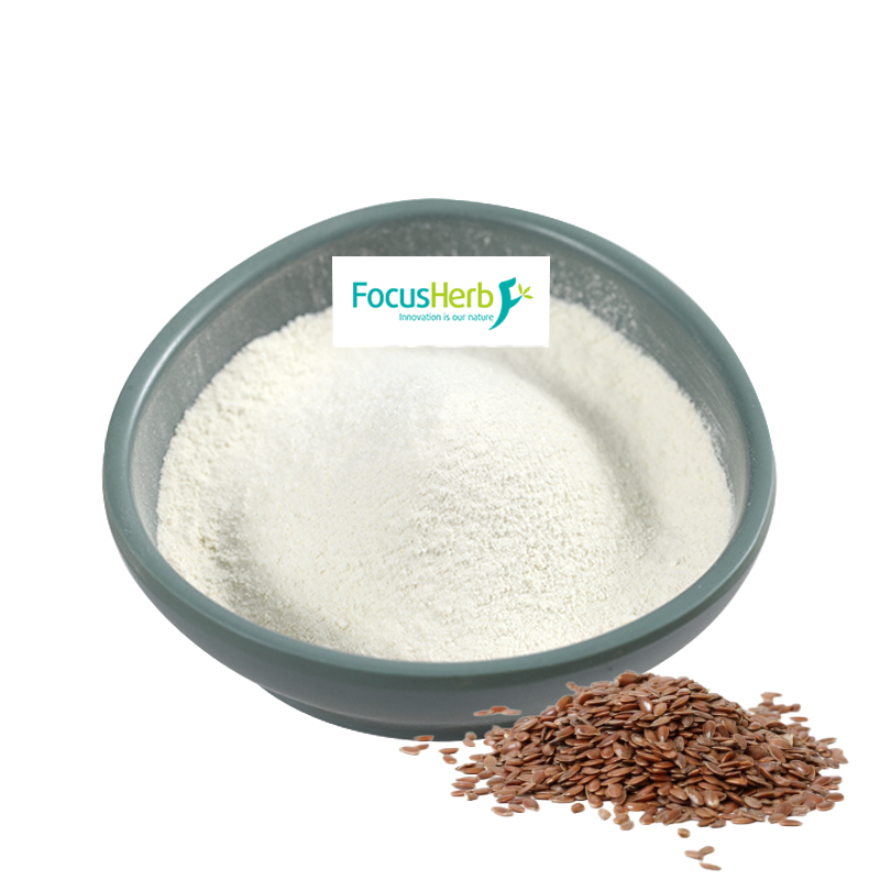
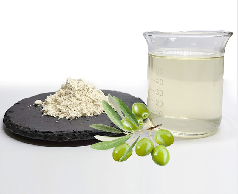
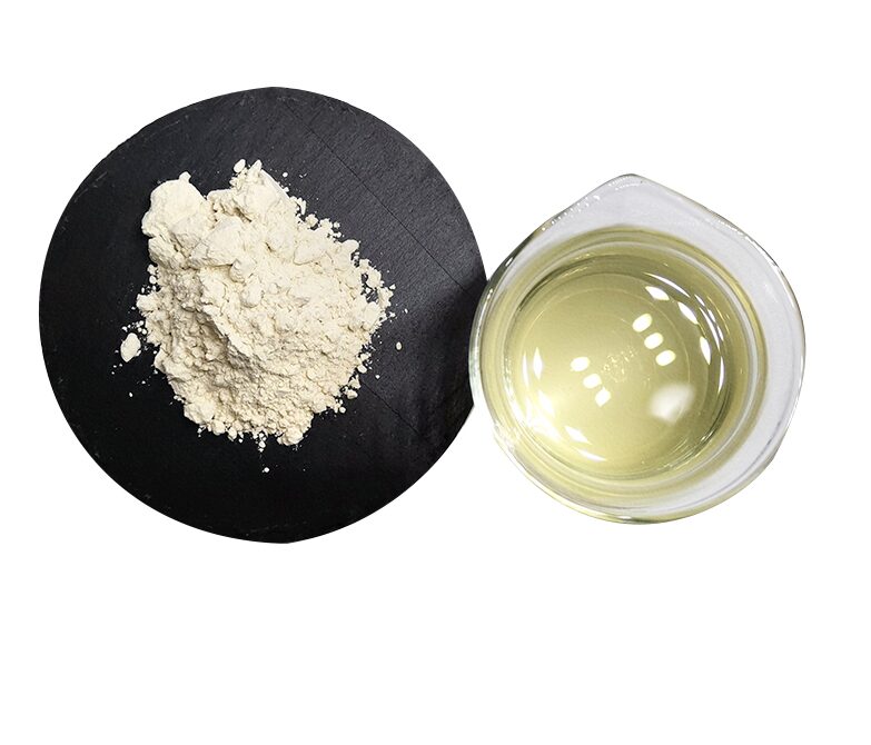
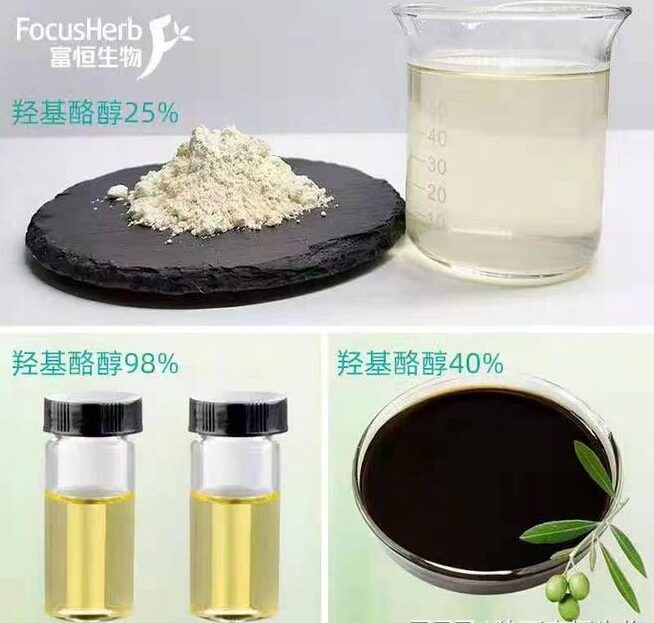
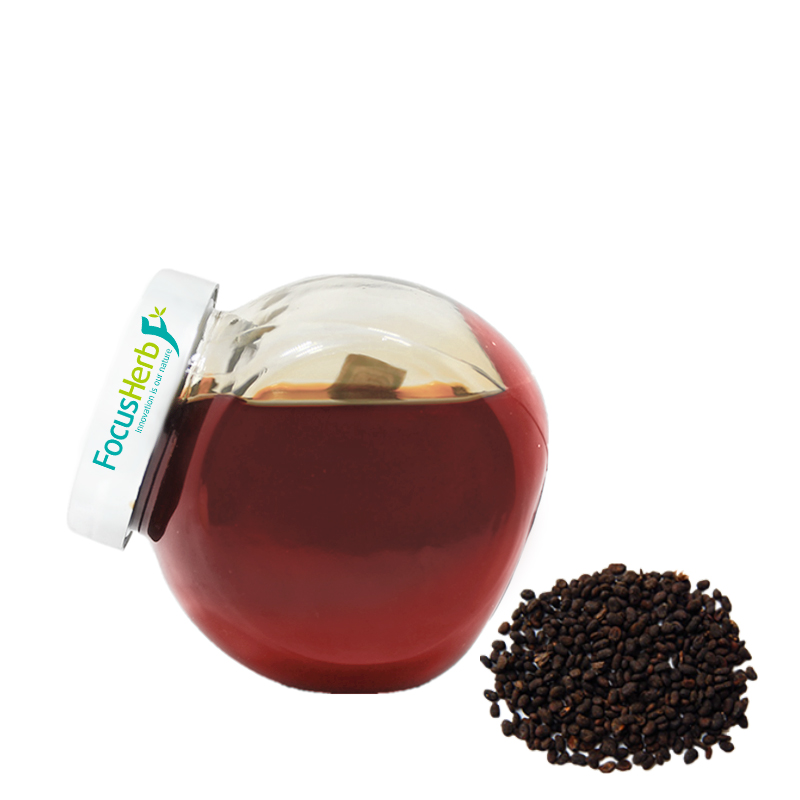
Hydroxytyrosol may be unfamiliar to many, but its benefits are significant. It’s the core active ingredient in olive oil and olive leaf extract, chemically known as 3,4-dihydroxyphenylethanol. Chemically, the hydroxytyrosol molecule contains a catechol group. This unique structure gives it exceptional hydrogen-donating capacity, making it one of nature’s most potent antioxidant polyphenols.

Hydroxytyrosol is unevenly distributed throughout olives. Its concentration in olive leaves is relatively high, ranging from 1.5% to 3%. In olive oil, hydroxytyrosol primarily exists as an ester. When we consume olive oil or products containing olive leaf extract, these esters undergo a series of metabolic processes in the intestines, ultimately releasing free hydroxytyrosol, which then exerts its various health benefits. For example, people in the Mediterranean region, who consume a high amount of olive oil in their diet, have a relatively low incidence of cardiovascular disease, partly due to the antioxidant and cardiovascular protective properties of hydroxytyrosol in olive oil.

As a small, fat-soluble ingredient, hydroxytyrosol offers unique bioavailability advantages. It efficiently penetrates cell membranes, like a tiny key, easily unlocking the cell’s door and gaining access to its inner workings. Furthermore, hydroxytyrosol can cross the blood-brain barrier, enabling it to exert antioxidant and neuroprotective effects in the brain, which is crucial for preventing and improving certain neurological diseases.

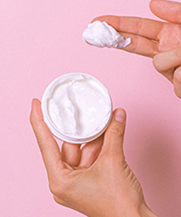
Research has shown that hydroxytyrosol accumulates in target sites such as the dermis and mitochondrial membranes. Compared to vitamin C, its bioavailability is over three times higher. Furthermore, hydroxytyrosol does not carry the dose-dependent toxicity risk common with traditional antioxidants, meaning that when used appropriately, it can provide antioxidant protection more safely. When added to skincare products, hydroxytyrosol is rapidly absorbed into the dermis, scavenging free radicals, reducing oxidative damage, and delaying skin aging, without the risk of adverse reactions related to dosage.

Core Antioxidant Mechanism: A Multi-Dimensional Free Radical Defense System

The “Molecular Sponge” Effect of Free Radical Scavenging

1. Broad-Spectrum Free Radical Capturing Ability

Free radicals are ubiquitous in our environment, like restless little monsters, constantly threatening cellular health. Toxic free radicals such as superoxide anions (O₂⁻), hydroxyl radicals (・OH), and peroxynitrites (ONOO⁻) are the primary culprits of cellular oxidative damage. Hydroxytyrosol, like a valiant “guardian,” exhibits powerful free radical scavenging capabilities thanks to its unique molecular structure.
Hydroxytyrosol’s catechol structure is its “secret weapon” against free radicals. This structure can simultaneously donate two active hydrogen atoms. Upon encountering a free radical, hydroxytyrosol rapidly donates its active hydrogen atoms, stabilizing the free radical and thereby interrupting the lipid peroxidation chain reaction. This reaction acts like a shackle on the free radical, preventing it from further damaging biomolecules such as lipids, proteins, and DNA within the cell. Compared to the traditional antioxidant vitamin E, hydroxytyrosol exhibits significantly higher free radical scavenging efficiency, with its IC₅₀ value being 40% lower than that of vitamin E. This means that under the same conditions, hydroxytyrosol can achieve the same free radical scavenging effect as vitamin E at a lower concentration. In a UV-induced skin oxidation model, hydroxytyrosol also performed remarkably well, reducing intracellular reactive oxygen species (ROS) levels by 65%, effectively mitigating UV-induced oxidative damage to skin cells.

2. Activation of Endogenous Antioxidant Pathways

In addition to directly scavenging free radicals, hydroxytyrosol can also activate endogenous antioxidant pathways within cells, fundamentally boosting their antioxidant capacity and essentially injecting a powerful boost into the cell’s antioxidant defense system.

The Nrf2/ARE signaling pathway is a crucial intracellular anti-oxidative stress response pathway. When cells are exposed to oxidative stress, Nrf2 is activated and translocates from the cytoplasm to the nucleus, where it binds to the antioxidant response element (ARE), triggering the expression of a series of antioxidant enzyme genes. Hydroxytyrosol can activate the Nrf2/ARE signaling pathway, upregulating the expression of antioxidant enzymes such as superoxide dismutase (SOD) and glutathione peroxidase (GSH-Px) by 30%-50%. These antioxidant enzymes act as intracellular “cleaners,” promptly removing free radicals generated within the cell and maintaining redox balance.

Hydroxytyrosol can also restore intracellular glutathione (GSH) levels. GSH is a key antioxidant in cells that directly participates in free radical scavenging reactions and regenerates other antioxidants, such as vitamins C and E. In a H₂O₂-damaged hepatocyte model, hydroxytyrosol reduced GSH depletion by 70%, effectively protecting hepatocytes from oxidative damage and maintaining normal liver function.

Targeted Mitochondrial Protection: From Energy Factories to Aging Regulation

1. Maintaining Respiratory Chain Complex Activity

Mitochondria, known as the “energy factories” of the cell, are crucial for normal cellular function. It produces ATP, the energy molecule needed by cells, through oxidative phosphorylation reactions via respiratory chain complexes. However, during this process, mitochondria also produce a large number of free radicals. When excessive free radicals accumulate, they damage mitochondria, impairing their function and accelerating cellular aging. Hydroxytyrosol acts as a “guardian” of mitochondria. It can reduce the generation of free radicals caused by electron leakage by increasing the activity of mitochondrial complexes I and II. In vitro experiments have shown that hydroxytyrosol treatment increased the activity of mitochondrial complexes I and II by 25%-35%, facilitating smoother electron transfer in the respiratory chain and reducing the generation of free radicals. Furthermore, hydroxytyrosol promotes the expression of PGC-1α protein, increasing its peak concentration by 205%. PGC-1α is a key regulator of cellular energy metabolism, inducing mitochondrial biogenesis and increasing the number and quality of mitochondria. In aged fibroblasts, hydroxytyrosol treatment increased mitochondrial DNA copy number by 40%, indicating that the number of mitochondria is replenished, improving the cell’s energy supply capacity and thus slowing the aging process.

2. Blocking Membrane Lipid Peroxidation

The mitochondrial membrane is composed of a lipid bilayer. These lipids are susceptible to free radical attack, leading to peroxidation reactions and damage to the membrane’s structure and function. Malondialdehyde (MDA) is one of the main products of lipid peroxidation, and its content reflects the extent of membrane lipid peroxidation.

Hydroxytyrosol, a lipid-soluble antioxidant, can be incorporated into the mitochondrial membrane lipid bilayer, forming a protective layer on the membrane. It inhibits the production of peroxidation products such as MDA, reducing MDA levels by 55%, effectively blocking membrane lipid peroxidation and maintaining the stability of mitochondrial membrane potential.

In the Caenorhabditis elegans model, researchers have found that when mitochondria are damaged, the lifespan of the nematodes is significantly shortened. However, treatment with hydroxytyrosol reduced the rate of lifespan reduction caused by mitochondrial damage by 30%. This strongly demonstrates that hydroxytyrosol has a positive impact on the aging process by protecting the mitochondrial membrane and maintaining mitochondrial function, providing strong support for delaying aging.

Multi-target anti-aging action network

Cellular Level: The Molecular Switch that Delays Aging

1. Telomeres and DNA Damage Protection

Telomeres, like the “safety caps” at the ends of chromosomes, are closely linked to cellular aging. As cells divide, telomeres gradually shorten. When telomere shortening reaches a certain level, cells enter a state of senescence. DNA damage is another major factor in cellular aging. Factors such as oxidative stress can cause DNA damage, impairing normal cellular function.
Hydroxytyrosol plays a key role in protecting telomeres and DNA. It inhibits the production of 8-OHdG, a marker of DNA oxidative damage, reducing its damage to DNA. Studies have shown that hydroxytyrosol can reduce 8-OHdG production by 45%, thereby slowing the rate of telomere shortening. In human umbilical cord mesenchymal stem cells, hydroxytyrosol increased telomerase activity by 22%, indicating that cells are better able to maintain telomere length. With increased telomerase activity, the number of cell passages increased by 25%, effectively delaying the process of replicative aging. This is like putting a lock on the cell’s “lifespan clock,” slowing down the rate of cellular aging.

2. Regulating the Autophagy-Apoptosis Balance

Autophagy and apoptosis are two important physiological processes within cells, crucial for maintaining cellular homeostasis and health. Autophagy acts as a cellular “cleaner,” removing waste from aging organelles, misfolded proteins, and other debris, keeping cells clean and functioning normally. Apoptosis, on the other hand, is a form of programmed cell death. When cells are severely damaged or beyond repair, they initiate apoptosis to prevent further damage to the body.

During cellular aging, the balance between autophagy and apoptosis is disrupted. Hydroxytyrosol can induce autophagy by activating the AMPK/mTOR pathway, promoting the clearance of senescence-associated β-galactosidase (SA-β-gal)-positive cells by up to 35%. Hydroxytyrosol also inhibits the expression of the pro-apoptotic protein Bax, reducing the occurrence of apoptosis. In UV-damaged keratinocytes, hydroxytyrosol reduced the apoptosis rate by 60%, demonstrating its ability to effectively protect cells, maintain the balance between autophagy and apoptosis, and delay cellular aging.

Skin Anti-Aging: Three-Dimensional Repair from the Dermis to the Epidermis

1. Collagen Network Remodeling and Wrinkle Improvement

Skin elasticity and firmness primarily depend on collagen fibers in the dermis. With aging and environmental influences such as UV exposure and pollution, collagen in the skin gradually depletes, damaging collagen fibers and leading to aging symptoms such as sagging and wrinkles.

Hydroxytyrosol plays a crucial role in remodeling the skin’s collagen network. It stimulates fibroblast type I collagen mRNA expression, increasing it by 60%, thereby increasing the density of collagen fibers in the dermis. In a double-blind clinical trial, 8 weeks of hydroxytyrosol supplementation increased skin elasticity by 28% and reduced the depth of forehead wrinkles by 15%. This is like injecting a “tightening force” into sagging skin, restoring its elasticity and reducing the appearance of wrinkles. Hydroxytyrosol also inhibits the activity of matrix metalloproteinases (MMP-1/MMP-3) by 40%. These MMPs are the primary culprits in collagen fibril breakdown. By inhibiting their activity, hydroxytyrosol blocks UV-induced collagen fibril breakdown. In a mouse model of photoaging, hydroxytyrosol reduced collagen degradation products in the dermis by 50%, effectively protecting the skin’s collagen network and slowing the aging process.

2. Breaking the Inflammation-Oxidation Vicious Cycle

Skin inflammation and oxidative stress are closely linked, mutually reinforcing each other and forming a vicious cycle. Inflammation leads to increased oxidative stress, generating a large number of free radicals, which in turn further exacerbate the inflammatory response, damage skin cells, and accelerate skin aging.

Hydroxytyrosol can inhibit the NF-κB pathway, reducing the release of pro-inflammatory cytokines such as IL-6 and TNF-α by 55%. This acts like a pause button on the inflammatory response, alleviating inflammatory skin conditions such as acne and seborrheic dermatitis. Hydroxytyrosol also reduces histamine-induced vascular permeability, alleviating skin redness and sensitivity, and reducing the area of ​​erythema by 30%. By interrupting the vicious cycle of inflammation and oxidation, hydroxytyrosol provides a healthy environment for the skin, helping to delay skin aging.

3. Synergistic Effects of Whitening and Moisturizing

Skin whitening and moisturizing are important aspects of skin health and beauty. Excessive melanin production can lead to dullness and dark spots, while dehydration can cause dryness, roughness, and a loss of radiance.

Hydroxytyrosol has a synergistic effect in whitening and moisturizing. Regarding whitening, it competitively binds to the active site of tyrosinase, inhibiting melanin synthesis and reducing tyrosinase activity by 40%. In a B16 melanoma cell model, hydroxytyrosol reduced melanin production by 65%, resulting in brighter, more translucent skin. In terms of moisturizing, hydroxytyrosol’s small molecular structure allows it to penetrate deep into the stratum corneum, increasing the expression of the aquaporin AQP3 by 35%. Aquaporins act like the skin’s “water pipelines.” Increased expression of aquaporins can increase skin hydration by 22% and reduce transepidermal water loss (TEWL) by 18%, keeping skin constantly hydrated.


Scientific Application Scenarios and Supplementation Strategies

Functional Foods and Dietary Supplements

1. Natural Sources and High-Efficiency Formulations

In their pursuit of health, more and more people are turning to functional foods and dietary supplements. Hydroxytyrosol, a natural antioxidant, is attracting significant attention for its sources and formulations.
Olive oil, especially extra virgin olive oil, is a key natural source of hydroxytyrosol, containing approximately 0.2-0.5 mg of hydroxytyrosol per kilogram. Consuming a moderate amount of olive oil in daily diets not only adds a unique flavor to dishes but also allows for the intake of hydroxytyrosol and its health benefits. For example, using extra virgin olive oil instead of regular cooking oil in salad vegetables preserves the nutrients while allowing for easy intake of hydroxytyrosol.
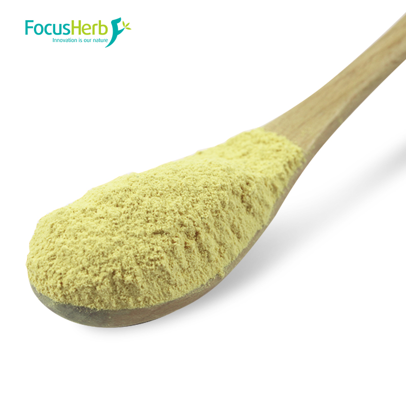
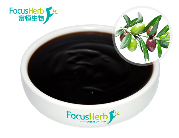
Olive leaf extract is a more concentrated source of hydroxytyrosol. Standardized olive leaf extract products can contain as much as 50-98% hydroxytyrosol. To enhance the bioavailability of hydroxytyrosol, researchers have employed nanoliposomal encapsulation technology. This technology acts like an invisible protective layer around hydroxytyrosol, allowing it to be better absorbed and utilized by the body once it enters. Its bioavailability is three times higher than traditional formulations.

For those seeking to consume hydroxytyrosol through dietary supplements, the recommended daily dose is 50-200 mg, which is roughly equivalent to 10-40 grams of olive leaf extract. When choosing a supplement, pay attention to the product’s ingredient list and production process, choosing one that utilizes advanced technology and offers reliable quality to ensure adequate hydroxytyrosol intake.

2. Suitable for Specific Populations

Different people have different physical conditions and needs, and hydroxytyrosol supplementation has specific benefits and suitable solutions for certain populations.

For those with dyslipidemia, which is a major risk factor for cardiovascular disease, an increase in oxidized low-density lipoprotein (ox-LDL) exacerbates the progression of atherosclerosis. Studies have found that a daily supplement of 100 mg of hydroxytyrosol can significantly reduce the level of ox-LDL oxidation by up to 25%. To further enhance cardiovascular protection, it is recommended to use hydroxytyrosol in conjunction with omega-3 supplements. Omega-3s are unsaturated fatty acids that can lower blood lipids and reduce inflammation. Working in conjunction with hydroxytyrosol, they can support cardiovascular health in multiple ways. For example, some fish oil supplements are rich in omega-3s. People with dyslipidemia can supplement with hydroxytyrosol and fish oil under a doctor’s guidance to improve their lipid profile and reduce the risk of cardiovascular disease.

For those with UV sensitivity, UV rays are one of the main external factors contributing to skin aging and damage. These individuals are more susceptible to UV damage, resulting in skin erythema and sunburn. Supplementing with 50 mg of hydroxytyrosol two hours before UV exposure, combined with a 0.5% hydroxytyrosol topical preparation, can reduce UVB-induced erythema by 40%. This is like putting on an invisible sunscreen, providing additional protection for those with UV sensitivity. Before outdoor activities, these individuals can take supplements containing hydroxytyrosol and apply sunscreen or skincare products containing hydroxytyrosol to effectively mitigate UV damage and maintain skin health.

Innovative Applications in Skincare Products

1. Active Ingredient Combinations

In the skincare market, hydroxytyrosol has become a favorite among many brands due to its outstanding antioxidant and anti-aging properties. To further enhance its effectiveness, researchers are continuously exploring combinations of hydroxytyrosol with other active ingredients.

In anti-wrinkle and firming formulas, combining 0.5%-2% hydroxytyrosol with 0.1% retinol can produce surprising synergistic effects. Retinol is a classic anti-wrinkle ingredient that promotes skin cell renewal and increases collagen production. The combined effect of hydroxytyrosol and retinol can boost collagen production by up to 1.8 times that of a single dose, while also reducing the potential irritation caused by retinol. They’re like a tacitly matched pair, each leveraging its own strengths while also complementing the other’s shortcomings. For consumers seeking firmer skin and wrinkle reduction, choosing skincare products containing this complex ingredient can more effectively achieve these anti-wrinkle and firming goals.

In a sunscreen booster formula, adding 0.3% hydroxytyrosol to a physical sunscreen neutralizes the free radicals generated by the sunscreen itself, increasing the sunscreen’s photostability by 30%. We all know that sun protection is a crucial part of skincare, and while sunscreen absorbs UV rays, it also generates free radicals, which can damage the skin. The addition of hydroxytyrosol acts like a protective shield for the sunscreen, enhancing its effectiveness while also reducing potential skin damage. When choosing a sunscreen, consider whether it contains hydroxytyrosol to achieve optimal sun protection and skincare benefits.

2. Clinically Proven Golden Concentration

The concentration of hydroxytyrosol in skincare products is crucial to its effectiveness. After extensive clinical research, researchers have identified the optimal concentration for optimal hydroxytyrosol performance in skincare products.

A cream containing 1% hydroxytyrosol increased the content of glycosaminoglycans (GAGs) in the dermis by 12% after four weeks of continuous use, effectively improving skin plumpness. Glycosaminoglycans are an important component of the skin’s extracellular matrix, helping to retain moisture and maintain firmness and elasticity. This cream injects a youthful energy into the skin, restoring its radiance. For those with dry, inelastic skin, choosing a cream containing 1% hydroxytyrosol and using it consistently for a period of time will yield noticeable improvements.

In a facial mask, a 0.8% concentration of hydroxytyrosol can increase the water content of the stratum corneum by 40% within 30 minutes, providing immediate repair. Facial masks are a skincare product that quickly replenishes nutrients and moisture. A 0.8% hydroxytyrosol mask is like a timely rain for the skin, instantly providing ample moisture and alleviating dryness and discomfort. When skin is dry and tired, using a mask containing 0.8% hydroxytyrosol can quickly restore moisture and vitality.


Research Frontiers and Future Outlook

Exploring New Targets for Disease Prevention

In medical research, hydroxytyrosol is emerging as a key molecule in the exploration of new targets for disease prevention.
A Phase II clinical study for Alzheimer’s disease has brought us exciting news. The study showed that hydroxytyrosol can reduce cerebrospinal fluid Aβ42 deposition by up to 22%, which is of great significance for improving mild cognitive impairment. Abnormal Aβ42 deposition is a key pathological feature of Alzheimer’s disease, leading to neuronal damage and death, and ultimately cognitive decline. By reducing Aβ42 deposition, hydroxytyrosol provides new insights and approaches for the prevention and treatment of Alzheimer’s disease.
Hydroxytyrosol has also demonstrated unique effects in diabetic models. By activating AMPK and promoting glucose uptake, it increased skeletal muscle insulin sensitivity by 18%. This discovery provides a new potential target for intervention in diabetes and related metabolic syndromes. AMPK is a key regulator of intracellular energy metabolism. Activating AMPK promotes glucose utilization and fatty acid oxidation, thereby improving insulin resistance and lowering blood sugar levels. Hydroxytyrosol, by activating AMPK, offers new hope for diabetic patients and is expected to become a new drug or adjunctive therapy for the treatment of diabetes.

Safety and Dose Optimization

Safety and dose optimization are crucial issues in the application of hydroxytyrosol. The safety threshold for long-term intake (over 6 months) of hydroxytyrosol has been determined to be 500 mg/day. Within this dose range, significant side effects are generally not observed in humans. This research finding provides important safety evidence for the long-term use of hydroxytyrosol, allowing people to use it with greater confidence for health maintenance.

However, there are some important considerations when using hydroxytyrosol. Taking hydroxytyrosol with iron supplements may impair the absorption of polyphenols. This is because the iron ions in the iron supplement interact with hydroxytyrosol, forming complexes that reduce its absorption efficiency. To avoid this, it is recommended to take hydroxytyrosol and iron supplements at least 2 hours apart to ensure that both can achieve the best effect.

Nervonic Acid: The core protection code of the nervous system

Nervonic Acid, scientifically known as cis-15-tetracosenoic acid, is a very-long-chain monounsaturated fatty acid with unique double bond placement within its carbon chain, which gives it unique physical and chemical properties. It was first discovered in mammalian neural tissue, hence its name. Over the course of biological evolution, neuraminic acid has gradually evolved a key role within neural tissue, becoming an essential core component of sphingolipids. Sphingolipids play a crucial role in the construction of nerve cell membranes, and neuraminic acid, as a key component, is directly involved in the formation of the myelin sheath. The myelin sheath acts like insulation for electrical wiring, ensuring efficient and accurate transmission of nerve signals along the nerve fiber, avoiding interference and attenuation. Furthermore, neuraminic acid plays a key role in maintaining the stability of nerve cell membranes, ensuring that nerve cells can function normally in this complex internal environment.

One of the most remarkable properties of neuraminic acid is its ability to cross the blood-brain barrier. The blood-brain barrier (BBB) ​​is the brain’s natural defense line, composed of tightly connected vascular endothelial cells, the basement membrane, and the end feet of astrocytes. It blocks most harmful substances and large molecules from entering the brain. Nervonic Acid, due to its unique molecular structure, can successfully cross this barrier and directly reach the central nervous system, enabling it to exert its neuroprotective and repairing effects.

When the nervous system is damaged, neuratomic acid activates pathways related to neuronal repair through a series of complex and sophisticated molecular mechanisms. Nervonic Acid plays a significant role in regulating the expression of neurotrophic factors. Neurotrophic factors are a class of proteins that play a key role in the survival, growth, differentiation, and maintenance of neurons. Nerve growth factor (NGF) and brain-derived neurotrophic factor (BDNF) are the most important members. Nervonic Acid promotes the expression of these neurotrophic factors, acting as a nutrient supply to injured neurons. After neuronal damage, NGF and BDNF bind to corresponding receptors on the neuronal surface, activating a series of intracellular signaling pathways and promoting axonal regeneration. Axons are crucial structures for neuronal signal transmission, and their regeneration is crucial for restoring neurological function. These neurotrophic factors also promote synaptic remodeling, reestablishing effective connections between neurons and thus restoring nerve signal transmission.

Oligodendrocyte precursor cells play a crucial role in the repair of myelin sheaths, and neuraminic acid can induce them to differentiate into mature oligodendrocytes. Oligodendrocytes produce myelin sheaths, which wrap around nerve fibers and repair the myelin sheath. In a demyelination model, researchers found that neuraminic acid treatment significantly increased the rate of myelin repair, by 40% compared to the untreated group. This result demonstrates that neuraminic acid effectively promotes myelin repair, thereby accelerating the conduction of nerve signals and improving the recovery of neurological function.

Nervonic acid’s Multidimensional Protective Effects on the Nervous System

Nerve Damage Repair and Degenerative Disease Intervention

1. Supporting the Regeneration of Damaged Nerve Fibers

Serious illnesses such as stroke and spinal cord injury can severely impact the human nervous system, often leading to nerve fiber rupture and, in turn, a series of severe functional impairments. Take stroke, for example, an acute cerebrovascular disease characterized by high morbidity, disability, and mortality. Once it occurs, it disrupts local brain tissue circulation, causing oxygen and blood deprivation to nerve cells, ultimately leading to nerve fiber damage and rupture. Patients may experience symptoms such as limb paralysis, speech impairment, and cognitive decline, severely impacting their quality of life. Spinal cord injury, on the other hand, typically results from trauma, disease, or other factors that damage the spinal cord structure and function, hindering the transmission of neural signals. Patients may suffer from conditions such as paraplegia and incontinence.
Neuronic acid demonstrates potent repair capabilities in addressing these nerve fiber damage issues. Its primary mechanism of action is through activation of the PI3K/Akt signaling pathway. The PI3K/Akt signaling pathway plays a key regulatory role in cell survival, proliferation, and differentiation. When neuratomic acid activates this signaling pathway, it inhibits neuronal apoptosis. Neuronal apoptosis is a common phenomenon following nerve injury, and excessive apoptosis can lead to further deterioration of neurological function. By inhibiting this process, neuratomic acid provides a stable environment for nerve fiber repair. Furthermore, neuratomic acid promotes the proliferation of Schwann cells. Schwann cells are important glial cells in the peripheral nervous system. They form myelin sheaths that surround nerve fibers and play a crucial role in nerve fiber regeneration and repair. Neuroatomic acid promotes Schwann cell proliferation, acting as a “building block” for nerve fiber regeneration and providing solid structural support for axonal regeneration.

Extensive preclinical studies have provided strong evidence for this efficacy of neuratomic acid. In one study in a nerve injury model, researchers found that treatment with neuratomic acid increased the length of regenerated axons by 35% compared to the control group. This significant difference strongly demonstrates that neuratomic acid can effectively promote the regeneration of damaged nerve fibers, offering hope for the restoration of neurological function.

2. Potential Intervention Targets for Alzheimer’s Disease

Alzheimer’s disease is the most common neurodegenerative disease, primarily affecting the elderly. Its incidence increases significantly with age. Globally, approximately 50 million people with Alzheimer’s disease are diagnosed, and this number is expected to triple by 2050. Key pathological hallmarks of the disease include the accumulation of Aβ amyloid protein and imbalanced tau protein phosphorylation. Aβ amyloid is a normal protein, but in Alzheimer’s patients, it abnormally aggregates to form amyloid plaques. These plaques accumulate in the brain, disrupting connections between neurons and impairing neural signaling. Tau, a protein that binds to microtubules, plays a crucial role in maintaining the normal structure and function of neurons. In Alzheimer’s patients, tau becomes hyperphosphorylated, reducing its ability to bind to microtubules. This leads to microtubule disassembly, ultimately forming neurofibrillary tangles, further impairing neuronal function. Nervonic acids have the potential to intervene in Alzheimer’s disease through multiple mechanisms of action. Firstly, they can reduce Aβ amyloid deposition, primarily by inhibiting β-secretase activity. β-secretase is a key enzyme in the production of Aβ amyloid. Inhibiting its activity can reduce Aβ amyloid production at its source, thereby reducing its accumulation in the brain and alleviating neuronal damage. Secondly, Nervonic acids can improve the imbalance in tau phosphorylation. They regulate the activity of related kinases and phosphatases, restoring tau phosphorylation levels to normal, preventing the formation of neurofibrillary tangles and protecting neuronal structure and function.

In a clinical study, neuratoic acid supplementation was administered to people with mild cognitive impairment. Results showed that after six months of neuratoic acid supplementation, memory scores improved by 12% compared to the control group. Memory scores are a key indicator of cognitive function, and this improvement suggests that neuratoic acid can effectively improve patients’ memory and slow cognitive decline. At the same time, cerebrospinal fluid (CSF) p-tau protein levels decreased by 18%. CSF p-tau protein levels are an important biomarker reflecting the pathological progression of Alzheimer’s disease. This decrease suggests that neuraminic acid can, to a certain extent, inhibit the hyperphosphorylation of tau protein, thereby intervening in the pathological progression of Alzheimer’s disease. These research results provide important clinical evidence for the use of neuraminic acid in the prevention and treatment of Alzheimer’s disease.

Brain Development and Cognitive Optimization

1. Essential Nutrients for Infant and Child Neurological Development

During infancy and childhood, the brain undergoes a critical period of rapid development, and the development of the nervous system plays a decisive role in a child’s future development. From birth to two years of age, the brain develops most rapidly, and during this period, the brain requires a large amount of nutrients to support its complex developmental processes. As a naturally occurring component in breast milk, neuraminic acid plays an irreplaceable role during this period.

The impact of neuraminic acid on brain development is primarily reflected in the myelination process. Myelination refers to the process by which nerve fibers are encased in myelin. The myelin sheath acts as an insulating coat, significantly increasing the speed and accuracy of nerve signal transmission. Nervonic acid accumulates most rapidly in the brain during the first two years of life, coinciding with the rapid development of myelination. A deficiency of Nervonic acid during this critical period can have serious consequences for the development of the nervous system. Studies have shown that a deficiency of Nervonic acid can lead to a 20% decrease in synaptic density. Synapses are key structures for transmitting information between neurons. A decrease in synaptic density means fewer connections between neurons and reduced efficiency of information transmission, which directly impacts a child’s cognitive development.

Conversely, timely supplementation with Nervonic acid can positively promote cognitive development in infants and young children. A related study, which supplemented infants with Nervonic acid, found that these infants had increased dendritic branching. Neuronal dendrites are crucial for receiving information, and increased branching means neurons can receive more information, thereby promoting the development of the brain’s neural network. Furthermore, cognitive development scores in these infants, as assessed by the MDI scale, improved by 15%. The MDI scale is a widely used tool for assessing infants’ and young children’s cognitive development. The improvement in scores clearly demonstrates the important role of neuratomic acid in promoting brain development and enhancing cognitive function in infants and young children. Therefore, neuratomic acid is an essential nutrient for the development of infants’ and young children’s nervous systems.

2. Maintaining and Improving Adult Brain Function

Although brain development is largely complete, adults still need to maintain good brain function to cope with the challenges of work, study, and life. With aging, increased stress, and the influence of unhealthy lifestyle habits, brain function gradually shows signs of decline, such as decreased memory, difficulty concentrating, and slower reaction speed.

Neurosolic acid plays a crucial role in maintaining and improving adult brain function. Its mechanism of action is primarily through enhancing glucose metabolism in the prefrontal cortex and improving synaptic plasticity in the hippocampus. The prefrontal cortex is a brain region closely associated with higher-level cognitive functions, involved in numerous important cognitive processes such as attention, decision-making, and working memory. Neurosolic acid can enhance glucose metabolism in the prefrontal cortex, providing it with more energy and enabling it to function more efficiently. The hippocampus is a brain region closely associated with learning and memory. Synaptic plasticity, the ability of synapses to change in response to changes in environment and experience, is crucial for the formation of learning and memory. Nervonic acids can improve synaptic plasticity in the hippocampus, strengthening the connections between neurons, thereby increasing memory encoding efficiency and information processing speed.

A study of healthy adults supplemented with Nervonic acids found that these individuals significantly improved their performance on working memory tasks. Working memory, a memory system that temporarily stores and processes information, plays a crucial role in daily life and work. The results showed that after supplementing with Nervonic acids, reaction times on working memory tasks decreased by 10%, meaning participants were able to process and respond to information more quickly. Error rates also decreased by 15%, indicating increased accuracy and fewer errors. These results strongly suggest that Nervonic acids can effectively maintain and enhance brain function in adults, helping them better cope with the challenges of life and work.

Emotion Regulation and Neurotransmitter Balance

1. The Auxiliary Regulatory Role of Depression

Depression is a common mental disorder that severely impacts patients’ quality of life and physical and mental health. According to the World Health Organization, approximately 350 million people suffer from depression worldwide, and the incidence rate is increasing annually. Key symptoms of depression include low mood, loss of interest, self-blame, sleep disturbances, and loss of appetite. These symptoms can cause significant distress to patients and even lead to serious consequences such as suicide.

The pathogenesis of depression is complex, with neurotransmitter imbalance and abnormal expression of brain-derived neurotrophic factor (BDNF) as key factors. Neurotransmitters are chemical substances that transmit information between neurons. Monoamines, such as serotonin, dopamine, and norepinephrine, play a key role in mood regulation. In patients with depression, levels of these neurotransmitters are often reduced, leading to an imbalance in the concentration of monoamines in the synaptic cleft, thus triggering mood disorders. BDNF is a protein that plays a crucial role in neuronal survival, growth, and differentiation. It promotes interneuronal connectivity and synaptic plasticity, and also plays a crucial role in mood regulation. In patients with depression, BDNF expression is often decreased, further exacerbating neuronal damage and dysfunction.

Neuronic acid plays a positive role in assisting the regulation of depression. It upregulates the serotonin transporter (SERT), increasing serotonin reuptake, thereby increasing serotonin concentrations in the synaptic cleft and resolving neurotransmitter imbalances. Furthermore, neuric acid increases the expression of brain-derived neurotrophic factor (BDNF), promoting neuronal growth and repair, strengthening interneuronal connectivity, and improving brain neuroplasticity, thereby alleviating depressive-like behaviors.

In animal studies, researchers administered neuric acid to animal models exhibiting depressive-like behaviors. The results showed that neuric acid reduced forced swimming immobility time by 25%. The forced swim test is a commonly used experimental method for assessing depressive-like behaviors in animals. A reduction in immobility time indicates that the animals’ depressive-like behaviors have been alleviated, and they are more proactive in the face of stress. Furthermore, sugar water preference increased by 30%. Sugar water preference measures an animal’s interest in rewards and experience of pleasure. Increased sugar water preference indicates an improved emotional state and enhanced sensitivity to pleasurable things. These experimental results strongly demonstrate the effectiveness of neuraminic acid in assisting the regulation of depression and provide new insights and approaches for its treatment.

2. Anxiety and Improved Sleep Quality

Anxiety and sleep problems are common health concerns for modern people. They not only affect daily life and work but also cause long-term damage to physical health. Anxiety is a psychological state characterized by emotions such as tension, uneasiness, and fear. Chronic anxiety can lead to a range of physical symptoms, such as palpitations, sweating, hand tremors, and difficulty breathing. Poor sleep quality manifests as difficulty falling asleep, shallow sleep, frequent dreams, and frequent awakenings. Chronic sleep deprivation can affect the body’s immune system, metabolic function, and cognitive function. Nervonic acids have a unique mechanism of action in improving anxiety and sleep quality. They work by modulating the activity of GABAA receptor chloride channels. GABA, a key inhibitory neurotransmitter, binds to GABAA receptors, opening chloride channels and allowing chloride ions to flow in, thereby inhibiting neuronal excitability. In patients with anxiety and sleep disorders, GABA levels in the brain are often reduced, leading to neuronal hyperexcitability and causing anxiety and sleep problems. Nervonic acids modulate GABAA receptor chloride channel activity, enhancing GABA’s inhibitory effects and suppressing overactivation of the amygdala. The amygdala, a brain region closely involved in emotion regulation, plays a key role in the development of anxiety. By inhibiting overactivation of the amygdala, Nervonic acid can reduce abnormal firing in anxiety-related brain regions (such as the dorsolateral prefrontal cortex), thereby alleviating anxiety.

In clinical observations, Nervonic acid intervention in individuals with anxiety and sleep problems has shown a 22% improvement in the Pittsburgh Sleep Quality Index (PSQI). The PSQI is a widely used scale for assessing sleep quality. A decrease in its score indicates a significant improvement in sleep quality. These individuals experienced significant improvements in sleep onset time, sleep depth, and sleep efficiency, waking up feeling more rested and less anxious. This demonstrates the significant effectiveness of neuraminic acid in improving anxiety and sleep quality, providing a safe and effective approach to addressing these health concerns faced by modern people.

Neurovascular Unit Protection and Metabolic Regulation

1. Structural Maintenance of the Blood-Brain Barrier

The blood-brain barrier (BBB) ​​is a critical line of defense for the brain. Composed of structures such as cerebral microvascular endothelial cells, the basement membrane, and the end feet of astrocytes, it effectively blocks harmful substances and large molecules from entering the brain, maintaining a stable internal environment and protecting neurons from external factors. However, in certain pathological conditions, such as cerebral ischemia-reperfusion injury and inflammation, the permeability of the BBB increases, allowing harmful substances to enter the brain, leading to a series of problems such as cerebral edema and neuronal cell damage.

Cerebral ischemia-reperfusion injury is a common cerebrovascular disease, commonly occurring in conditions such as stroke and cardiac arrest. When blood flow to the brain is suddenly interrupted and then restored, a complex series of pathophysiological processes are triggered, leading to damage to the blood-brain barrier. During this process, the tight junctions between brain microvascular endothelial cells are disrupted, increasing the permeability of the blood-brain barrier. Plasma proteins, water, and other substances can leak into the brain tissue, causing cerebral edema, further compressing neural tissue and exacerbating brain damage.
Neuronic acid plays a key role in maintaining the structure and function of the blood-brain barrier. It achieves this by enhancing the expression of tight junction proteins (ZO-1 and claudin-5) in brain microvascular endothelial cells. ZO-1 and claudin-5 are key proteins that form the tight junctions of the blood-brain barrier. They tightly connect adjacent brain microvascular endothelial cells, preventing the blood from leaking out.

Nervonic acids can promote the expression of these tight junction proteins, strengthening intercellular connections and thus reducing the permeability of the blood-brain barrier.

In a cerebral ischemia-reperfusion model, researchers found that treatment with Nervonic acid reduced Evans blue extravasation by 30%. Evans blue is a commonly used dye for measuring blood-brain barrier permeability. Reduced extravasation indicates reduced permeability of the blood-brain barrier, reducing the amount of large molecules in plasma entering the brain tissue, thereby alleviating brain edema damage. This experimental result fully demonstrates the protective effect of Nervonic acids on the blood-brain barrier and provides an important theoretical basis for the prevention and treatment of cerebrovascular disease.

2. Coordinated Regulation of Lipids and Energy Metabolism

Dyslipidemia and energy metabolism disorders are common metabolic problems that not only increase the risk of cardiovascular disease but also have adverse effects on the nervous system. High blood lipids increase blood viscosity and slow blood flow, impairing blood supply to the brain, which in turn affects the nutrient supply to nerve cells and the excretion of metabolic waste. At the same time, disrupted energy metabolism can lead to insufficient energy supply to neurons, impairing their normal function and accelerating their aging and death.

Neuronic acid exhibits unique benefits in synergistically regulating lipid and energy metabolism. It promotes fatty acid β-oxidation by activating the PPARγ receptor. The PPARγ receptor is a nuclear receptor that plays a key role in regulating lipid metabolism and energy balance. When activated by neuric acid, it promotes the entry of fatty acids into mitochondria for β-oxidation, breaking them down into carbon dioxide and water while generating energy. This process not only lowers serum low-density lipoprotein cholesterol (LDL-C) levels, reduces cholesterol deposition in blood vessels, and reduces the risk of cardiovascular disease, but also provides more energy for neurons.

Studies have shown that neuric acid can reduce serum LDL-C levels by 15%, demonstrating its significant lipid-regulating effects. Furthermore, neuric acid can increase the activity of mitochondrial complex IV in the brain. Mitochondria are the “energy factories” of cells, and mitochondrial complex IV is a key enzyme in the mitochondrial respiratory chain. It participates in the final step of cellular respiration, converting oxygen to water and generating ATP. Nervonic acid increases the activity of mitochondrial complex IV, enhancing mitochondrial energy metabolism. This provides sufficient energy for nerve cells, improves neuronal energy supply, and slows nerve cell aging. By synergistically regulating blood lipids and energy metabolism, Nervonic acid provides comprehensive protection for the health of the nervous system.

Targeted Application Scenarios for Special Populations

Scientific Additions to Infant Formula

Infancy is a golden period for brain development. As a crucial nutrient for brain development, the scientific addition of neuraminic acid to infant formula has far-reaching significance. The study “Neuraminic Acid and Brain Health” shows that many European and American countries have approved neuraminic acid as a nutritional fortifier for infant formula. This initiative is well-supported by scientific evidence. Adding neuraminic acid to premature infant formula significantly promotes their development. Research data shows that the addition of neuraminic acid can improve the developmental quotient (DQ) by 9% at 18 months of age. The DQ is a key indicator of an infant’s mental development, encompassing gross motor skills, fine motor skills, language, adaptability, and social behavior. This 9% improvement indicates improved development in all of these areas, laying a solid foundation for future growth.

In terms of specific abilities, improvements in visual tracking and language comprehension are particularly significant. Visual tracking is a crucial foundation for infants’ understanding of the world, enabling them to better observe their surroundings and acquire information. Language comprehension is a key component of language development, and good language comprehension helps infants and young children communicate effectively with others. Infants and young children supplemented with neuraminic acid were able to follow the movement of objects more accurately and quickly in visual tracking tasks, and they also understood verbal commands more acutely and responded more quickly. This suggests that neuraminic acid can effectively promote the development of the brain’s nervous system and enhance cognitive abilities.

Preventive Supplementation for the Elderly

With aging, the nervous system function of the elderly gradually declines, and the risk of cognitive impairment increases accordingly. Preventive supplementation with neuraminic acid is particularly important for those over 65. Studies have found that daily supplementation with 200-300mg of neuraminic acid can reduce the conversion rate to mild cognitive impairment by 28%. Mild cognitive impairment is a transitional state between normal aging and dementia, and without intervention, it can easily progress to dementia. Neuroaminic acid supplementation can effectively reduce this conversion rate and slow the progression of cognitive decline.

To enhance the neuroprotective effects, neuraminic acid is recommended for use in conjunction with omega-3 and B vitamins. Omega-3 fatty acids, such as DHA and EPA, also play a crucial role in brain health. They regulate cell membrane fluidity, improve the transmission of nerve signals, and possess anti-inflammatory and antioxidant properties, reducing inflammation and oxidative stress in the brain and protecting nerve cells from damage. B vitamins, including vitamins B1, B6, and B12, play a key role in the metabolism of the nervous system. They participate in the synthesis of neurotransmitters and maintain the integrity of the myelin sheath, making them crucial for normal neurological function. Nervonic acids work synergistically with omega-3 and B vitamins to protect and repair the nervous system in multiple ways, providing comprehensive support for brain health in the elderly, helping them maintain good cognitive function and improve their quality of life.


Safe Use and Scientific Supplementation Strategies

Natural Sources and Formulation Development

Natural sources of neuraminic acid primarily include Acer truncatum seed oil and shark brain extract. Acer truncatum seed oil, a unique plant oil with a neuraminic acid content of 6% to 8%, is currently a key botanical resource for neuraminic acid. Acer truncatum is a tree species endemic to China, and its kernels are rich in oil. Advanced pressing and extraction techniques allow the extraction of neuraminic acid-rich Acer truncatum seed oil from Acer truncatum seeds. This source is not only sustainable but also boasts a relatively high neuraminic acid content compared to other plant oils, providing strong support for the industrial production and application of neuraminic acid. Shark brain extract is also an important source of neuraminic acid, as it is rich in neuraminic acid. However, due to the need to protect shark resources and the high cost of extraction, shark brain extract currently accounts for a relatively small proportion of neuraminic acid supply.
In formulation development, the application of modern technologies has enabled the efficient utilization of neuraminic acid. Nanoliposome delivery technology is a leading approach, encapsulating neuraminic acid within nano-sized liposomes. Liposomes are tiny vesicles composed of a phospholipid bilayer, offering excellent biocompatibility and targeting. Encapsulating neuraminic acid within liposomes effectively increases its intestinal absorption rate, reaching three times that of traditional formulations. The small particle size of nanoliposomes allows them to more easily penetrate the intestinal mucosa and enter the bloodstream, thereby enhancing the bioavailability of neuraminic acid. Furthermore, nanoliposomes protect neuraminic acid from degradation by gastrointestinal enzymes, ensuring its maximum efficacy within the body. Studies have shown that neuraminic acid bioavailability is optimal when taken on an empty stomach, providing scientific guidance for the clinical use and daily supplementation of neuraminic acid.

Dosage and Contraindications

1. Recommended Supplementation Dosage

For adults, the dosage of neuraminic acid supplementation should be adjusted appropriately based on the intended use. For the prevention of neurological diseases, the recommended dosage is 100-200 mg/day. This dosage range effectively maintains normal nervous system function, enhances nerve cell vitality, and prevents the onset of neurodegenerative diseases. For example, for those experiencing high work pressure and chronic mental stress, supplementing with 100-200mg of neuraminic acid daily can help alleviate neural fatigue, improve memory and concentration, and maintain a positive mental state.

When adjunctive intervention is needed for neurological disorders, the dosage should be appropriately increased, generally to 300-600mg/day, and it is recommended to be taken in two divided doses. This dosage and administration method ensures that neuraminic acid continues to work in the body, promoting the repair of nerve damage and the recovery of neural function. In clinical studies, patients with mild cognitive impairment who received 300-600mg/day of neuraminic acid supplementation showed significant improvements in cognitive function, including significant increases in memory and executive function, after a period of intervention.

Neuraminic acid supplementation should also be scheduled scientifically and rationally. It is generally recommended to continue supplementing for 3-6 months. This is because the repair and improvement of nervous system function is a relatively slow process, requiring time for the effects to accumulate. During this process, neuraminic acid continuously provides nutrients to nerve cells, promoting their regeneration and repair, and gradually improving nervous system function. For example, in the treatment of some Alzheimer’s patients, after six months of neuraminic acid supplementation, their condition was effectively controlled, and their cognitive abilities and ability to care for themselves improved to a certain extent.

2. Contraindications and Precautions

Pregnant and breastfeeding women should exercise extreme caution when using neuraminic acid and must use it under the strict guidance of a physician. This is because pregnant and breastfeeding women are in a unique physiological state, and the nutrient requirements and metabolism of the developing fetus or infant are unique. While neuraminic acid plays an important role in the development and repair of the nervous system, its metabolism in pregnant and breastfeeding women and its effects on the fetus or infant are not fully understood. Indiscriminate supplementation of neuraminic acid may pose potential risks to the development of the fetus or infant. Therefore, during this period, the decision regarding whether to supplement with neuraminic acid, as well as the dosage and method of supplementation, should be made by a physician based on a comprehensive evaluation of the pregnant or breastfeeding woman’s specific circumstances.

When used in combination with anticoagulants, coagulation parameters should be closely monitored. Neuroaminic acid may interact with anticoagulants, affecting their efficacy or increasing the risk of bleeding. Anticoagulants inhibit blood clotting and prevent thrombosis, but certain mechanisms of action of neuraminic acid may interfere with the effects of anticoagulants. For example, neuraminic acid may affect platelet function, resulting in either synergistic or antagonistic effects with anticoagulants. Therefore, when neuraminic acid and anticoagulants are used together, regular monitoring of coagulation parameters, such as prothrombin time (PT) and international normalized ratio (INR), is essential to facilitate timely dosage adjustments and ensure safe use.

Before using neuraminic acid, individuals with allergies are advised to check for polymorphisms in genes involved in erucic acid metabolism (FADS1/FADS2). Individuals with allergies have a more sensitive immune system and may experience allergic reactions to certain substances. Erucic acid is a metabolite of neuraminic acid, and polymorphisms in the FADS1/FADS2 genes can affect the body’s ability to metabolize erucic acid. Certain mutations in the FADS1/FADS2 genes may lead to abnormal erucic acid metabolism, increasing the risk of allergic reaction to neuraminic acid. Through genetic testing, we can understand the individual’s genetic condition, assess the risk of allergies in advance, avoid allergic reactions caused by the use of neuraminic acid, and ensure the safe use of neuraminic acid.


Research Progress and Future Outlook

Currently, research interest in neurological diseases continues to grow, offering broad prospects for its future clinical applications and the development of the health industry. In Parkinson’s disease research, researchers are focusing on the protective mechanisms of neuratomic acid on dopaminergic neurons in the substantia nigra. Numerous studies in cell models and animal models have demonstrated that neuratomic acid can significantly inhibit oxidative stress and reduce the apoptosis rate of dopaminergic neurons. In cell experiments, treatment with neuratomic acid increased the survival rate of dopaminergic neurons under oxidative stress by 30%. In animal models, supplementation with neuratomic acid significantly improved behavioral symptoms in Parkinson’s disease models, with their time spent in the rotarod test increased by 25%, demonstrating that neuratomic acid can effectively alleviate motor symptoms of Parkinson’s disease and improve motor function.

For amyotrophic lateral sclerosis (ALS), research focuses on neuratomic acid’s protective effects on motor neurons and its ability to slow disease progression. In a preclinical study, neuratomic acid treatment of ALS mice demonstrated a 15% increase in survival and a slowed rate of motor decline. Further mechanistic studies have revealed that neuraminic acid can regulate neurotransmitter balance, reduce glutamate excitotoxicity, and mitigate motor neuron damage. This research provides a potential new strategy for the treatment of ALS, potentially improving patients’ quality of life and prolonging survival.

The combined use of neuraminic acid and stem cell therapy is also a hot topic in current research. Stem cell therapy possesses powerful regenerative and repair capabilities, capable of differentiating into various types of neural cells to replace damaged neurons. Neuroaminic acid, on the other hand, provides a favorable microenvironment for stem cell differentiation and neural cell growth, promoting stem cell differentiation into neurons and enhancing neural cell survival and function. In animal studies, the combination of neuraminic acid and stem cells in a brain injury model demonstrated significantly greater neurological recovery compared to stem cell therapy alone, with neurological function scores increasing by 20%. This finding demonstrates that the combined use of neuraminic acid and stem cell therapy has a synergistic effect, further promoting neurological recovery and offering new hope for the treatment of neurological diseases.

In the future, neuraminic acid has the potential to be developed as a “neuroregenerative nutrient” for precision medicine. With the advent of the era of precision medicine, in-depth research into the mechanism of action of neuraminic acid will provide a solid theoretical foundation for its precise application. By analyzing individual genetic, metabolic, and other multi-omics data, neuratomic acid supplementation regimens can be customized to improve therapeutic efficacy and minimize adverse reactions. For example, for patients with neurodegenerative diseases carrying specific gene mutations, the dosage and duration of neuratomic acid supplementation can be precisely determined based on their genetic profile, enabling targeted treatment.

Nervonic acids may also become an important strategy for early intervention in neurodegenerative diseases. Early intervention is crucial for the treatment of neurodegenerative diseases, slowing disease progression and improving patients’ quality of life. Due to its favorable safety profile and neuroprotective properties, neuratomic acid supplementation in the early stages of disease can effectively delay neuronal damage and death, providing patients with more time for treatment. However, the long-term safety and efficacy of neuratomic acid still require further high-quality clinical data. Large-scale, multicenter, long-term clinical trials are needed to further clarify the optimal dosage, duration, and safety of neuratomic acid and provide a solid scientific basis for its widespread use.

Nervonic acids regulate neural structure and function through multiple targets, demonstrating a lifelong protective effect on the nervous system, from early myelin formation in development to neuroprotection during aging. Scientifically supplementing with neuraminic acid is becoming a cutting-edge option for proactively maintaining brain health. We believe that in the future, with continued in-depth research and technological advancements, neuraminic acid will play an even more important role in the prevention, treatment, and health management of neurological diseases, making greater contributions to human health and well-being.

Six core benefits of Spermidine: Decoding the biological regulatory network of polyamine molecules

In the microscopic world of cells, autophagy is a diligent “scavenger,” constantly maintaining a clean and stable intracellular environment. Spermidine is the core engine that powers this “scavenger”‘s efficient work. Through unique molecular mechanisms, it activates the autophagy pathway and safeguards cellular health.

(I) Molecular Activation Mechanism of the Autophagy Pathway

Spermidine’s activation of the autophagy pathway is a delicate and complex molecular regulatory process. Like a precise conductor, it orchestrates the melody of autophagy activation by regulating the expression of a series of autophagy-related genes. Among these, genes such as ATG5/ATG7 show significant upregulation in response to spermidine. These genes act as key components of autophagosome assembly, their increased expression providing the necessary building blocks for autophagosome formation.

For example, the LC3 protein undergoes a remarkable transformation from LC3-I to LC3-II under the influence of spermidine. LC3-I originally exists in a soluble form in cells. During spermidine-induced autophagy, it binds to phosphatidylethanolamine and converts to the membrane-bound form LC3-II. This conversion acts as a specialized “coat” for the LC3 protein, enabling it to participate in the elongation and maturation of the autophagosome membrane. Ultimately, the autophagosome successfully encapsulates damaged mitochondria, abnormal protein aggregates, and other cellular “waste,” paving the way for subsequent clearance.

The 2016 Nobel Prize in Physiology and Medicine’s in-depth research on the mechanisms of autophagy further confirmed the importance of spermidine as a natural autophagy activator. Studies have shown a strong positive correlation between intracellular spermidine concentration and autophagy efficiency. Elevated intracellular spermidine concentrations significantly increase the number of autophagosomes generated and enhance autophagic activity, leading to more efficient clearance of harmful substances from the cell. This discovery provides strong scientific evidence for the role of spermidine in regulating autophagy.

(II) Metabolic Waste Removal and Cellular Homeostasis

During the life cycle of a cell, various metabolic waste products and damaged cellular structures are inevitably produced. If these “waste products” are not promptly removed, they will accumulate within the cell, impairing normal cell function and even leading to cell aging and death. The cellular autophagy mechanism activated by spermidine acts like an efficient waste disposal system for the cell, effectively removing these metabolic waste products and maintaining cellular homeostasis.

Studies in yeast, nematode, and mammalian cell models clearly demonstrate the remarkable performance of spermidine in metabolic waste removal. When spermidine was administered to these cell models, the number of cells positive for senescence-associated β-galactosidase (SA-β-gal) was significantly reduced, by as much as 30%-40%. SA-β-gal-positive cells are a key hallmark of senescent cells, and their reduction indicates that spermidine can effectively delay cellular aging.

Spermidine also significantly reduces the accumulation of lipofuscin in cells. Lipofuscin is a pigment granule composed of oxidized proteins and lipids that gradually accumulates within cells as they age. This accumulation not only occupies intracellular space but also causes oxidative stress, further damaging the cells. Spermidine promotes autophagy, accelerating the clearance of lipofuscin and mitigating its harmful effects on cells.

Furthermore, spermidine plays a key role in maintaining the integrity of organelle function. Damaged organelles, such as mitochondria and the endoplasmic reticulum, are promptly cleared or repaired through spermidine-induced autophagy. This provides an optimal environment for various metabolic reactions within the cell, ensuring the normal functioning of cellular processes such as biosynthesis and energy conversion. Much like a factory, efficient production is only possible if the equipment remains functioning properly. The proper functioning of organelles within cells maintains normal physiological functions and provides the metabolic space and material foundation necessary for cell repair and regeneration.

Delaying the Aging Process: The Secret to Longevity from Model Organisms to Humans

Aging is a complex yet inevitable physiological process, like the chisel of time, leaving its mark on every aspect of life. From cellular senescence and apoptosis to the decline of tissue and organ function, the impact of aging is pervasive. However, the emergence of spermidine offers new hope for delaying the aging process. Numerous studies have shown that spermidine acts at multiple levels, delaying the aging process by activating autophagy, regulating epigenetics, and protecting telomeres, providing strong support for organisms’ path to longevity.

(I) Cross-species Lifespan-Extension Effects

Spermidine’s lifespan-extending effects have been widely demonstrated in multiple species, from simple model organisms to mammals. It is like a magical key, unlocking the door to longevity.

In experiments with fruit flies, when spermidine was added to their drinking water, these tiny flies exhibited astonishing changes, extending their lifespan by 15%-20%. As a commonly used model organism, fruit flies have a short lifespan and rapid reproduction rate, allowing scientists to observe the effects of spermidine on lifespan in a relatively short period of time. This result initially reveals the potential of spermidine in delaying aging.

Spermidine also demonstrated its potent effects when applied to middle-aged mice. Middle-aged mice are equivalent to 50-60 years old in humans, experiencing a period of accelerated aging. When these mice were supplemented with spermidine, their median lifespan was extended by 12%. This significant change demonstrates that even in middle-aged organisms, where the aging process has already accelerated, spermidine can still effectively delay aging and extend lifespan.

Spermidine’s lifespan-extending effects are not accidental; they are driven by profound molecular mechanisms. Research has found that these effects rely on the activation of the autophagy pathway and the improvement of mitochondrial function. The autophagy pathway acts as a “garbage removal system” within the cell, clearing out harmful substances such as damaged organelles and abnormal protein aggregates, maintaining a stable intracellular environment. By activating the autophagy pathway, spermidine enhances the cell’s self-repair and cleanup capabilities, thereby delaying cellular aging.

Mitochondria are the cell’s “energy factories,” and their proper function directly impacts the cell’s energy supply and metabolic levels. Spermidine improves mitochondrial function, increasing energy production efficiency and reducing cellular damage caused by oxidative stress. It’s like upgrading a factory’s equipment to enable more efficient operation. This not only provides cells with ample energy but also helps maintain normal physiological functions and slow the aging process.

Mechanistically, spermidine inhibits mTOR kinase activity, initiating a remarkable journey that affects the fate of cells. mTOR kinase is a key regulator of cell growth and metabolism. When its activity is inhibited, cells receive a unique signal and begin adjusting their metabolic patterns. This activates the expression of the Sirtuins family of “longevity genes.” Sirtuins act as “longevity guardians” within cells, regulating numerous crucial processes, including metabolism, stress response, and DNA repair. Under the influence of spermidine, the expression of Sirtuins increases, actively working to reset the cellular aging clock, slowing the aging process and laying the foundation for longevity.

(II) Epigenetics and Telomere Protection

In the microscopic world of cells, epigenetics and telomeres act as two guardians of cellular youth, and spermidine is a powerful ally. By regulating epigenetic modifications and protecting telomeres, it plays a key role in delaying cellular aging.
Epigenetic modification refers to a mechanism that regulates gene expression without altering the DNA sequence. DNA methylation is a key mechanism of epigenetic regulation. With aging, DNA methylation patterns change. Changes in methylation levels in some aging-related genes, such as p16INK4a, can lead to abnormal gene expression, accelerating cellular aging. The emergence of spermidine has brought a new twist to this process. Research has found that spermidine can regulate DNA methylation, restoring the normal expression of aging-related genes like p16INK4a. Like a precise “gene regulator,” spermidine, by adjusting the methylation status of genes, inhibits the overexpression of aging-related genes, thereby slowing the rate of cellular aging. Telomeres are specialized structures at the ends of chromosomes, acting like caps. They play a crucial role in maintaining chromosome stability and integrity. As cells divide, telomeres gradually shorten. When telomere shortening reaches a certain level, cells enter a state of senescence or apoptosis. Therefore, telomere length is considered a key marker of cellular aging. Spermidine has demonstrated remarkable telomere protection. In experiments with human fibroblast cultures, spermidine treatment of the cells produced surprising changes. The rate of telomere attrition decreased by 25%, indicating a significant slowing of telomere shortening. Furthermore, the number of cell passages increased by 30%, indicating enhanced cell proliferation and a delayed aging process. These results provide direct and compelling evidence for spermidine’s anti-aging effects, highlighting its crucial role in protecting telomeres and delaying cellular aging.


Cardiovascular Protection: Multidimensional Regulation of Vascular Rejuvenation

Cardiovascular disease is one of the leading global threats to human health, with high morbidity and mortality rates. From a pathophysiological perspective, the development and progression of cardiovascular disease involves a complex process involving endothelial dysfunction, atherosclerosis, and thrombosis. Spermidine, a naturally occurring polyamine, has recently demonstrated multidimensional regulatory effects in cardiovascular protection, offering new hope for the prevention and treatment of cardiovascular disease.

(I) Endothelial Cell Repair and Function Maintenance

Endothelial cells act like a protective membrane lining the vascular lining. They not only serve as a physical barrier between blood and the vessel wall but also secrete a variety of bioactive substances, playing a vital role in maintaining normal vascular function. When endothelial cells are damaged, it’s like a breach in a wall, triggering a chain reaction that impairs vasodilation and increases the risk of cardiovascular disease.

Spermidine plays a key role in promoting endothelial cell repair and maintaining its function. In in vitro cell experiments, researchers surprisingly found that spermidine significantly promoted endothelial cell proliferation. Specifically, spermidine-treated cells showed a 40% increase in the number of Ki67-positive cells. Ki67 is a marker closely associated with cell proliferation. An increase in Ki67-positive cells indicates that more cells have entered a proliferative state, providing a sufficient source of cells for repairing damaged vascular endothelial cells.

Spermidine also upregulates the expression of nitric oxide synthase (eNOS). Nitric oxide synthase acts like a “factory” for nitric oxide; its increased expression means that more nitric oxide can be produced. Nitric oxide, as a key vasodilator, has a potent vasodilatory effect. It rapidly diffuses into vascular smooth muscle cells, activates guanylate cyclase, and increases intracellular cGMP levels, causing vascular smooth muscle relaxation, reducing peripheral vascular resistance and improving vasodilation. This process is like “loosening” tight blood vessels, allowing blood to flow more smoothly.

Numerous clinical and epidemiological studies have also provided strong evidence for the protective effects of spermidine on cardiovascular health. Studies have shown that insufficient spermidine intake is significantly associated with a 35% increased risk of coronary heart disease and a 28% increased risk of stroke. This means that when spermidine levels are low, the cardiovascular system loses a protective shield, making it more vulnerable to disease. Appropriate spermidine supplementation can potentially reduce the risk of these cardiovascular diseases and safeguard the health of our heart and blood vessels.

(II) Anti-atherosclerosis and Lipid Metabolism Regulation

Atherosclerosis is the primary pathological basis of cardiovascular disease, and its development is closely linked to inflammation and lipid metabolism disorders. During this process, monocytes adhere to vascular endothelial cells and subsequently differentiate into macrophages. Macrophages ingest large amounts of oxidized low-density lipoprotein (ox-LDL) and gradually transform into foam cells. These foam cells accumulate, ultimately forming atherosclerotic plaques.

Spermidine effectively inhibits the development and progression of atherosclerosis by inhibiting the NF-κB inflammatory pathway. NF-κB is a key inflammatory regulator. When activated, it triggers a series of inflammatory responses and promotes the expression of monocyte adhesion molecules (such as VCAM-1). Spermidine acts like a brake, inhibiting NF-κB activity and thereby reducing the expression of monocyte adhesion molecules. This makes it difficult for monocytes to adhere to vascular endothelial cells, preventing the formation of foam cells and slowing the progression of atherosclerosis.

Spermidine also plays a key role in regulating lipid metabolism. It promotes reverse cholesterol transport mediated by hepatic ABCA1. ABCA1 acts as a transporter, transporting excess cholesterol out of the cell where it binds to apolipoprotein A-I to form high-density lipoprotein (HDL). HDL then transports the cholesterol to the liver for metabolism and excretion. In mouse studies, spermidine supplementation reduced arterial plaque size by 22% and decreased serum ox-LDL levels by 18%. This result shows that spermidine reduces the deposition of lipids in blood vessel walls by promoting reverse cholesterol transport, effectively inhibiting the development of atherosclerosis and restoring blood vessels to normal state.


Immunomodulation: A Two-Way Controller of Inflammatory Balance

In the human immune system, inflammation is a double-edged sword. Appropriate inflammation is a crucial defense mechanism against pathogen invasion, but excessive or persistent inflammation can damage the body and lead to various diseases. Spermidine plays a key role in immune regulation. Like a precise two-way controller, it skillfully modulates the inflammatory response according to the body’s immune status, maintaining immune balance and safeguarding the body’s health.

(I) Dynamic Balance of Pro-inflammatory and Anti-inflammatory Signaling

Spermidine’s modulation of pro-inflammatory and anti-inflammatory signaling is like a delicate symphony, with each note working in harmony to maintain the dynamic balance of the inflammatory response. In a lipopolysaccharide (LPS)-induced inflammation model, spermidine demonstrated potent anti-inflammatory properties. It inhibited the release of pro-inflammatory cytokines such as TNF-α and IL-6 by as much as 40%-50%. TNF-α and IL-6 are like the vanguards of the inflammatory response. Their excessive release can trigger a strong inflammatory response and lead to tissue damage. The presence of spermidine acts like a rein for these vanguards, effectively inhibiting their activity and thus reducing the intensity of the inflammatory response.

Spermidine can also enhance the secretion of anti-inflammatory factors such as IL-10. IL-10 is a key anti-inflammatory cytokine that inhibits inflammatory cell activation and reduces the release of inflammatory mediators, thereby exerting its anti-inflammatory effects. By enhancing IL-10 secretion, spermidine further strengthens the body’s anti-inflammatory capacity, thereby better controlling the inflammatory response.

In immunocompromised models, spermidine has an immune-boosting effect. It can promote T cell proliferation and optimize the CD4+/CD8+ ratio. T cells are key players in the immune system. CD4+ T cells primarily participate in immune regulation and helper functions, while CD8+ T cells are cytotoxic and can directly kill pathogen-infected cells or tumor cells. Spermidine enhances the immune system’s cellular immunity by promoting T cell proliferation and optimizing its proportion, thereby improving the body’s resistance.

Spermidine also boosts the phagocytic activity of macrophages, increasing the phagocytic index by 30%. Macrophages are the “scavengers” of the immune system, engulfing and clearing pathogens, senescent cells, and other foreign matter. By enhancing macrophage phagocytic activity, spermidine enables them to more effectively eliminate harmful substances from the body, thereby maintaining the body’s health. This bidirectional regulatory effect on immune cells under different immune states makes spermidine indispensable in maintaining immune balance.

(II) Curbing Age-Associated Inflammation

With aging, the body inevitably develops age-associated inflammation, a chronic “inflammatory storm” that silently damages various tissues and organs. The senescent cell secretory phenotype (SASP) is a key hallmark of age-associated inflammation. These senescent cells secrete large amounts of inflammatory factors, such as IL-8 and MCP-1, which trigger inflammatory responses in surrounding tissues, leading to an increase in systemic inflammatory burden. Spermidine acts like a fire extinguisher in this “inflammatory storm,” curbing age-related inflammation through a unique mechanism. Spermidine has the ability to eliminate senescent cells (senolysis). The accumulation of senescent cells in the body is a major cause of age-related inflammation. Spermidine can identify and eliminate these senescent cells, reducing their impact on surrounding tissues and thereby reducing the severity of the inflammatory response.

Spermidine also inhibits the release of SASP factors. By modulating intracellular signaling pathways, it reduces the secretion of inflammatory factors such as IL-8 and MCP-1, thereby curbing the development and progression of age-related inflammation at the source. In a mouse model, supplementation with spermidine reduced age-related chronic inflammation scores by 35%, a significant change that fully demonstrates spermidine’s effectiveness in curbing age-related inflammation. By reducing the systemic inflammatory burden in elderly individuals, spermidine helps maintain immune balance, slow the aging process, and improve their quality of life.


Neuroprotection: Multi-Target Defense of the Central Nervous System

The brain, as the “headquarters” of the human body, is crucial for its health. However, with aging and the influence of various external factors, the brain faces numerous challenges, including neurodegeneration and oxidative stress. Spermidine, with its unique multi-target defense mechanism, provides comprehensive protection for brain health and has become a research hotspot in the field of neuroprotection.

(I) Anti-Neurodegeneration

Neurodegenerative diseases, such as Alzheimer’s disease and Parkinson’s disease, pose a serious threat to human health and quality of life. The development and progression of these diseases are often accompanied by abnormal protein aggregation and nerve cell damage. For example, Alzheimer’s disease is characterized by the deposition of Aβ amyloid protein and the hyperphosphorylation of tau protein. These pathological changes lead to nerve cell death and impaired synaptic function, ultimately causing cognitive impairment and memory loss.

In studies using Alzheimer’s disease models in mice, spermidine demonstrated significant anti-neurodegenerative properties. It was able to reduce Aβ amyloid protein deposition by up to 25%. This effect acts like a powerful boost to the brain’s “garbage disposal system,” promptly clearing out these abnormal protein deposits and reducing their toxic effects on nerve cells.

Spermidine also inhibits tau hyperphosphorylation, effectively pressing a pause button on the abnormal “modification” of tau, maintaining its normal function. Under normal conditions, tau promotes the assembly and stability of microtubules, maintaining the structure and function of nerve cells. However, when tau becomes hyperphosphorylated, it dissociates from microtubules, destabilizing them and impairing their normal function. By inhibiting tau hyperphosphorylation, spermidine protects microtubule stability, ensuring the normal function of nerve cells.

Spermidine’s effects have also been impressive in behavioral experiments. Mice treated with spermidine showed a 20% reduction in latency in the Morris water maze test. The Morris water maze test is a commonly used method for assessing spatial memory in animals. A shortened latency means that mice are able to find the platform hidden in the water more quickly, indicating that spermidine effectively improves spatial memory in mice.

Mechanistically, spermidine’s anti-neurodegenerative effects are closely related to autophagy-mediated protein clearance and maintenance of synaptic plasticity. Autophagy is an intracellular self-degradation and recycling mechanism that removes damaged organelles, misfolded proteins, and pathogens, maintaining a stable intracellular environment. By activating autophagy, spermidine promotes the clearance of Aβ amyloid and hyperphosphorylated tau, alleviating their toxic effects on neurons.

Spermidine also upregulates PSD-95 protein expression by 15%. PSD-95 is an important postsynaptic density protein that plays a key role in synaptic plasticity. Synaptic plasticity refers to the ability of synaptic structure and function to change in response to neuronal activity and is the neurobiological basis of learning and memory. Spermidine enhances synaptic stability and function by upregulating PSD-95 protein expression, promoting signaling between neurons and thereby improving cognitive function.

(II) Oxidative Stress and Blood-Brain Barrier Protection

Oxidative stress is a significant issue in the normal physiological activities of the brain. When the brain is exposed to stimuli such as ischemia-reperfusion injury and inflammation, it produces large amounts of reactive oxygen species (ROS), such as superoxide anions and hydroxyl radicals. These ROS are like a double-edged sword. Under normal circumstances, they participate in physiological processes such as cellular signaling and immune defense. However, when ROS production exceeds the cell’s antioxidant defense capacity, oxidative stress results, damaging intracellular biomolecules such as lipids, proteins, and DNA, leading to apoptosis and death of neurons.

Spermidine plays an important role in combating oxidative stress. It can elevate glutathione (GSH) levels, a key intracellular antioxidant that reacts with ROS, converting them into harmless substances, thereby reducing ROS damage to cells. Spermidine also inhibits the production of ROS in neuronal mitochondria. Mitochondria are the “energy factories” of cells and a major source of ROS. By inhibiting mitochondrial ROS production, spermidine mitigates the threat of oxidative stress to neurons at the source.

The blood-brain barrier (BBB), composed of brain microvascular endothelial cells, basement membranes, and astrocytes, is a crucial line of defense for the brain. It prevents harmful substances from entering the brain and maintains a stable internal environment. Damage to the BBB is like a breach in a wall, allowing inflammatory cells, pathogens, and toxins to enter the brain, triggering neuroinflammation and neuronal damage.

Spermidine enhances the expression of tight junction proteins (such as ZO-1) in brain microvascular endothelial cells. Tight junction proteins act as the “glue” that connects brain microvascular endothelial cells. Increased expression of these proteins strengthens the connections between endothelial cells, thereby maintaining the integrity of the BBB. In a model of ischemia-reperfusion, spermidine’s protective effect was particularly pronounced, reducing the rate of neuronal apoptosis by 40%. This suggests that spermidine protects the blood-brain barrier, reduces the damage to nerve cells caused by harmful substances, effectively reduces the apoptosis rate of neurons, and provides strong support for brain health.


Regulation of Reproductive Health: From Gametogenesis to Maintenance of Fertility Potential

Reproductive health is a crucial cornerstone of human reproduction and individual health. It encompasses the normal development of the reproductive system, the production and maturation of gametes, the conception process, and the maintenance of health throughout the reproductive cycle. In this complex and delicate physiological process, spermidine plays an indispensable role. Like a silent guardian, it meticulously regulates reproductive health at multiple levels, safeguarding the birth and continuation of life.

(I) Protection of Male Germ Cells

In the male reproductive system, sperm quality and function are directly related to fertility. High concentrations of spermidine in semen act like a sturdy “protective shield” for sperm, playing a crucial role in protecting their health and function.

Spermidine’s antioxidant effect is one of its key mechanisms for protecting sperm. During sperm production and maturation, sperm are inevitably exposed to various oxidative stresses, resulting in the production of excessive amounts of reactive oxygen species such as hydrogen peroxide (H2O2). These reactive oxygen species are like a double-edged sword. In moderate amounts, they participate in physiological processes such as cell signaling, but in excess, they can cause severe damage to sperm. H2O2 is a highly oxidizing agent, attacking biomolecules within sperm, such as DNA, proteins, and lipids. This can lead to DNA fragmentation, protein denaturation, and cell membrane damage, thus impairing normal sperm function.

Spermidine acts like a heroic antioxidant, rapidly reacting with excess H2O2 within sperm, converting it into harmless water and oxygen. This effectively eliminates these reactive oxygen species and mitigates the damage caused by oxidative stress to sperm. Studies have shown that when spermidine in semen exerts its antioxidant properties, H2O2 levels in sperm are significantly reduced, significantly preserving the integrity of sperm DNA. Specifically, sperm DNA fragmentation rates decreased by 22%, meaning more sperm possess intact DNA, enabling better transmission of genetic information and improving fertilization success rates.

Spermidine also plays a key role in regulating sperm motility. Sperm motility is a key factor in its ability to successfully reach the egg and complete fertilization. Male fruit flies deficient in spermidine are like a boat without power; their sperm motility is significantly reduced, resulting in a 50% drop in fertilization rate. This phenomenon clearly demonstrates the importance of spermidine for sperm motility. However, when these male fruit flies were supplemented with spermidine, their sperm motility was significantly restored, and their fertilization rate returned to wild-type levels. This is like refueling a boat without power, allowing it to sail smoothly. Studies on human sperm have also shown that spermidine regulates sperm flagellar motility, providing sufficient power for sperm to swim, enabling them to more quickly and accurately reach the egg and complete fertilization.

(II) Maintenance of Female Reproductive Reserve

In the female reproductive system, the ovary is like a “treasure house of life,” storing numerous follicles, each containing a potential egg. Spermidine, like a loyal guardian of this “treasure trove,” plays a vital role in maintaining the health of ovarian granulosa cells and the normal development of follicles, effectively delaying the depletion of female reproductive reserves and prolonging the reproductive cycle.

In the ovary, granulosa cells surround the egg, providing essential nutrients and support for its development and maturation. However, with aging, increased stress, and various adverse environmental factors, granulosa cells are susceptible to damage and apoptosis. Once granulosa cells undergo apoptosis, the normal development and function of the follicles is compromised, leading to follicle depletion and, in turn, shortening the female reproductive cycle.

Spermidine inhibits apoptosis in ovarian granulosa cells, providing a favorable environment for the development and survival of follicles. Studies have shown that spermidine can regulate the expression of apoptosis-related proteins within cells, increasing the Bcl-2/Bax ratio by 30%. Bcl-2 is an anti-apoptotic protein that inhibits apoptosis; Bax, a pro-apoptotic protein, has the opposite effect of Bcl-2. An elevated Bcl-2/Bax ratio indicates enhanced anti-apoptotic signaling within cells, inhibiting apoptosis. Under the action of spermidine, Bcl-2 expression in ovarian granulosa cells increases, while Bax expression decreases. This effectively inhibits granulosa cell apoptosis, delays follicular exhaustion, and effectively protects against female reproductive failure.

Clinical studies have more clearly demonstrated the close connection between spermidine and female reproductive health. Polycystic ovary syndrome (PCOS) is a common gynecological endocrine disorder, and patients often face ovulatory disorders and infertility. Studies have found that spermidine levels in the follicular fluid of PCOS patients are positively correlated with egg quality (r = 0.62, P < 0.01). This suggests that higher spermidine levels in follicular fluid are associated with better egg quality. Spermidine acts as a barometer of egg quality; its levels directly influence egg development and maturation, and thus, female fertility. By supplementing with spermidine, it is possible to improve the quality of eggs in women with PCOS, increase their chances of fertility, and bring new hope to women who desire to become mothers.


Scientific Supplementation and Research Outlook

Spermidine’s six major benefits, including autophagy, anti-aging, cardiovascular protection, immune regulation, neuroprotection, and reproductive health regulation, reveal its enormous potential for maintaining human health, slowing the aging process, and preventing and treating various diseases. These benefits do not exist in isolation but rather are interconnected and synergistic, forming a complex and intricate biological regulatory network.

The efficacy of spermidine depends on its dose and tissue distribution. The current recommended dietary intake is 0.3-1.5 mg/day. This dosage range is based on extensive scientific research and practical experience and is designed to ensure that spermidine maximizes its health-promoting effects while maintaining safety. Low doses may not achieve the desired health benefits, while high doses may cause adverse reactions, such as gastrointestinal discomfort.

Wheat germ, aged cheese, soybeans, and mushrooms are all good food sources of spermidine. Wheat germ, for example, contains up to 243mg of spermidine per kilogram. Aged cheese also contains significant amounts, reaching 128mg per kilogram. In daily life, we can meet our body’s spermidine needs by adjusting our diet and increasing our intake of these foods. For those who, for specific reasons, cannot get enough spermidine from food, spermidine supplements may be considered. However, before use, it is important to consult a doctor or nutritionist to ensure safety and effectiveness.
Currently, clinical research on spermidine in areas such as metabolic syndrome and sarcopenia is actively underway. Metabolic syndrome is a complex group of metabolic disorders, including obesity, hypertension, hyperglycemia, and dyslipidemia, which pose a serious threat to human health. Sarcopenia, on the other hand, is a condition characterized by a decrease in muscle mass and strength with aging, which can lead to a decline in quality of life in the elderly and increase the risk of falls and fractures. As a “multi-functional anti-aging molecule,” spermidine has shown potential application in the prevention and treatment of these diseases. By activating autophagy, regulating metabolism, and providing anti-inflammatory and antioxidant benefits, spermidine is expected to provide new strategies and approaches for the treatment of metabolic syndrome and sarcopenia.

With continued research, the potential applications of spermidine in more disease prevention and treatment areas will gradually be revealed. We believe that in the near future, spermidine will make even greater contributions to human health, becoming a powerful weapon in maintaining health and delaying aging.

Spermidine: A versatile cell repair agent and a new anti-aging target

Spermidine, also known as spermidine, is a naturally occurring polyamine compound that plays a key role in life. It is a colorless liquid at room temperature and soluble in water, ether, and ethanol. It was first discovered in human semen in the 17th century, and centuries of research have gradually clarified its structure and function. Spermidine is widely distributed throughout the biological world, from bacteria and plants to mammals and even humans. In plants, spermidine exists in free form or bound to fatty acids and cinnamic acid in plants such as oats, tobacco, tomatoes, and soybeans. In animals, spermidine can be detected in the heart, sperm, and eggs. Everyday foods such as wheat germ, corn, mushrooms (especially shiitake mushrooms), blue cheese, aged cheddar cheese, milk, some nuts, and fermented foods (such as sauerkraut and miso) are all good sources of spermidine. Spermidine levels in the human body decrease with aging, which has led to in-depth research on spermidine supplementation for maintaining health.

Cells, as the fundamental units of life, are constantly challenged by various internal and external factors, such as UV radiation, chemical damage, and the accumulation of metabolic products. These factors can damage cellular structure and function. To maintain normal physiological functions, cells have evolved a complex and sophisticated repair mechanism, of which autophagy and regeneration are core components.

Autophagy is a highly conserved self-degradation process within cells, acting as a “cleaning system” for the cell. It plays a key role in maintaining cellular homeostasis, responding to stress, and delaying aging. Autophagy is activated when cells sense abnormalities such as nutrient deficiency, oxidative stress, protein aggregation, or organelle damage. The autophagy process begins with the formation of the autophagosome. Autophagy-related proteins (ATGs) participate in the extension and maturation of the autophagosome membrane, encapsulating damaged proteins, organelles, and other substances. The autophagosome then fuses with the lysosome to form the autophagolysosome. Hydrolytic enzymes within the autophagosome degrade the encapsulated substances, and the degradation products can be reused by the cell, providing raw materials for cellular metabolism and repair. The 2016 Nobel Prize in Physiology or Medicine was awarded to the scientists who discovered the mechanism of autophagy, highlighting the importance of autophagy in the life sciences.

Cell regeneration, based on autophagy’s ability to clear out “waste,” activates related genes and signaling pathways to achieve cell proliferation and differentiation, replenish damaged or dead cells, and restore tissue and organ function. This process involves multiple complex events, including cell cycle regulation, growth factor signaling, and extracellular matrix remodeling. For example, in skin wound healing, after injury, local cells undergo apoptosis, inflammatory cells accumulate, and an immune response is initiated. Subsequently, fibroblasts are activated, proliferating and secreting extracellular matrix components such as collagen, promoting wound healing. Simultaneously, epidermal cells proliferate and migrate to cover the wound surface, completing the repair and regeneration of skin tissue.


Spermidine’s Targeted Effects in Critical Tissue Repair

Skin Repair and Rejuvenation: From Autophagy to Structural Remodeling

1. Activating Autophagy to Delay Skin Aging

As the body’s largest organ, the skin is directly exposed to the external environment, suffering multiple insults from UV rays, pollutants, and oxidative stress. Furthermore, with aging, its own aging process gradually becomes apparent. Autophagy plays a central role in maintaining the normal function of skin cells and regulating aging. Normally, skin cells use autophagy to remove “waste” such as damaged organelles and misfolded proteins, maintaining a stable intracellular environment and ensuring normal cellular function. However, with aging, the autophagic capacity of skin cells significantly decreases, and this “waste” accumulates, leading to cellular metabolic disorders and accelerating skin aging.
Spermidine acts like a “starter,” effectively activating autophagy in skin cells. At the molecular level, spermidine inhibits the activity of acetyltransferases (such as EP300), deregulating autophagy-related genes (such as ATG5 and ATG7), thereby initiating the autophagy pathway. Specifically, spermidine activates mitochondrial autophagy, precisely eliminating dysfunctional mitochondria. Mitochondria, the “energy factories” of cells, are also a major source of reactive oxygen species (ROS). Excessive ROS produced by damaged mitochondria can attack collagen fibers in the dermis, accelerating skin aging. Skin cells treated with spermidine efficiently degrade misfolded proteins, such as abnormal keratin and denatured collagen that accumulate in aging skin, promoting their degradation and recycling, reducing interference with cellular function. In keratinocytes, spermidine activates autophagy, accelerating the metabolism of dead skin cells, resulting in a smoother, more translucent epidermis and improved skin tone. In vitro studies have shown that spermidine-treated skin fibroblasts exhibited a 2-3-fold increase in autophagosomes, a 40% increase in the degradation rate of damaged proteins, and a significant increase in cell viability, laying the foundation for skin rejuvenation at the cellular level.

2. Promotes Collagen Synthesis and Maintains Elasticity

Collagen is a major component of the dermis, contributing to its elasticity and firmness. Type I and type III collagen are the most critical, forming a fibrous network that supports the skin’s structure. With aging, UV exposure, and oxidative stress, fibroblasts’ ability to synthesize collagen declines. Simultaneously, increased activity of collagen-degrading enzymes such as matrix metalloproteinases (MMPs) accelerates collagen breakdown, leading to a loss of skin elasticity and the appearance of wrinkles, sagging, and other aging symptoms.

Spermidine has a positive effect on the synthesis and maintenance of collagen. It directly stimulates fibroblasts, regulating the expression of cell cycle-related proteins (such as cyclin D1 and CDK4), driving fibroblasts from the G1 phase to the S phase (DNA synthesis), accelerating cell proliferation, and providing more “producers” for collagen synthesis. Furthermore, spermidine upregulates the gene expression of type I and type III collagen in fibroblasts, increasing protein synthesis and thickening the dermis. Spermidine, through its anti-inflammatory properties, inhibits the NF-κB inflammatory pathway, reducing the secretion of pro-inflammatory factors such as tumor necrosis factor (TNF-α) and interleukin (IL-6), thereby reducing the activity of MMPs and delaying the breakdown of existing collagen. Clinical data show that skincare products containing spermidine can increase skin elasticity by 20%, significantly improving wrinkles and sagging, and restoring a firmer, radiant complexion.

3. Strengthening the Antioxidant Barrier

In daily life, the skin is constantly attacked by free radicals produced by external factors such as ultraviolet rays and environmental pollution. These free radicals trigger oxidative stress, damaging the lipids, proteins, and DNA of skin cells, leading to damage of the skin barrier, cellular aging, and accelerated photoaging. Antioxidant enzymes such as superoxide dismutase (SOD) and glutathione peroxidase (GSH-Px) are important components of the skin’s antioxidant defense system. They can promptly scavenge free radicals and protect skin cells from oxidative damage. Spermidine has excellent antioxidant capacity. On the one hand, it can directly neutralize free radicals, reducing their attack on skin cells. On the other hand, it can upregulate the expression of antioxidant enzymes such as SOD and GSH-Px in skin cells, enhancing the skin’s own antioxidant defenses. Furthermore, spermidine promotes glutathione (GSH) synthesis, further enhancing the antioxidant capacity of skin cells and reducing lipid peroxidation, such as by lowering malondialdehyde (MDA) levels. In a UV-induced skin photoaging model, spermidine pretreatment reduced skin erythema by 30% and collagen fiber fragmentation by 50%, demonstrating its ability to effectively protect against exogenous aging damage, strengthen the skin’s antioxidant barrier, and maintain skin health.

Maintaining Vascular Health: Endothelial Repair and Elasticity Regulation

1. Supporting the Regeneration of Vascular Endothelial Cells

Vascular endothelial cells are a layer of flat epithelial cells lining the inner surface of blood vessels. They not only serve as a physical barrier between blood and the vessel wall but also play key endocrine and paracrine roles in maintaining vascular homeostasis. Normal vascular endothelial cells secrete vasoactive substances such as nitric oxide (NO), which regulate vascular tone, inhibit platelet aggregation and leukocyte adhesion, and prevent thrombosis and inflammation. However, adverse factors such as hypertension, hyperlipidemia, hyperglycemia, smoking, and oxidative stress can easily damage endothelial cells, disrupting their integrity and causing functional disturbances. This is the initial step in the development of cardiovascular diseases such as atherosclerosis.

Spermidine has a positive effect on endothelial cell regeneration, promoting their proliferation and migration. In cell culture experiments, the addition of spermidine significantly accelerated endothelial cell proliferation, upregulated the expression of cell cycle-related proteins, and increased the number of cells transitioning from quiescence to division. Furthermore, spermidine enhances endothelial cell migration, enabling them to more quickly colonize damaged vascular areas and accelerate endothelial repair. This support for endothelial cell regeneration helps maintain endothelial integrity, restore its normal function, and prevent the formation of atherosclerotic plaques in the first place.

2. Smooth Muscle Cell Proliferation Inhibition and Vascular Remodeling

Vascular smooth muscle cells (VSMCs) are the primary component of the medial layer of the vascular wall. Their normal contraction and relaxation functions are crucial for maintaining vascular elasticity and blood pressure stability. However, under pathological conditions, such as hypertension and atherosclerosis, VSMCs undergo abnormal proliferation and migration, migrating from the medial layer to the subintima. This leads to vascular wall thickening, lumen narrowing, and decreased vascular elasticity, further exacerbating the development of hypertension and cardiovascular disease.

Spermidine can effectively inhibit the abnormal proliferation and migration of VSMCs. Studies have shown that spermidine can modulate intracellular signaling pathways, inhibiting the expression of genes and proteins related to proliferation and migration, such as reducing the activity of cyclin-dependent kinases (CDKs) and matrix metalloproteinases (MMPs). Furthermore, spermidine promotes the release of nitric oxide (NO) from endothelial cells. NO, a key vasodilator, relaxes vascular smooth muscle, dilates blood vessels, lowers blood pressure, and reduces mechanical load on the vascular wall, helping to maintain normal vascular structure and elasticity. Furthermore, spermidine can regulate the balance between synthesis and degradation of the extracellular matrix in the vascular wall, promoting the proper remodeling of extracellular matrix components such as elastic fibers and collagen, further improving vascular elasticity.

3. Potential Target for Cardiovascular Disease Prevention

Cardiovascular disease is one of the major global threats to human health, encompassing hypertension, coronary heart disease, myocardial infarction, and stroke. Its development and progression are closely related to vascular aging and atherosclerosis. With aging, the vascular wall gradually hardens and thickens, reducing elasticity, impairing endothelial function, and increasing inflammation and oxidative stress. These factors combine to significantly increase the risk of cardiovascular disease.

Spermidine has shown great potential in the prevention of cardiovascular disease. Preclinical studies have shown that spermidine can reduce cholesterol deposition in the blood and reduce the oxidative modification of low-density lipoprotein cholesterol (LDL-C), while increasing high-density lipoprotein cholesterol (HDL-C). HDL-C has the function of reverse cholesterol transport and promotes cholesterol clearance, thereby reducing the formation of atherosclerotic plaques. Spermidine also inhibits platelet aggregation and adhesion, reduces blood viscosity, and reduces the risk of thrombosis, potentially preventing cardiovascular diseases such as hypertension and coronary heart disease. In particular, spermidine demonstrates promising interventional potential in age-related vascular sclerosis through multiple mechanisms, including regulating endothelial function, inhibiting abnormal smooth muscle cell proliferation, and improving vascular elasticity. It is expected to become a new target for the prevention and treatment of cardiovascular diseases.

Immune System Regulation: Inflammatory Balance and Cellular Homeostasis

1. Bidirectional Immune Regulation Mechanism

The immune system is a crucial defense system for the human body to resist pathogen invasion and maintain internal environmental stability. It is composed of immune organs, immune cells, and immune molecules. Under normal circumstances, the immune system accurately identifies and eliminates foreign pathogens while maintaining tolerance to self-tissues, maintaining immune balance. However, when the body is affected by factors such as infection, trauma, and aging, the immune system may become dysfunctional, manifesting as either immune deficiency or immune overactivation, leading to various diseases. When the immune system is weakened, the body is vulnerable to pathogen invasion, increasing the risk of infectious diseases. Overactivation of the immune system, on the other hand, can lead to uncontrolled inflammatory responses, triggering autoimmune diseases, chronic inflammation, and tissue and organ damage.

Spermidine has a unique bidirectional regulatory effect on the immune system. In immunocompromised states, spermidine can promote the proliferation and differentiation of T and B lymphocytes, enhance the phagocytic and bactericidal activity of macrophages, and improve the body’s immune defenses. For example, in infection models, supplementation with spermidine significantly enhanced immune cell activity, improved pathogen clearance, and alleviated infection symptoms. Conversely, in conditions of immune overactivation or inflammation, spermidine can inhibit the release of inflammatory factors. It primarily does this by inhibiting the activation of inflammatory signaling pathways such as nuclear factor-κB (NF-κB), reducing the secretion of pro-inflammatory factors such as tumor necrosis factor (TNF-α) and interleukin-6 (IL-6), thereby alleviating inflammatory responses and maintaining immune balance. This bidirectional regulatory mechanism enables spermidine to precisely modulate immune function based on the body’s immune status, reducing tissue damage caused by chronic inflammation.

2. Regulation of Age-Associated Inflammation

With aging, the body gradually enters a state of senescence, and the immune system undergoes a series of changes. One key characteristic is the emergence of age-associated inflammation. Senescent cells accumulate in the body and secrete large amounts of inflammatory factors, forming the senescence-associated secretory phenotype (SASP), triggering a chronic, low-grade inflammatory response. This inflammatory state is known as age-associated inflammation (inflammaging). Inflammaging not only accelerates immune aging but is also closely linked to the development and progression of various age-related diseases, such as cardiovascular disease, neurodegenerative diseases, and diabetes.

Spermidine plays a key role in regulating age-associated inflammation. On the one hand, spermidine can activate autophagy, eliminate senescent cells from the body, reduce SASP secretion, and curb the inflammatory response at the source. Studies have found that in senescent cell models, spermidine treatment significantly reduces the number of senescent cells and the secretion of inflammatory factors. Spermidine, on the other hand, can inhibit inflammatory signaling pathways and block the inflammatory cascade triggered by the SASP. By inhibiting the activation of key inflammatory signaling molecules such as NF-κB and reducing the transcription and translation of pro-inflammatory factors, it can alleviate age-related systemic inflammation. Clinical studies have also shown that spermidine supplementation can reduce the levels of inflammatory markers in the blood of the elderly, slow the decline of multiple organ function, and improve quality of life.


Spermidine Application Scenarios and Scientific Supplementation Strategies

Innovative Applications in Functional Skincare Products

1. Core Ingredient for Anti-Aging Skincare Products

In the pursuit of youthful skin, consumers are increasingly demanding the efficacy of skincare products, and anti-aging has become a core focus of skincare research and development. Spermidine, with its exceptional cell-repairing and anti-aging properties, has distinguished itself from other skincare ingredients and become a key ingredient in high-end anti-aging skincare products. Spermidine creams from renowned brands, for example, are ingeniously formulated using advanced microencapsulation technology to stably encapsulate spermidine within tiny particles, ensuring its activity is maintained throughout the product’s shelf life. During use, the microcapsules gradually release spermidine from the skin’s surface. Through transdermal absorption, spermidine reaches the dermis, where it precisely targets key skin cells such as fibroblasts. Clinical trial data shows that after three months of continuous use of spermidine creams, skin elasticity is significantly improved, wrinkle depth is reduced by an average of 15%-20%, and skin firmness is noticeably improved, resulting in a more youthful and plump appearance. In addition to face creams, spermidine serums are also highly sought after. These serums typically have a higher concentration and are synergistically formulated with ingredients like small-molecule peptides and hyaluronic acid. They deeply nourish the skin, stimulate skin metabolism, and leave it radiant and radiant, becoming a powerful aid for many consumers in combating the signs of aging.

2. Development of Efficient Transdermal Delivery Systems

To further enhance the efficacy of spermidine in skin care, researchers are committed to developing efficient transdermal delivery systems to address the challenge of low transdermal absorption. Liposome encapsulation technology has achieved significant success in the development of spermidine masks. Liposomes are bilayer vesicles composed of lipid-like materials such as phospholipids. Their structure is similar to that of skin cell membranes, resulting in excellent biocompatibility and targeting. Encapsulating spermidine within liposomes creates a spermidine liposome mask that effectively protects spermidine from environmental damage while enhancing its affinity for skin cells. When a spermidine mask is applied, the liposomes fuse with the skin cell membranes, efficiently delivering spermidine into the cells in a process similar to endocytosis, increasing spermidine’s transdermal absorption rate by 3-5 times. This highly efficient delivery system allows for intensive skin repair in a short period of time. After a single use of a spermidine mask, skin radiance can increase by 30-40%, with noticeable improvement in smoothness and texture. This delivers immediate, visible skincare benefits, meeting the demand for effective skincare in today’s fast-paced lives and becoming a new favorite in the skincare market.

Dietary Supplementation and Health Management

1. Natural Food Sources and Daily Intake

In daily life, a balanced diet is the fundamental way to obtain spermidine. Wheat germ is a spermidine “gold mine,” containing 3-5mg of spermidine per 100g. Consuming 30g of wheat germ daily can easily provide approximately 1-2mg of spermidine. Adding spermidine to oatmeal or yogurt for breakfast is simple and nutritious. Shiitake mushrooms are also a good source of spermidine. Every 100g of shiitake mushrooms (dry weight) contains approximately 2-3mg of spermidine, and 50g of shiitake mushrooms provide about 1-2mg of spermidine. Whether using shiitake mushrooms in stir-fries or soups, they offer a delicious way to supplement spermidine while enjoying a delicious meal. In addition to wheat germ and shiitake mushrooms, grains such as corn, soybeans, and oats, as well as some nuts and fermented foods, also contain certain amounts of spermidine. A balanced and diverse diet can help maintain normal spermidine levels in the body.

For middle-aged and elderly individuals, due to declining bodily functions, their ability to synthesize spermidine decreases, and a regular diet alone may not be able to meet their needs. Fortified foods or dietary supplements can be effective supplements. Some food manufacturers have introduced fortified foods such as bread and milk powder with spermidine for easier consumption by middle-aged and elderly individuals. When choosing dietary supplements, it is recommended to select high-purity, reliable products with a recommended dosage of 0.3-0.4mg/day. Excessive supplementation may cause adverse reactions such as gastrointestinal discomfort. During supplementation, a gradual approach should be followed to allow the body to gradually adapt and ensure safe and effective spermidine supplementation.

2. Contraindications for Special Populations

Although spermidine is safe and beneficial for most people, special populations require extra caution when using it. Pregnant and lactating women, due to their unique physiological conditions, may experience unknown effects of spermidine supplementation on the growth and development of the fetus or infant. Currently, there is insufficient research data to support its safety, so casual supplementation is not recommended. Patients with autoimmune diseases (such as systemic lupus erythematosus and rheumatoid arthritis) have highly sensitive and dysfunctional immune systems. Spermidine’s immunomodulatory effects may interfere with their condition and aggravate symptoms. Therefore, supplementation should be avoided until the condition is effectively controlled.

For patients with liver cancer, it is particularly important to note that spermidine derivatives such as N1-acetylspermidine may participate in tumor cell proliferation and metabolism, promoting tumor growth. Therefore, supplements or foods containing these derivatives should be avoided. Before using spermidine supplementation, special populations must consult a professional physician and undergo a comprehensive physical evaluation to eliminate potential risks and ensure safety. At the same time, attention should be paid to potential interactions between spermidine and other medications (such as anticoagulants and immunosuppressants) to avoid affecting drug efficacy or causing adverse reactions.

Future Research and Clinical Translation

Currently, spermidine continues to attract increasing attention in the biomedical field, with numerous research teams focusing on its mechanisms of action in complex diseases such as neurodegenerative diseases, muscle regeneration, and metabolic syndrome. In the area of ​​neurodegenerative diseases, such as Alzheimer’s disease, studies have found that spermidine can regulate autophagy-related proteins, promote the degradation of abnormal proteins such as β-amyloid, and reduce their accumulation in the brain, thereby protecting neurons and improving cognitive function. However, further large-scale, long-term clinical studies are still needed to verify its therapeutic efficacy and safety in humans, clarify the optimal dosage and treatment course, and provide solid evidence-based clinical treatment.

In the field of muscle regeneration, spermidine promotes muscle protein synthesis by activating satellite cells, enhancing muscle strength and endurance, and addressing sarcopenia, a common condition affecting the elderly. However, many challenges remain to be addressed, from laboratory research to clinical application. For example, how to optimize spermidine’s delivery method and improve its targeting and bioavailability in muscle tissue for more effective treatment. Regarding metabolic syndrome, spermidine’s regulatory effects on blood sugar and lipid metabolism are beginning to emerge. Animal studies have shown that it can improve insulin resistance and lower blood lipid levels. However, clinical research in humans is still in its infancy. Further research is needed to explore its mechanisms of action and potential applications, opening up new avenues for the prevention and treatment of metabolic syndrome.


Recommendations for Safe Use and Optimizing Effectiveness

Principle of Individualized Application

Due to individual differences in physical condition and physiological functions, reactions to spermidine vary. For those with sensitive skin, it is important to perform a patch test before using skincare products containing spermidine. To do this: Apply a small amount of product to a delicate area, such as behind the ear or on the inner arm. Cover with breathable medical tape and leave it on for 48-72 hours. During this period, closely monitor the applied area for any allergic reactions, such as redness, swelling, itching, or stinging. If significant discomfort occurs, discontinue use immediately and clean the applied area. Avoid direct skin contact with high-concentration spermidine products. Choose milder, lower-concentration products and use them sparingly to gradually build skin tolerance.

When taking oral spermidine supplements, individual gastrointestinal tolerance should also be considered. Different people have varying intestinal flora and digestive functions, resulting in varying absorption and tolerance of spermidine. It’s recommended to start with a low dose, such as 0.1-0.2mg per day, and observe for 1-2 weeks to see if gastrointestinal symptoms such as nausea, vomiting, abdominal pain, and diarrhea occur. If the body adapts well, gradually increase the dose by approximately 0.1mg each time, until the recommended dose of 0.3-0.4mg/day is reached. If gastrointestinal discomfort occurs during the dosage adjustment process, pause the dose increase and maintain the current dose for a period of time. Once the body has adapted, gradually increase the dose again to ensure safe and effective spermidine supplementation.

Comprehensive Interventions to Enhance Repair Efficiency

To fully utilize the cellular repair benefits of spermidine and form a multifaceted anti-aging plan, a combination of regular exercise, caloric restriction (simulating a fasting effect), and an antioxidant diet can be used. Regular exercise promotes blood circulation, enhances cellular metabolism and repair capacity, and can also regulate the immune system and reduce inflammation. It’s recommended to engage in at least 150 minutes of moderate-intensity aerobic exercise per week, such as brisk walking, jogging, and swimming. Incorporating strength training, such as weightlifting and push-ups, can help increase muscle mass and boost your basal metabolic rate.

Calorie restriction or mimicking fasting can activate cellular autophagy and enhance the cellular repair effects of spermidine. Intermittent fasting, such as the 16:8 fasting method, which involves fasting for 16 hours daily and eating within an 8-hour window, puts the body into a “starvation state,” triggering autophagy to eliminate damaged cells and metabolic waste. Additionally, a diet rich in antioxidants should be supplemented with plenty of fresh vegetables, fruits, and whole grains, such as blueberries, broccoli, and oatmeal. The antioxidants in these foods, such as vitamin C, vitamin E, and flavonoids, work synergistically with spermidine to reduce free radical damage, enhance cells’ antioxidant defenses, promote cell repair and regeneration, and slow the aging process. (III) Risk Warning and Dosage Control

Although spermidine has numerous health benefits, excessive supplementation can cause a range of adverse reactions, the most common of which is gastrointestinal discomfort. Because spermidine is a mild irritant, excessive intake can overwhelm the gastrointestinal tract, leading to symptoms such as nausea, vomiting, stomach pain, and indigestion. To avoid these discomforts, when using spermidine supplements, it is important to strictly follow the dosage recommended in the product instructions or by a doctor. Furthermore, care should be taken to avoid combining spermidine with other polyamines (such as spermine), as their metabolic pathways overlap in the body. Combined use may increase metabolic burden, affect liver and kidney function, and, in the long term, increase the risk of cardiovascular disease.

Spermidine regulates cell repair and the aging process through multiple pathways, demonstrating broad protective effects in the skin, blood vessels, immune system, and other systems. From basic research to applied practice, its scientific value is gradually being translated into practical health management strategies, providing new targets and approaches for delaying aging and improving age-related diseases. In the future, with continued in-depth research and technological advancements, spermidine is expected to play a significant role in even more areas, bringing greater benefits to human health.

Flaxseed Gum: The Health Code of Natural Dietary Fiber

In today’s pursuit of a healthier lifestyle, people’s focus on food has shifted from simply taste and satiety to a deeper exploration of its nutritional and functional properties. Flaxseed Gum, a natural ingredient derived from flaxseed, has quietly evolved from a traditional food additive to become a research hotspot in the health field, thanks to its unique polysaccharide structure and bioactivity. Flaxseed gum not only improves food texture and stability but also demonstrates significant potential for intestinal health, metabolic regulation, immune protection, and other aspects, offering new perspectives and options for modern health management.

Core Health Functions: Systemic Regulation from the Intestines to the Whole Body

Guardian of Intestinal Health

Lubricates the Intestines and Improves Intestinal Motility

Constipation, a global intestinal problem, is becoming a hidden threat to quality of life. According to the World Health Organization (WHO), approximately 16% of adults worldwide suffer from constipation, and this rate rises as high as 33% among the elderly. Flaxseed gum, as a natural intestinal motility booster, offers new hope for those suffering from constipation. Its rich content of water-soluble dietary fiber acts like a “sponge” in the intestines. Upon contact with water, it rapidly expands to form a highly viscous gel. This gel not only adds bulk to stool, making it softer and easier to pass, but also acts like a gentle massager, stimulating intestinal peristalsis and significantly shortening bowel movement. Clinical studies have strongly confirmed this benefit of flaxseed gum. A daily intake of 5-10g of flaxseed gum can increase bowel movement frequency by 30% in constipated patients, restoring a sense of smooth bowel movement.

Regulating the Intestinal Microbiome and Building a Balanced Microbiome

Our intestines are home to trillions of microorganisms, forming a complex and delicate ecosystem known as the intestinal microbiome. This small ecosystem plays a vital role in our health, from nutrient digestion and absorption to immune system regulation. However, factors such as poor dietary habits and long-term antibiotic use in modern life often disrupt this ecological balance, leading to the proliferation of harmful bacteria and a decrease in beneficial bacteria, causing various intestinal problems. Flaxseed gum is like a wise ecosystem manager. As a prebiotic, it specifically provides a rich “food” for beneficial bacteria like Bifidobacteria and Lactobacilli, helping them thrive while inhibiting the overgrowth of harmful bacteria like Escherichia coli. In vitro studies have shown promising results, showing that flaxseed gum promotes the production of short-chain fatty acids, such as butyrate and acetic acid. These short-chain fatty acids are not only a vital energy source for intestinal cells, but also strengthen the intestinal barrier function and reduce the risk of intestinal inflammation. For those experiencing dysbiosis caused by antibiotic use, flaxseed gum is an ideal choice for restoring the balance of the intestinal microbiome.

Protects the Intestinal Mucosa and Relieves Digestive Discomfort

Common digestive tract diseases such as gastritis and gastric ulcers often cause patients to suffer from symptoms such as heartburn and nausea. Flaxseed gum, with its unique viscosity, forms a tough, physical protective layer on the surface of the gastric mucosa, acting like a protective layer, effectively reducing damage from irritants like gastric acid and alcohol. This protective layer not only relieves inflammation but also provides a favorable environment for the repair of damaged mucosa, helping to alleviate the various discomforts caused by gastritis and gastric ulcers, restoring comfort and peace to the digestive tract.

A Natural Aid in Metabolic Regulation

1. Blood Sugar Regulator: Delaying Sugar Absorption

Globally, the incidence of diabetes is increasing at an alarming rate, becoming a major public health issue threatening human health. According to the International Diabetes Federation (IDF), the number of people with diabetes worldwide reached 537 million in 2021, and this number is projected to reach 783 million by 2045. For people with diabetes, controlling blood sugar levels is a long and arduous battle. Flaxseed gum, as a natural blood sugar regulator, provides powerful support in this battle. It slows gastric emptying, allowing food to remain in the stomach longer, thereby slowing the rate at which sugar enters the bloodstream. It also inhibits α-amylase activity, reducing starch breakdown and further reducing post-meal blood sugar spikes. Animal studies have shown encouraging results. Adding 0.7% flaxseed gum to the diet of diabetic rats reduced postprandial blood glucose levels by 25%, making flaxseed gum an ideal supplemental ingredient in the dietary management of diabetic patients.

2. Blood Lipid and Cholesterol Management: Dual Regulatory Mechanisms

High blood lipids, a significant risk factor for cardiovascular disease, are gaining increasing attention. Excessive levels of triglycerides and low-density lipoprotein (LDL-C) in the blood act like “garbage” in the blood vessels, gradually accumulating on the vessel walls and forming atherosclerotic plaques, increasing the risk of cardiovascular and cerebrovascular diseases. Flaxseed gum, acting as a “scavenger” for the blood vessels, helps maintain healthy blood lipid and cholesterol levels through a dual regulatory mechanism. On the one hand, it binds tightly to bile acids, promoting intestinal cholesterol excretion and reducing bile acid reabsorption, thereby lowering blood cholesterol levels. On the other hand, it inhibits intestinal absorption of dietary fat, lowering blood triglyceride levels. Human trials have shown that consuming 10g of flaxseed gum daily can reduce total cholesterol by 8%-12%. This significant effect is particularly beneficial for those with high blood lipids, safeguarding their health.

3. Aids Weight Management: Low Calories, High Satiety

In an era of pursuit of health and beauty, weight management has become a focus for many. However, the hunger pangs and cravings that come with dieting often derail weight loss plans. Flaxseed gum, as a natural weight management aid, offers a scientific and healthy approach. Its high fiber content quickly absorbs water and swells upon entry, creating a lasting feeling of fullness, allowing us to enjoy delicious food while reducing our intake of regular meals. Furthermore, flaxseed gum has an extremely low energy density, adding virtually no additional caloric burden to the body. Animal studies have shown that supplementing with flaxseed gum can effectively inhibit obesity induced by a high-fat diet and reduce adipose tissue accumulation, allowing us to achieve our weight management goals in a relaxed and enjoyable manner.

Immunity and Antioxidant Protection

1. Nutrient Carrier: Multi-nutrient Supplementation

In the fast-paced modern world, people often suffer from nutrient deficiencies due to unbalanced diets and high work pressure, which in turn affects their immunity. Flaxseed gum, a nutrient-rich natural substance, is a treasure trove of nutrients, rich in linoleic acid, vitamin E, minerals such as calcium, iron, and zinc, and various amino acids, among other essential nutrients. These nutrients work synergistically to support normal body function and enhance immunity. For those experiencing malnutrition or recovering from surgery, flaxseed gum is an ideal nutritional supplement, helping them quickly recover and boosting immunity.

2. Antioxidant Stress: Eliminating Free Radical Damage

With aging and the effects of external factors such as environmental pollution and ultraviolet radiation, our bodies continuously produce free radicals. These free radicals act as disruptors within the body, damaging cellular DNA and membranes, triggering oxidative stress, accelerating the aging process, and increasing the risk of various diseases, including cardiovascular disease and neurodegenerative disorders. Flaxseed gum, with its potent antioxidant capacity, is a “killer” of free radicals. It effectively scavenges free radicals in the body by chelating metal ions and inhibiting lipid peroxidation. In vitro data show that flaxseed gum has a superoxide anion scavenging rate of up to 72.93%, significantly reducing free radical damage to cells, delaying aging, and providing a strong defense against aging.

Special Physiological Functions: Heavy Metal Chelation and Barrier Protection

1. Promotes Lead Excretion: Heavy Metal Detoxification

In the process of modern industrialization, environmental pollution is becoming increasingly serious, and heavy metal pollution has become a major threat to human health. Once heavy metals such as lead and mercury enter the human body, they accumulate and cause serious damage to the nervous system, blood system, and other organs. Flaxseed gum, with its unique polysaccharide structure, contains galacturonic acid groups that act like “claws,” specifically binding to heavy metal ions like lead and mercury, forming stable complexes that are excreted in feces, thereby reducing the risk of heavy metal accumulation in the body. For those chronically exposed to environmental pollution, flaxseed gum is undoubtedly a powerful daily defense.

2. Skin Health Support: Internal and External

Skin, the body’s largest organ, is not only a showcase for our outward appearance but also a vital barrier protecting our internal tissues and organs. With aging, increased stress, and environmental irritation, skin often develops dryness, dullness, and loss of elasticity. Flaxseed gum offers unique benefits for skin health, serving as a “guardian” of the skin. When taken internally, the antioxidants in flaxseed gum inhibit melanin production, reducing the formation of dark spots, while promoting collagen synthesis and increasing skin elasticity for a healthy glow. When applied topically, it acts as a natural moisturizer, forming a thin protective film on the skin’s surface, strengthening the barrier function of the stratum corneum, reducing water loss, and keeping the skin hydrated and smooth. For those suffering from dry dermatitis and photoaging, flaxseed gum has tremendous potential for repair, offering new hope for improving skin conditions.

An In-Depth Analysis from Molecular Structure to Physiological Effects

(I) Physical Properties: The Functional Basis of Viscous Gel

The unique health benefits of flaxseed gum stem from its complex and sophisticated molecular structure and physical properties. At the molecular level, flaxseed gum is primarily composed of acidic and neutral polysaccharides. The backbone comprises various monosaccharides, such as xylose and galacturonic acid, interconnected by 1,4- and 1,6-glycosidic bonds, forming a highly hydrophilic network. When flaxseed gum comes into contact with water, these hydrophilic groups rapidly bind to water molecules, causing it to expand rapidly and form a highly viscous gel. This physical property is the key foundation for flaxseed gum’s numerous physiological functions.

In the intestines, the viscous gel formed by flaxseed gum performs multiple functions. It acts as a lubricant, increasing the moisture and bulk of stool, facilitating easier passage through the intestines and effectively preventing and relieving constipation. Furthermore, the gel forms a protective barrier on the intestinal mucosa, preventing harmful substances from coming into direct contact with the mucosa, reducing inflammation, and protecting the integrity of the mucosa. Flaxseed gum’s thickening and emulsifying properties are also being fully utilized in food systems. It can increase food viscosity, enhance food stability, prevent food separation and sedimentation, and improve food taste and texture, making it a valuable additive in a wide range of food products, including dairy products, meat products, and beverages.

(II) Biological Activity: Interactions between Polysaccharides and the Intestine

In addition to its physical properties, flaxseed gum possesses a wealth of biological activities, enabling complex interactions with intestinal cells and microorganisms, thereby comprehensively regulating health. Studies have found that flaxseed gum can activate intestinal endocrine cells (L cells), prompting them to secrete GLP-1 (glucagon-like peptide-1). GLP-1 is an important intestinal hormone that regulates glucose and lipid metabolism through multiple pathways. On the one hand, GLP-1 can stimulate insulin secretion and increase insulin sensitivity, thereby lowering blood sugar levels. On the other hand, it can inhibit gastric emptying, reduce appetite, and reduce energy intake, thereby aiding weight management. Clinical studies have shown that consuming foods rich in flaxseed gum significantly increases blood GLP-1 levels and significantly improves postprandial blood sugar and insulin responses.

Flaxseed gum also has a positive regulatory effect on the intestinal immune system. It stimulates immune cells in the intestine, enhancing their activity and boosting intestinal immunity. Flaxseed gum also promotes mucus secretion by goblet cells, which not only lubricates the intestine but also forms a physical barrier to prevent pathogen invasion and protect the intestine from infection. For those with dysbiosis, flaxseed gum can help restore the balance of the intestinal microbiome by regulating the composition and metabolism of intestinal microbes, promoting the growth of beneficial bacteria and inhibiting the proliferation of harmful bacteria.


Diverse Practices from Food to Health

(I) Functional Food Additives

1. Dietary Supplements: A New Choice for Healthy Foods

Amid the wave of innovation in healthy foods, flaxseed gum, thanks to its unique physical and chemical properties and health benefits, has become a favorite among many food companies and is widely used in various dietary supplement products. Adding an appropriate amount of flaxseed gum to yogurt not only makes the yogurt thicker, creamier, and smoother, but also increases the dietary fiber content, allowing consumers to enjoy a delicious flavor while also benefiting from better intestinal health. Some brands have introduced probiotic yogurts enriched with flaxseed gum, combining the prebiotic effects of flaxseed gum with the intestinal regulating properties of probiotics to provide consumers with more comprehensive intestinal health care.
Flaxseed gum also proves useful in snack foods such as jelly. It not only replaces traditional gelling agents, giving jelly greater elasticity and toughness, but also imparts certain health benefits, making snacks a healthy alternative. Flaxseed gum is also increasingly used in meat products. For example, adding flaxseed gum to products like ham and sausage improves water retention and stability, reduces fat release, and reduces the starchiness of the sausage. It also increases dietary fiber content, making the meat products healthier and more nutritious.

To meet the modern consumer’s demand for convenient nutritional supplements in their fast-paced lives, flaxseed gum is also available in solid beverages or meal replacement powders. These products are typically rich in various nutrients, such as protein, dietary fiber, vitamins, and minerals, providing a quick boost of energy and nutrition. Flaxseed gum thickens, stabilizes, and enhances nutrition, improving solubility and taste. Some brands of meal replacement powders incorporate flaxseed gum, which not only increases satiety but also helps regulate blood sugar and lipid levels, making it an ideal choice for dieters and those seeking a healthier diet.

2. Special Dietary Applicants: Targeted Adaptation

For special populations, such as those with diabetes, those with high blood lipids, and infants, flaxseed gum, due to its natural safety and unique health benefits, is a preferred ingredient in special diets. For people with diabetes, controlling blood sugar is a top priority. Flaxseed gum, with its low glycemic index (GI), can slow the digestion and absorption of carbohydrates, effectively controlling the rise in blood sugar after a meal. Some food companies have developed low-GI foods, such as bread and biscuits, that incorporate flaxseed gum, offering healthier options for people with diabetes. These products not only taste good but also help them better manage their blood sugar levels and improve their quality of life.

People with high blood lipids are at risk for cardiovascular disease and need to strictly control their blood lipid levels. Flaxseed gum can lower blood cholesterol and triglyceride levels, reducing the risk of cardiovascular disease. Therefore, flaxseed gum is widely used in low-fat foods for people with high blood lipids. Some brands have introduced flaxseed gum-enriched oatmeal, nut bars, and other products. These products are not only rich in dietary fiber and healthy fats, but also effectively lower blood lipids, making them an ideal daily choice for people with high blood lipids.

Flaxseed gum also plays an important role in infant formula. As a natural prebiotic, flaxseed gum can promote the growth of beneficial bacteria in infants’ intestines, regulate intestinal flora balance, and enhance intestinal immunity. Some infant formula and complementary foods contain an appropriate amount of flaxseed gum to help establish a healthy intestinal microecological environment and promote nutrient digestion and absorption. Flaxseed gum can also improve the stability and taste of formula foods, making them more suitable for infants and young children.

(II) Health Products and Pharmaceuticals

1. Dietary Supplements: Diversified Dosage Forms

With increasing health awareness, the dietary supplement market is booming. Flaxseed gum, as a natural health ingredient, is also emerging in the dietary supplement sector. Currently, flaxseed gum is available directly as a dietary supplement in capsules and tablets, making it convenient for consumers to take. These products typically undergo rigorous production processes and quality control to ensure the purity and activity of the flaxseed gum. Consumers can choose the appropriate flaxseed gum dietary supplement for their daily needs. To meet the needs of diverse consumers, flaxseed gum is often combined with other nutrients, such as probiotics and vitamins, to develop multifunctional dietary supplements. Some products combine flaxseed gum with probiotics to regulate intestinal flora, promote intestinal motility, and improve intestinal health. Others combine flaxseed gum with antioxidants such as vitamin C and vitamin E to enhance its antioxidant capacity, helping consumers fight free radical damage and slow aging. These combined products offer comprehensive nutritional support, meeting the health needs of diverse populations and are highly sought after by consumers.

2. Medical Aids: Potential Clinical Value

In the pharmaceutical field, flaxseed gum’s gelling properties hold great potential as a drug carrier. Research has shown that flaxseed gum can serve as a sustained-release carrier for drugs, controlling their release rate, prolonging their duration of action, and enhancing their efficacy. Formulating flaxseed gum into microspheres or nanoparticles and encapsulating drugs within them allows for targeted drug delivery, minimizing damage to normal tissues. This novel drug delivery system offers new insights and approaches for drug development and treatment, and holds broad application prospects.

Flaxseed gum also demonstrates unique advantages in wound repair. It forms a protective film on the wound surface, protecting it from bacterial infection and promoting wound healing. For patients with oral ulcers, lozenges containing flaxseed gum can relieve pain and promote ulcer healing. For patients undergoing gastrointestinal surgery, flaxseed gum preparations can protect the gastrointestinal mucosa, reduce inflammation, and promote wound repair. The use of flaxseed gum in wound repair provides a safe and effective adjunct to clinical treatment.


Safety and Scientific Intake Recommendations

(I) Suitable Populations and Contraindications

1. Recommended Use

Flaxseed gum’s numerous health benefits make it an ideal choice for maintaining health for many people. For those suffering from chronic constipation, flaxseed gum’s laxative properties can effectively improve intestinal function and facilitate smoother bowel movements. Diabetics and those with hyperlipidemia can benefit from flaxseed gum’s blood sugar and lipid-regulating effects, helping them better manage their conditions and reduce the risk of complications. For those with intestinal dysbiosis caused by long-term antibiotic use or poor dietary habits, flaxseed gum can help regulate their intestinal flora and restore a balanced intestinal microbiome. For those living in areas with severe environmental pollution, flaxseed gum’s heavy metal chelation properties can help eliminate heavy metals from their bodies, reducing the health risks of pollution. For those pursuing a healthy diet and valuing a balanced diet, flaxseed gum is a natural and healthy food additive that can add nutrition and functionality to their diet.

2. Precautions

While flaxseed gum offers numerous health benefits, it’s not suitable for everyone to consume in large quantities. For people with weak spleens and stomachs, excessive intake of flaxseed gum may cause indigestion symptoms such as bloating and diarrhea due to their relatively weak digestive function. Therefore, it is recommended that such people limit their daily flaxseed gum intake to less than 15g and carefully monitor their body’s reactions. People with allergies should exercise caution when consuming flaxseed gum. Although flaxseed gum allergies are relatively rare, some people may be allergic to certain components in flaxseed. When first consuming foods containing flaxseed gum, it is important to try a small amount and observe for allergic reactions such as rash, itching, or difficulty breathing. If allergic symptoms occur, discontinue consumption immediately and seek medical attention.

(II) Usage Instructions

To fully realize the health benefits of flaxseed gum, proper consumption is crucial. It is recommended that flaxseed gum be consumed with meals. This allows it to mix better with food, slowing gastric emptying and promoting a sense of fullness, while also facilitating nutrient absorption. When consuming flaxseed gum, it is important to consume it with plenty of water to help it form a gel in the intestines, providing a laxative effect. Generally speaking, it’s recommended to drink 200-300ml of water for every gram of flaxseed gum consumed.

There are many simple ways to incorporate flaxseed gum into your daily diet. When cooking oatmeal or millet porridge, add a small spoonful of flaxseed gum to not only thicken the porridge but also enrich its taste and nutritional value. When making baked goods like bread and cookies, adding a moderate amount of flaxseed gum improves the texture, making it softer and more delicious while also increasing the dietary fiber content. Flaxseed gum can be mixed with yogurt or salad dressing to create a healthy dip for fruits and vegetables, adding both flavor and nutrition.

Future Outlook: From Natural Ingredients to Precision Health Solutions

With in-depth research into structural modifications of flaxseed gum (such as oligosaccharide degradation and metal ion chelation), its potential for applications in targeted delivery and disease prevention will be further unlocked. Combining synthetic biology and nanotechnology to develop highly bioavailable functional formulations is expected to elevate flaxseed gum from a food additive to a core ingredient for lifecycle health management, providing a new path for precision nutrition and personalized health care.

In the food sector, flaxseed gum is poised to develop in a greener and more efficient direction. Researchers will continue to explore new extraction technologies to improve the extraction rate and purity of flaxseed gum and reduce production costs. The application of flaxseed gum in novel food systems will also become a research hotspot. For example, in plant-based foods and 3D-printed foods, flaxseed gum is expected to play a unique role in improving food texture and taste, and enhancing its nutritional value.

In the pharmaceutical field, research on flaxseed gum as a drug carrier will continue to deepen. By structurally modifying flaxseed gum, it can better load drugs, achieving precise delivery and controlled release, improving drug efficacy, and reducing side effects. Flaxseed gum’s applications in tissue engineering and regenerative medicine are also promising, providing new avenues for disease treatment and recovery.

In the healthcare sector, flaxseed gum will be deeply integrated with other natural ingredients to develop more synergistic health products. Combining probiotics and prebiotics with other ingredients could lead to the development of products that comprehensively regulate the intestinal microbiome. Combining them with antioxidants, vitamins, and other ingredients could lead to the development of products with enhanced antioxidant and immune-modulating properties. As demand for health management continues to grow, the market prospects for flaxseed gum in the healthcare sector are expected to expand.

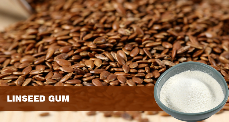
Flaxseed gum, with its natural properties and diverse health benefits, redefines the role of dietary fiber in modern health. From regulating the intestinal microbiome to optimizing systemic metabolism, its scientific value is increasingly being explored, serving as a bridge between daily diet and targeted healthcare. Embracing the principle of “prevention is better than cure,” this natural ingredient offers a safe and effective new option for health management.

Hydroxytyrosol: Natural protective shield for cellular oxidative damage

Within the microscopic world of our bodies, cells constantly carry out complex and orderly metabolic activities, a process that relies on oxygen. However, like a coin with two sides, while oxygen provides cells with energy, it also produces some unruly byproducts—reactive oxygen species (ROS), including superoxide anions and hydroxyl radicals. Under normal circumstances, cells possess a sophisticated antioxidant defense system that maintains a dynamic balance between ROS production and clearance, ensuring normal cellular function. However, when the body is attacked by adverse external factors, such as long-term exposure to environmental pollution and ultraviolet radiation, severe mental stress, or unhealthy lifestyle habits such as staying up late, smoking, excessive alcohol consumption, and an unhealthy diet, this balance can be disrupted, leading to excessive accumulation of ROS within cells.

Excessive ROS are extremely chemically active, acting like a swarm of rampaging “little demons,” attacking various biological macromolecules within the cell. Lipids are the first to be affected. Cell membranes are primarily composed of lipids. ROS react with lipids in a peroxidative reaction, disrupting the structural integrity of the cell membrane. The previously stable and orderly phospholipid bilayer becomes riddled with holes. This not only affects the cell membrane’s selective permeability, leading to an imbalance in the exchange of substances inside and outside the cell, but can also disrupt cellular signaling. Proteins are also vulnerable. ROS attack the amino acid residues of proteins, altering their spatial structure and rendering many enzymes inactive. Once enzymes involved in key physiological processes such as cellular metabolism, synthesis, and signaling are deactivated, normal cellular metabolism is disrupted. More seriously, ROS can directly affect the cell’s genetic material, DNA, causing DNA strand breaks, base modifications, and gene mutations. This is like scribbling on the cell’s “blueprint of life.” If the repair mechanism cannot correct these errors in a timely manner, it can lead to cell cancer or other serious genetic diseases.

Long-term oxidative stress can make the body like a dilapidated machine, with a variety of problems ensue. From an external perspective, skin aging is the most visible manifestation. The constant attack of free radicals on skin cells damages collagen and elastin fibers, leading to a gradual loss of skin elasticity and increasingly noticeable signs of aging, such as wrinkles, sagging, and dullness. Internally, the cardiovascular system bears the brunt of this damage. Oxidative damage triggers inflammation and lipid peroxidation, which contribute to the development of atherosclerosis. Oxidative modification of low-density lipoprotein cholesterol in the blood makes it more likely to deposit on blood vessel walls, forming plaques that block blood vessels and increase the risk of cardiovascular and cerebrovascular diseases such as coronary heart disease, myocardial infarction, and stroke. The nervous system is also severely affected. In neurodegenerative diseases such as Alzheimer’s and Parkinson’s, oxidative stress leads to nerve cell damage and death, gradually impairing cognitive and motor function. Furthermore, oxidative damage is closely linked to the development and progression of numerous chronic diseases, including diabetes and cancer, making it a silent threat to modern human health, quietly eroding our quality of life and lifespan.

Hydroxytyrosol: The Rise of a Natural Antioxidant

In the fierce battle against cellular oxidative damage, nature has provided us with a powerful ally: hydroxytyrosol. A polyphenolic compound extracted from a variety of plants, including olives and blueberries, hydroxytyrosol, while relatively low-profile in the plant kingdom, has gradually caught the attention of researchers thanks to its unique molecular structure and exceptional antioxidant properties, becoming a shining star in the antioxidant field.

Hydroxytyrosol’s molecular structure resembles a precisely designed antioxidant weapon. Its core structure is catechol, with two adjacent phenolic hydroxyl groups acting as powerful free radical scavengers, each with an exceptionally high hydrogen-donating capacity. When ROS wreak havoc within cells, hydroxytyrosol rapidly reacts with them, donating hydrogen atoms from its phenolic hydroxyl groups to the free radicals, stabilizing them by gaining electrons. Hydroxytyrosol then transforms into a relatively stable quinone compound, effectively interrupting the free radical chain reaction. This acts like a cold shower on the raging oxidative “flame,” effectively curbing the further spread of oxidative damage. This unique molecular structure endows hydroxytyrosol with exceptional free radical scavenging capabilities, making it stand out from traditional antioxidants. Research data shows that its antioxidant activity is over 10 times that of vitamin E and comparable to that of vitamin C. It performs exceptionally well in experiments scavenging a variety of free radicals, demonstrating its potent antioxidant efficacy.

In addition to its exceptional antioxidant capacity, hydroxytyrosol also boasts a significant advantage: high bioavailability. When consumed from foods or supplements rich in hydroxytyrosol, it is well absorbed in the gastrointestinal tract, enters the bloodstream, and is efficiently transported to various tissues and cells throughout the body, precisely targeting the sites of oxidative damage to exert its antioxidant effects. Furthermore, hydroxytyrosol’s low toxicity makes it safer and more reliable for use, unlike some synthetic antioxidants, which can carry potential side effects while delivering their effectiveness. It is precisely these excellent properties that make hydroxytyrosol show great application potential in many fields such as food, health products, cosmetics and even medicine. It has attracted many scientific researchers to conduct in-depth research on its mechanism of action and application value, and has become a cutting-edge research hotspot in the field of cellular oxidative protection, bringing us new hope and possibilities for fighting oxidative damage and maintaining physical health.


Multi-dimensional protection mechanism of hydroxytyrosol: precise protection from molecular to cellular level

Free radical capture: Cut off the “start bond” of the oxidation chain reaction​

Free radicals, as the “initiator” of oxidative damage, have extremely active chemical properties. Once produced in the cell, they will be like a ignited fuse, triggering a series of chain reactions, causing serious damage to the normal structure and function of the cell. With its unique molecular structure, hydroxytyrosol plays a key role as a “free radical capturer” in this antioxidant battle.​
In the molecular structure of hydroxytyrosol, phenolic hydroxyl groups are like “ammunition depots” that are on standby at any time, containing rich hydrogen atoms. When free radicals such as superoxide anions and hydroxyl radicals appear in the cells, hydroxytyrosol can react quickly, and its phenolic hydroxyl groups generously provide hydrogen atoms to bind to these radicals. This combination is not a simple combination, but a “neutralization” process. After the free radical obtains hydrogen atoms, its activity is greatly reduced, becoming relatively stable, and no longer has the ability to attack other molecules. After providing hydrogen atoms, hydroxytyrosol itself will be converted into relatively stable quinone compounds. This conversion process is like a clever “chemical magic”, which not only successfully eliminates free radicals, but also ensures that it does not produce new harmful substances, thus effectively blocking the occurrence of free radical chain reactions.​
Numerous in vitro experiments provide strong evidence for this powerful free radical capture capability of hydroxytyrosol. In common DPPH radical scavenging experiments, after a period of reaction, the detection results were surprising: its scavenging rate of DPPH radicals is as high as more than 85%. This means that in the experimental system, most of the DPPH radicals are successfully captured and neutralized by hydroxytyrosol, causing the originally strong oxidative free radicals to lose their activity. In the experiment of induced oxidative damage with H₂O₂, when hydroxytyrosol was added to the cell culture medium, advanced fluorescence probe technology detection found that the ROS level in the cells was significantly reduced. This intuitively shows that hydroxytyrosol can efficiently remove excessive ROS in cells, curbing the onset of oxidative damage from the source, just as when a fire just ignited, it will extinguish the flame in time to avoid the spread of the fire, thereby protecting the cells from the invasion of free radicals and maintaining the stability of the intracellular environment.​

Cell membrane protection: building a “antioxidation barrier” for lipid bilayers​

Cell membranes, as an important line of defense between cells and the external environment, play an indispensable role in maintaining the normal physiological function of cells. It is mainly composed of phospholipids, and the unsaturated fatty acids rich in phospholipids are of great significance to the fluidity and function of the cell membrane, but it also makes the cell membrane a “weak link” in ROS attack. When ROS meets unsaturated fatty acids on the cell membrane, a dangerous chain reaction of lipid peroxidation starts quietly.​
In the initial stage of the lipid peroxidation reaction, superoxide anions, hydroxyl radicals, etc. in ROS will take the lead in attacking unsaturated fatty acids, seizing hydrogen atoms from fatty acid molecules, and converting unsaturated fatty acids into lipid radicals. These lipid radicals have extremely high activity. They will quickly bind to the surrounding oxygen molecules to form peroxidized lipid radicals, which will continue to attack other unsaturated fatty acid molecules, and thus repeat, forming a constantly amplified chain reaction. During this process, peroxidized lipids will further decompose, producing a series of toxic products, among which malondialdehyde (MDA) is a representative harmful substance. The large-scale generation of MDA will cause serious damage to the structure of the cell membrane, the originally ordered phospholipid bilayer becomes chaotic, and the fluidity of the cell membrane will drop significantly. This not only affects the normal transport function of the cell membrane to substances, making it impossible for cells to normally ingest nutrients and excrete metabolic waste, but also interferes with the signaling pathways in the cells, leading to disorders in cell functions.​
Hydroxytyrosol, as the “loyal guardian” of the cell membrane, can block this dangerous lipid peroxidation process through dual mechanisms, building a solid “antioxidation barrier” for the cell membrane.​
Multiple phenolic hydroxyl groups in the hydroxytyrosol molecule play a key role. These phenolic hydroxyl groups are like powerful “free radical scavengers”. When ROS rages on the surface of the cell membrane, hydroxytyrosol can quickly approach and react with it, directly providing hydrogen atoms on the phenolic hydroxyl groups to ROS, thereby neutralizing superoxide anions, hydroxyl radicals, etc., and reducing their initial attack on membrane lipids. This direct removal of ROS inhibits the initiation of lipid peroxidation from the source, just like blocking the enemy’s attack at the first time, killing the danger in the cradle.​
Hydroxytyrosol can also take advantage of its fat-soluble characteristics to be successfully embedded in the lipid bilayer of the cell membrane. Once inside the cell membrane, it will closely bind to intermediate products produced during lipid peroxidation, such as lipid oxygen radicals. This binding effectively terminates the diffusion of the chain reaction and avoids large-area oxidation of membrane lipids. Because lipoxygen radicals are the key factor in the continuous progress of lipid peroxidation chain reaction, when hydroxytyrosol binds to lipoxygen radicals, it is equivalent to cutting off the “chain” of the chain reaction, making the reaction unable to continue to spread, thus protecting most areas of the cell membrane from oxidative damage.​
A large amount of experimental data strongly confirms the protective effect of hydroxytyrosol on cell membranes. In the oxidative stress model, researchers found through precise detection methods that the content of the lipid peroxidation product MDA in the cell membrane treated with hydroxytyrosol has significantly reduced, with a decrease of 40%-60%, and the membrane lipid hydroperoxide level has also decreased by more than 50%. These data intuitively show that hydroxytyrosol can significantly reduce the oxidative damage of membrane lipids, maintain the structural integrity and fluidity of the cell membrane, ensure that the cell membrane can perform its physiological functions normally, and provide a stable environment for the normal metabolism and life activities of cells.​

DNA and protein protection: a “molecular scalpel” that repairs oxidative damage

In the microscopic world of cells, DNA and proteins are the two “pillars” that maintain the normal function of cells. DNA, as a carrier of genetic information, accurately encodes various life activities instructions of cells, just like a detailed “life blueprint”, guiding the key processes of cell growth, division, and differentiation. Proteins are widely involved in various physiological and biochemical reactions in cells. From the synthesis and metabolism of substances to the transmission and regulation of signals, almost every cell activity is inseparable from the participation of proteins. However, when cells are threatened by oxidative stress, these two key molecules are also hard to survive and become targets of ROS attacks.​
When ROS meets DNA, it triggers a series of complex chemical reactions, one of the most common forms of damage is the production of the DNA oxidation product 8-hydroxydeoxyguanosine (8-OHdG). The emergence of 8-OHdG is like leaving a wrong mark on the “blueprint of life” of DNA, which interferes with the normal replication and transcription process of DNA. During DNA replication, DNA polymerase may misidentify 8-OHdG as a normal base, resulting in base mismatch, causing gene mutations in newly synthesized DNA strands. If this gene mutation cannot be repaired in time, as the cells divide, wrong genetic information will be continuously transmitted, which may cause serious consequences such as cell carcinoma and aging.​
Proteins are also severely affected by ROS. ROS attacks the amino acid residues of a protein and changes the spatial structure of the protein. The spatial structure of a protein is closely related to its function. Once the structure is destroyed, many enzyme proteins will lose their activity. For example, those enzymes involved in key steps in cell metabolism, such as hexokinases involved in sugar metabolism, lipases involved in lipid metabolism, etc., once inactivated, the energy supply and material metabolism of the cells will fall into chaos. Some proteins responsible for signaling will also be unable to transmit signals normally due to structural changes, resulting in abnormal cell responses to changes in internal and external environments, which will affect the normal physiological function of cells.​
Fortunately, hydroxytyrosol can repair and protect damaged DNA and proteins like an accurate “molecular scalpel”.​
Hydroxytyrosol exhibits strong protection against oxidative damage. It can reduce the attack on DNA by inhibiting the activity of DNA oxidase, thereby reducing the production of oxidative products such as 8-OHdG. It was like putting on a layer of “protective clothing” for DNA, preventing further damage to ROS. Hydroxytyrosol can also regulate some repair mechanisms in cells and promote the repair of damaged DNA. It can activate the activity of certain DNA repair enzymes, allowing cells to timely identify and repair damage sites on the DNA strand, ensure the accuracy and completeness of genetic information, and maintain the normal genetic function of cells.​
In terms of protein protection, hydroxytyrosol also has a unique mechanism of action. It regulates the expression of heat shock protein (HSP). Heat shock proteins are an important class of proteins in cells. They are like the “first aid team” of cells. When proteins are damaged, HSPs can respond quickly and bind to damaged proteins, helping them restore the correct spatial structure and regain their activity. Hydroxytyrosol enhances the cell’s ability to repair damaged proteins by upregulating HSP expression. For those severely damaged proteins that cannot be repaired, hydroxytyrosol also promotes their degradation and prevents these oxidative protein aggregates from accumulating in the cells. Because oxidative protein aggregates not only occupy space in the cell and affect the functions of other normal proteins, they may also trigger inflammatory responses in the cell, further damaging cell health. By removing these “garbage” proteins in a timely manner, hydroxytyrosol effectively maintains the homeostasis of proteins in the cell, ensures the stability of the functions of biological macromolecules in the cell, and ensures the normal progress of various physiological activities of the cell.​

Regulation of oxidative stress pathway: Activate endogenous antioxidant defense system​

In the cell’s antioxidant defense system, in addition to exogenous antioxidants like hydroxytyrosol, the cell itself also has a precise endogenous antioxidant defense system. This system is like a “remaining army” inside the cells, always protecting the cells from the threat of oxidative damage. The magic of hydroxytyrosol is that it can not only directly eliminate free radicals, but also enhance the cell’s own antioxidant ability by activating key signaling pathways in the cells, and build a dual protection system of “exogenous scavenging + endogenous enhancement” to provide cells with all-round protection.​
Among the antioxidant-related signaling pathways in many cells, the Nrf2/ARE signaling pathway plays a core role and can be called the “commander” of the intracellular antioxidant defense system. Nrf2 is a key transcription factor that usually binds to the Keap1 protein, is inactive, and is “bound” in the cytoplasm. When cells are stimulated by oxidative stress, oxidants such as ROS react with the Keap1 protein, causing its structure to change, thereby releasing Nrf2. The released Nrf2 is like a warrior untied, quickly entering the nucleus and combining with the antioxidant reaction element (ARE). This binding is like a key inserted into the correct keyhole, initiating the transcription and expression of a series of antioxidant enzyme genes.​
Superoxide dismutase (SOD), which can convert superoxide anions into oxygen and hydrogen peroxide, effectively eliminate superoxide anions in cells, is the first line of defense for cells to resist oxidative damage. Glutathione peroxidase (GSH-Px) can use reduced glutathione (GSH) to reduce hydrogen peroxide to water, further eliminating oxidative products in cells and protecting cells from toxic damage to hydrogen peroxide. There is also catalase (CAT), which can effectively decompose hydrogen peroxide and maintain the redox balance in the cells. These antioxidant enzymes work together in the cell to form a powerful antioxidant defense line, which can promptly remove ROS produced in the cell and maintain the stability of the intracellular environment.​
And hydroxytyrosol is the “key key” to activate the Nrf2/ARE signaling pathway. When hydroxytyrosol enters the cell, it can interact with some molecules in the cell, triggering a series of signaling cascades, which ultimately leads to Nrf2 being released from the constraints of the Keap1 protein and entering the nucleus to bind to ARE, thereby upregulating the gene expression of antioxidant enzymes such as SOD and GSH-Px. The results of preclinical studies are exciting. Experimental data show that the intracellular GSH level of cells treated with hydroxytyrosol has increased by more than 30%. As an important antioxidant substance in cells, its increased levels mean that the antioxidant capacity of cells is significantly enhanced. More GSH can provide adequate substrate for antioxidant enzymes such as GSH-Px, allowing it to more effectively remove ROS in cells. The enhanced activity of antioxidant enzymes such as SOD and GSH-Px also enables them to catalyze redox reactions more efficiently and eliminate harmful substances such as superoxide anions and hydrogen peroxide in the cells in a timely manner.​
By activating the Nrf2/ARE signaling pathway, hydroxytyrosol not only enhances the cell’s own antioxidant ability, but also forms a synergistic effect with its own exogenous antioxidant effects. In the face of oxidative stress, on the one hand, hydroxytyrosol directly exerts its free radical scavenging ability to quickly neutralize the excessive ROS produced in the cells; on the other hand, it activates the endogenous antioxidant defense system in the cells, allowing the cells themselves to continuously produce antioxidant enzymes and continuously eliminate ROS, forming a comprehensive and multi-level antioxidant protection network. The construction of this dual protection system greatly improves the ability of cells to resist oxidative damage and provides more reliable guarantees for the health of cells. It is like building a solid fortress for cells, allowing them to remain stable in the “storm” of oxidative stress and maintain normal physiological functions and life activities.


Multi-dimensional verification of hydroxytyrosol antioxidant damage

In vitro experiments: from cellular models to molecular evidence​

In the field of scientific research, in vitro experiments are an important cornerstone for exploring the mechanism of action of substances, and research on the antioxidant damage ability of hydroxytyrosol is no exception. Researchers have carefully constructed a variety of cell models to deeply analyze the antioxidant effects of hydroxytyrosol from different angles, providing conclusive molecular evidence for its protective effect at the cellular level.​
In the classic HEK – 293 cell model experiment, researchers cleverly used hydrogen peroxide (H₂O₂) to induce oxidative stress in cells, mimicking the oxidative damage state of cells when they are attacked by adverse external factors. Subsequently, different concentrations of hydroxytyrosol were added to the cell culture medium for pretreatment. The experimental results are amazing. With the increase in hydroxytyrosol concentration, the reactive oxygen species (ROS) levels in cells show a significant dose-dependent decrease trend. When the hydroxytyrosol concentration reaches 50μM, its inhibition rate on H₂O₂-induced ROS is as high as 55%. This data visually shows that hydroxytyrosol can efficiently remove excessive ROS in cells and curb the occurrence of oxidative damage from the source. The researchers were also surprised to find that the content of intracellular reduced glutathione (GSH) after hydroxytyrosol treatment increased significantly, with an increase of 25%. As an important antioxidant substance in cells, its content increases means that the antioxidant capacity of cells has been significantly enhanced, which can better resist the attack of ROS and maintain the stability of the intracellular environment. This series of experimental results fully demonstrate the direct antagonism of hydroxytyrosol on oxidative stress, providing strong support for its application in cell oxidative protection.​
In order to further explore the protective effect of hydroxytyrosol on skin cells, researchers have constructed a more complex three-dimensional skin model. This model can more realistically simulate the structure and physiological functions of human skin, providing an ideal platform for studying the antioxidant effect of hydroxytyrosol in deep skin cells. In the experiment, a three-dimensional skin model was treated for 4 weeks using a preparation containing 0.5% hydroxytyrosol. Through advanced detection technology analysis, it was found that the DNA oxidative damage markers in epidermal cells were significantly reduced, with a decrease of 35%. This shows that hydroxytyrosol can effectively protect the DNA of skin cells from oxidative damage and maintain the stability of genetic material. In the dermis, the degree of oxidative fracture of collagen fibers is also significantly reduced, reducing by 40%. Collagen fibers are the key structure for maintaining skin elasticity and firmness. The inhibitory effect of hydroxytyrosol on its oxidative breakage fully demonstrates its protective effect on deep skin cells, which can fundamentally delay the aging process of skin and provide solid theoretical basis and experimental support for the development of new antioxidant skin care products.​

Human body research: Quantification of the effects of skin care to health intervention​

In vitro experiments provide preliminary evidence for the antioxidant capacity of hydroxytyrosol, but rigorous human research is also needed to truly evaluate its efficacy in the human body. In recent years, researchers have carried out a series of carefully designed human experiments around hydroxytyrosol. From clinical experiments in the field of skin care to health intervention research on sub-healthy people, they have gradually quantified the antioxidant effect of hydroxytyrosol in the human body, allowing us to more intuitively recognize its important value in maintaining human health.​
In the field of skin care, a representative clinical trial focused on the repair of skin photoaging damage by 1% hydroxytyrosol. A group of subjects with obvious signs of photoaging on the facial skin were selected and allowed to use the essence continuously for 8 weeks. During the experiment, the researchers conducted detailed monitoring of various indicators of the subject’s facial skin through professional skin detection instruments. The results were delightful, and after 8 weeks of use, the malondialdehyde (MDA) level in the subject’s facial skin decreased significantly, reaching 28%. As a product of lipid peroxidation, the reduction in its level directly reflects that the oxidative damage suffered by skin cells is effectively reduced. Antioxidant enzyme activity in the stratum corneum has also been significantly improved, increasing by 20%, indicating that the skin’s own antioxidant defense ability has been enhanced. The depth of fine lines on the subject’s face also improved significantly, with an average reduction of 15%. These quantitative data fully confirm the repair effect of hydroxytyrosol on photoaging damage. It can effectively improve the oxidative state of the skin and enhance the antioxidant ability of the skin, thereby achieving the effect of delaying skin aging and improving skin texture, bringing new hope to the majority of people who pursue skin health and beauty.​
In addition to the skin care field, hydroxytyrosol has also shown significant results in health intervention in sub-healthy people. Researchers conducted a study in sub-healthy populations, where subjects were given daily supplements of 50mg of hydroxytyrosol for 3 months. After the experiment, the subject’s blood samples were comprehensively analyzed. The results showed that oxidative stress indicators in the serum, such as thiobarbituric acid reactant (TBARS), decreased significantly, by 18%. TBARS is an important indicator to measure the degree of lipid peroxidation in the body. The decline in its level indicates that hydroxytyrosol can effectively inhibit the oxidative stress response in the body and reduce the occurrence of lipid peroxidation. The stability of the erythrocyte membrane has also been significantly improved, increasing by 22%. The stability of the red blood cell membrane is crucial to maintaining the normal function of red blood cells. Hydroxytyrosol can enhance the stability of the red blood cell membrane, indicating that it can protect red blood cells from oxidative damage and maintain the normal physiological function of the blood system. These research results fully demonstrate the systemic effect of hydroxytyrosol in the body’s antioxidant. It can enhance the antioxidant capacity of various tissues and organs in the body by regulating the redox balance in the body, thereby improving the sub-health state and improving the overall health level of the human body, providing new ideas and methods for preventing and treating various chronic diseases caused by oxidative stress.​

Comparative advantages with traditional antioxidants​

In the large family of antioxidants, hydroxytyrosol has shown many significant advantages compared with traditional antioxidants such as vitamin C and vitamin E, making it stand out in the field of antioxidants and become the focus of attention of scientific researchers and consumers.​
From the perspective of fat-soluble antioxidant ability, hydroxytyrosol has a strong membrane-targeted protection effect, which is a significant feature that distinguishes it from traditional antioxidants. The cell membrane is mainly composed of lipids, it is the first line of defense for cells to resist external oxidative damage, and it is also the main target of ROS attack. The lipid solubility of hydroxytyrosol allows it to easily integrate into the lipid bilayer of the cell membrane and closely bind to the membrane lipid, thereby providing comprehensive protection to the cell membrane. When ROS strikes, hydroxytyrosol can quickly capture free radicals, block the chain reaction of lipid peroxidation, and effectively prevent the structure and function of the cell membrane from being destroyed. As a water-soluble antioxidant, vitamin C is mainly found in the extracellular fluid and the water-soluble environment in the cells, and it is difficult to directly protect the cell membrane; although vitamin E has a certain degree of fat-soluble, when exerting antioxidant effects, it often needs to work with other antioxidants to achieve the best effect, and its membrane-targeted protection ability alone is relatively weak.​
In terms of free radical scavenging rate, hydroxytyrosol performs well, and can quickly react with free radicals in a nanosecond time to scavenge them. This rapid reaction rate allows hydroxytyrosol to quickly capture free radicals when they are just produced, avoiding further damage to the cells by free radicals. By contrast, vitamin C free radical scavenging rate is milliseconds, while vitamin E takes seconds to react with free radicals. In the emergency situation of oxidative stress, the nanosecond rapid response ability of hydroxytyrosol is particularly important and can provide more timely and effective protection for cells.​
Bioavailability is also one of the important indicators to measure the effectiveness of antioxidants. Hydroxytyrosol has obvious advantages in this regard. Its absorption rate in the intestine is as high as more than 90%. It can be absorbed and utilized by the human body quickly, enter the blood circulation system, and transported to various tissues and cells of the body to exert its antioxidant effect. The bioavailability of vitamin C is affected by various factors such as dosage form and intake dose, and fluctuates greatly in different situations; vitamin E is a fat-soluble vitamin, and its absorption requires the participation of fat. If the intake of fat in the diet is insufficient, it will affect its absorption effect, resulting in a lower bioavailability.​
In addition to the above direct antioxidant performance advantages, hydroxytyrosol also has significant anti-inflammatory synergistic effects. During the inflammatory response, the nuclear factor-κB (NF-κB) pathway plays a key regulatory role, and its activation will lead to the release of a series of inflammatory factors, aggravating oxidative damage to cells. Hydroxytyrosol can reduce the production of inflammatory factors by inhibiting the activation of the NF-κB pathway, thereby reducing the damage to cells by the inflammatory response. This anti-inflammatory synergistic effect cooperates with its antioxidant effects to form a more comprehensive cell protection mechanism. Although vitamin C and vitamin E also have certain antioxidant and anti-inflammatory effects, their ability to regulate inflammatory signaling pathways is relatively limited, and they are mainly based on a single free radical scavenging effect.​
Overall, hydroxytyrosol has shown superior performance than traditional antioxidants in many aspects such as fat-soluble antioxidant capacity, free radical scavenging rate, bioavailability, and anti-inflammatory synergistic effects. These advantages make hydroxytyrosol have huge application potential in the field of antioxidant. Whether in skin care products, health products or medicine, it is expected to play an important role, providing people with more efficient and comprehensive antioxidant protection, and becoming a powerful “guardian” to protect human health.


Full-scene layout from precise skin care to disease prevention​

Functional skin care products: Create a new benchmark for “cell-level anti-aging”

On the road to pursuing youthful skin, consumers’ expectations for the efficacy of skin care products have been rising, from basic moisturizing and whitening to the core of fighting skin aging – cell oxidation damage. With its excellent antioxidant properties, Hydroxytyrosol is leading the functional skin care industry to a new journey of “cell-level anti-aging”, bringing consumers an unprecedented skin care experience.​
Antioxidant essence: High concentration of hydroxytyrosol (1%-2%) combined with hyaluronic acid, deep penetration repairs ultraviolet-induced oxidative damage, suitable for photoaging people.​
For people who have been exposed to the sun for a long time, ultraviolet rays are the number one “culprit” for skin aging. UVA and UVB in ultraviolet rays can penetrate the skin surface and stimulate the production of a large amount of reactive oxygen species (ROS) in cells. These ROSes will attack the DNA, proteins and lipids of skin cells, causing photoaging such as sagging, wrinkles, and tints. The antioxidant essence rich in 1%-2% high concentration of hydroxytyrosol is like creating a strong “antioxidant shield” for the skin. When the essence is applied to the skin, hydroxytyrosol quickly binds to ROS in the cells with its powerful free radical capture ability, blocks the oxidative chain reaction and reduces the damage of UV light to the skin at its root. The addition of hyaluronic acid is even more powerful. It has super moisturizing ability and can form a moist environment inside the skin, promote deep penetration of hydroxytyrosol, so that it can reach damaged cells more accurately and repair ultraviolet-induced oxidative damage. After long-term use of this essence, the photoaging skin can gradually restore elasticity, the wrinkles become lighter, the spots fade, and the brilliance will be reappear.​
Barrier Repair Cream: 0.5% hydroxytyrosol combines ceramide to enhance cell membrane stability and improve redness and dryness caused by oxidative stress of sensitive skin.​
People with sensitive skin are often troubled by problems such as redness, dryness, and tingling of the skin. This is mainly due to the damage to the skin barrier, the resistance to external stimulation is reduced, and the oxidative stress is intensified, further destroying the healthy state of the skin. Barrier repair cream containing 0.5% hydroxytyrosol brings good news to sensitive skin. Hydroxytyrosol can regulate the redox balance in cells, reduce the damage to the cell membrane by oxidative stress, and enhance the stability of the cell membrane. Ceramide, as an important part of the skin barrier, works in concert with hydroxytyrosol, can fill the “loopholes” of the skin barrier and repair damaged skin barriers. When face cream is applied to sensitive skin, it can quickly form a protective film on the surface of the skin to block external stimulation, and at the same time penetrate deep into the bottom layer of the skin, promote self-repair of skin cells, improve skin redness and dryness, and gradually restore the sensitive skin to a healthy and stable state.​
Sunscreen synergist: Added to sunscreen products to jointly resist UVA/UVB-induced ROS bursts and reduce photooxidation damage.​
In the field of sun protection, traditional sun protection products mainly absorb and reflect ultraviolet rays through physical or chemical means. However, even if sun protection products are used, it is still difficult for the skin to completely avoid the damage of ultraviolet rays, especially photooxidation damage caused by UVA and UVB. Adding hydroxytyrosol to sunscreen products has brought new ideas for sunscreen. Hydroxytyrosol can work in concert with sunscreens, not only enhances the protection of UV light, but also effectively resists ROS bursts caused by UVA/UVB. When UV rays hit the skin, hydroxytyrosol quickly exerts its antioxidant effect, clears ROS caused by UV excitation, reduces photooxidation damage, and provides the skin with more comprehensive sun protection. This sunscreen product containing hydroxytyrosol can not only prevent sunburn and tanning, but also fundamentally delay the skin’s photoaging process, allowing the skin to remain healthy and young in the sun.​

Health products and functional foods: Construct an antioxidant defense line in the body​

In the fast-paced modern life, people face increasing pressure, environmental pollution and bad living habits. These factors have led to the continuous increase in the level of oxidative stress in the body, posing a potential threat to physical health. As a natural antioxidant, hydroxytyrosol has injected new vitality into the fields of health products and functional foods with its excellent antioxidant properties, becoming a key force in building an antioxidant line in the body.​
Dietary supplements: Use microencapsulation technology to improve stability, develop 50-100mg/particle hydroxytyrosol capsules to target high-pressure people, smokers and other high-risk groups for oxidative stress.​
For high-pressure people and smokers, their bodies are in a state of stress for a long time. The harmful substances and mental stress in cigarettes will cause a large amount of free radicals to be produced in the body, causing a sharp increase in the level of oxidative stress, increasing the risk of chronic diseases such as cardiovascular diseases and cancer. In order to meet the urgent need for anti-oxidation in this group of people, researchers have developed 50-100mg/particle hydroxytyrosol capsules using advanced microencapsulation technology. Microencapsulation technology is like putting a “protective clothing” on hydroxytyrosol, which can effectively improve its stability in the gastrointestinal tract, prevent it from being degraded during digestion, and ensure that hydroxytyrosol can be absorbed into the blood circulation system intact. When high-pressure people or smokers take this capsule, hydroxytyrosol quickly acts to eliminate excessive free radicals in the body, regulate signal pathways related to oxidative stress, reduce the damage caused by oxidative stress to the body, help them maintain their health, and reduce various health risks caused by oxidative stress.​
Functional food: Added to olive oil, nut products or beverages to achieve preventive intervention of “antioxidation and rejuvenation” through daily diet.​
In addition to dietary supplements, adding hydroxytyrosol to functional foods provides people with a more convenient and natural way to antioxidant. Increasing the content of hydroxytyrosol in olive oil not only improves the antioxidant quality of olive oil, but also allows people to easily intake hydroxytyrosol during daily cooking and consumption, and enjoy the antioxidant benefits it brings. Nut products are rich in a variety of nutrients. After the addition of hydroxytyrosol, their antioxidant ability is further enhanced and they have become a high-quality choice for people’s healthy snacks. Incorporating hydroxytyrosol into the beverage, whether it is juice, tea or functional beverages, provides consumers with the possibility of antioxidant at any time and anywhere. By integrating hydroxytyrosol into these daily diets, people can unconsciously achieve “antioxidation and youthful” preventive intervention, protect their health from every bit of daily life, reduce the risk of various chronic diseases caused by oxidative stress, and let health and deliciousness go hand in hand.​

Medical field: Exploration from assisted treatment to disease prevention​

In the field of medicine, hydroxytyrosol is gradually moving from the role of auxiliary treatment to the forefront of disease prevention with its strong antioxidant and cell protection capabilities, bringing new hope to overcome modern medical problems such as cardiovascular and neurodegenerative diseases. Through in-depth research on the mechanism of action of hydroxytyrosol, researchers found that it plays an important role in multiple key physiological processes, providing a solid theoretical basis for the development of new therapeutic strategies and preventive measures.​
Cardiovascular protection: Hydroxytyrosol improves nitric oxide (NO) bioavailability and reduces the risk of atherosclerosis by inhibiting oxidative damage to vascular endothelial cells. Related clinical studies have shown that it can reduce serum ox-LDL levels in patients with coronary heart disease by 12%.​
Cardiovascular disease is one of the major killers threatening human health worldwide, with atherosclerosis being the key pathological basis for its onset. Oxidative stress plays an important role in the occurrence and development of atherosclerosis. It can cause damage to vascular endothelial cells, promote inflammatory responses and lipid peroxidation, and then form atherosclerotic plaques, block blood vessels, and cause serious cardiovascular events such as coronary heart disease and myocardial infarction. As a powerful antioxidant, hydroxytyrosol can directly act on vascular endothelial cells, remove excess reactive oxygen species (ROS) in cells, inhibit the damage to cells by oxidative stress, and maintain the normal function of vascular endothelial cells. It also regulates the bioavailability of nitric oxide (NO), an important vasodilator that can dilate blood vessels, lower blood pressure, and improve blood circulation. Hydroxytyrosol effectively reduces the risk of atherosclerosis by promoting the synthesis and release of NO, enhancing the diastolic function of blood vessels, reducing platelet aggregation and thrombosis. The relevant clinical research results are exciting. In the study of patients with coronary heart disease, the level of oxidized low-density lipoprotein (ox-LDL) in the patients’ serum decreased by 12% after supplementing with hydroxytyrosol. ox – LDL is an important risk factor for atherosclerosis. The reduction in its level means that the risk of cardiovascular events has been effectively reduced in patients. This fully demonstrates the significant efficacy of hydroxytyrosol in cardiovascular protection and opens up new avenues for the prevention and treatment of cardiovascular diseases.​
Neuroprotection: For Alzheimer’s disease, it inhibits neuronal oxidative apoptosis by clearing β-amyloid-induced ROS in the brain, and animal experiments have been shown to improve memory cognitive function.​
As a common neurodegenerative disease, Alzheimer’s disease seriously affects the quality of life and cognitive function of patients, and brings a heavy burden to the family and society. At present, the pathogenesis of Alzheimer’s disease has not been fully understood, but a large number of studies have shown that oxidative stress plays a key role in its onset. Abnormal aggregation of amyloid beta in the brain will induce the production of a large number of ROS, which will attack neurons, leading to oxidative damage and apoptosis of neurons, and thus trigger symptoms such as memory loss and cognitive impairment. Hydroxytyrosol has shown great potential in neuroprotection, which can cross the blood-brain barrier and enter the brain, directly clearing ROS induced by beta amyloid induced, alleviating the damage to neurons by oxidative stress. By regulating signaling pathways in cells, hydroxytyrosol can also inhibit oxidative apoptosis of neurons and protect their survival and function. In animal experiments, researchers supplemented hydroxytyrosol to mice with Alzheimer’s disease model. After a period of observation, the memory and cognitive function of the mice was significantly improved, and their performance in the maze experiment was better and they could find an outlet faster. This shows that hydroxytyrosol can effectively alleviate the symptoms of Alzheimer’s disease and provides new hope for the treatment and prevention of Alzheimer’s disease. Although the application of hydroxytyrosol in the medical field is still in the research and exploration stage, with the continuous deepening of research and the continuous advancement of technology, I believe that in the near future, it will bring good news to more patients and become a powerful weapon for the treatment and prevention of these diseases.

Redefine cell-grade antioxidant protection

The emergence of hydroxytyrosol marks the shift of antioxidant research from “single radical scavenging” to “cellular structure-function-pathways”. Through a multi-dimensional mechanism targeting cell membranes, DNA, and signaling pathways, it not only delays visible aging characteristics, but also fundamentally builds a line of defense for cells to resist oxidative damage. With the advancement of synthetic biology and delivery technology, this natural active ingredient is taking science as the cornerstone, opening up new possibilities from skin health to overall anti-aging, providing a promising solution to the challenges of oxidative stress in modern society.

Bakuchiol: From Collagen Protection to a New Paradigm for Anti-Aging

In the long quest for youthful-looking skin, natural active ingredients are gradually emerging as shining stars in the skincare industry. Bakuchiol, a natural polyphenol compound derived from plants such as Epimedium and Psoralea corylifolia, is a rising star, sparking a wave of interest in the anti-aging field thanks to its unique advantages.

Compared to traditional retinoids, bakuchiol is significantly less irritating, making it a boon for those with sensitive skin. Those with sensitive skin often exercise caution when choosing skincare products, fearing they might cause skin irritation. The emergence of bakuchiol allows them to pursue anti-aging benefits with confidence. Furthermore, bakuchiol’s high bioactivity allows it to more effectively exert its anti-aging effects, repairing and improving the skin from multiple perspectives.

From its mechanism of action, bakuchiol forms a comprehensive anti-aging system. It penetrates deep into the dermis, strengthening and repairing the collagen network. Collagen fibers in the dermis act as the “reinforcement” of the skin, providing support and elasticity. Bakuchiol promotes collagen synthesis, increasing the density and integrity of collagen fibers, thereby increasing the thickness of the dermis, plumping up the skin, and reducing the appearance of static lines. It also improves the function of elastic fibers, enhancing skin elasticity and toughness, allowing the skin to more quickly recover and smooth out after muscle contraction, effectively alleviating dynamic lines. Furthermore, bakuchiol optimizes the epidermal barrier by promoting normal differentiation of keratinocytes, maintaining the integrity of the stratum corneum, and reducing water loss. It also upregulates the expression of natural moisturizing factors, increasing stratum corneum hydration, making the skin softer and smoother, and reducing the appearance of dryness-related fine lines.

As we age, our skin gradually loses its radiance and elasticity, leading to wrinkles and sagging. The core reason behind this is a structural crisis in collagen fibers.

The collagen fibers in the skin primarily consist of type I and type III collagen, which are key components for maintaining skin structure and elasticity. However, over time and due to environmental influences such as UV radiation, pollution, and stress, the collagen fibers in the skin begin to gradually disappear. Decreased fibroblast activity leads to reduced collagen synthesis, while increased matrix metalloproteinase activity accelerates collagen degradation. This shift in activity reduces the supporting capacity of the dermis, leading to sagging skin and increasingly pronounced static lines like nasolabial folds and frown lines.

At the same time, elastic fibers are damaged in this process. Made of elastin, elastic fibers give the skin its elasticity and resilience. When these fibers are damaged, the skin cannot recover quickly enough from repeated muscle contraction, leading to the deepening of dynamic lines like crow’s feet and forehead wrinkles. Furthermore, the epidermal barrier function weakens with aging. Insufficient hydration in the stratum corneum leads to dry, rough skin, further accentuating fine lines, forming dryness-related fine lines and making the skin appear older.

Facing this severe aging challenge, bakuchiol has stepped up to the plate and launched a “defense” campaign for collagen fibers. It precisely addresses multiple collagen fiber injuries through a triple pathway of “synthesis-protection-repair.” Bakuchiol activates collagen synthesis signaling pathways in fibroblasts, such as the ERK/MAPK pathway, directly upregulating collagen gene expression, promoting collagen synthesis, and increasing collagen fiber density in the dermis. Simultaneously, it inhibits the activity of collagen-degrading enzymes (MMPs), reducing the breakdown of existing collagen and achieving a positive balance of “synthesis > degradation.” This strengthens the collagen network, fills the skin’s “support gaps,” and reduces the appearance of static lines. To protect and repair elastic fibers, bakuchiol utilizes its antioxidant properties to reduce free radical oxidative damage to elastin. It also upregulates proteins associated with elastic fibers, such as lysyl oxidase, promoting cross-linking and stabilization of elastic fibers, enhancing skin’s “resilience” and alleviating the appearance of dynamic lines. In addition, bakuchiol improves the epidermal barrier function, promotes the normal differentiation of keratinocytes, maintains the integrity of the stratum corneum, reduces water loss, upregulates the expression of natural moisturizing factors, increases the water content of the stratum corneum, reduces dry fine lines, and restores the skin to smoothness and fineness.

Targeting Collagen Fibers for Anti-Aging: From Molecular to Structural Repair

Bakuchiol’s anti-aging mechanism is like a sophisticated “molecular repair symphony,” targeting collagen fibers from multiple dimensions to achieve comprehensive protection from the molecular level to structural repair.

Strengthening the Collagen Network: Reshaping the “Supporting Framework” of the Dermis

1. Promoting Collagen Synthesis and Reversing Collagen Loss

Bakuchiol acts as a “vital stimulator” for fibroblasts, precisely activating the ERK/MAPK signaling pathway in fibroblasts. This signaling pathway acts as a “production instruction channel” within the cell. Once activated, it rapidly upregulates the expression of the COL1A1 and COL3A1 genes. These genes act as the “blueprints” for collagen synthesis, and their increased expression significantly promotes the synthesis of type I and type III collagen. Clinical research data is impressive, showing that after a period of using skincare products containing bakuchiol, collagen density in the dermis increased by over 30%. This is like adding more “reinforcement” to the skin’s “supporting skeleton,” making it stronger and more plump.

At the same time, bakuchiol acts as a “protector” of collagen fibers, inhibiting the activity of matrix metalloproteinases (MMP-1 and MMP-3). These enzymes are collagen destroyers. Under normal circumstances, their activity remains within a certain range, maintaining collagen metabolism. However, with aging and environmental influences, their activity increases, accelerating collagen degradation. The presence of bakuchiol effectively curbs this destruction, causing collagen synthesis to outpace degradation, creating a positive balance of “synthesis > degradation.” As a result, static lines that deepen due to collagen loss, such as nasolabial folds and frown lines, are seemingly lifted from within, becoming less noticeable.

2. Optimizing Collagen Fiber Arrangement and Improving Skin Plumpness

Bakuchiol not only focuses on collagen synthesis and degradation, but also strives to optimize collagen fiber arrangement. Like a meticulous designer, it strengthens the orderly cross-linking of collagen fibers, making the connections between them tighter and more orderly. This orderly arrangement strengthens the structure of the dermis and increases its thickness. Skin elasticity parameters, such as the R2 elasticity index, are significantly improved, resulting in a firmer, more plump complexion. Skin that once appeared sagging due to collagen loss is now revitalized, and deep wrinkles are gradually diminished, restoring a youthful glow.

Protecting Elastic Fibers: Restoring Skin’s “Resilience”

1. Antioxidant Protection, Blocking Elastin Damage

In daily life, our skin is constantly threatened by free radicals. Ultraviolet radiation, environmental pollution, and unhealthy lifestyle habits can all contribute to the production of free radicals within the skin. These free radicals act like a raging “destroyer,” attacking elastic fibers and causing oxidative breakdown. Bakuchiol, however, is a valiant “antioxidant warrior,” effectively scavenging free radicals like superoxide anions, building a strong “protective wall” for elastic fibers. Bakuchiol also inhibits the activity of elastin-degrading enzymes, such as MMP-9. MMP-9 is a natural enemy of elastin, breaking down elastin and causing damage to elastic fibers. Bakuchiol’s inhibition of MMP-9 effectively reduces damage to elastic fibers from external stimuli such as UV rays and pollution. This allows the skin to better maintain its elasticity in the face of various external challenges, slowing the progression of dynamic lines such as crow’s feet and forehead wrinkles, resulting in a more natural and youthful smile.

2. Promotes Elastic Fiber Repair and Cross-linking

In addition to providing protection, bakuchiol actively participates in the repair and cross-linking of elastic fibers. It does so by upregulating the expression of lysyl oxidase (LOX), providing critical support for the repair and stabilization of elastic fibers. LOX acts as the “glue” of elastic fibers, promoting the cross-linking and maturation of elastin precursors (tropoelastin) and enhancing the stability of the elastic fiber network.

When we make various facial expressions, our skin is repeatedly subjected to muscle contractions. If elastic fibers are damaged, the skin struggles to recover quickly after facial movements, and dynamic lines gradually transform into static lines. Bakuchiol helps the skin recover more quickly after repeated facial movements, reducing the transition from dynamic lines to static lines. This allows the skin to maintain its resilience, ensuring wrinkles are never a problem, whether you’re laughing heartily or raising your eyebrows in surprise.

Epidermal Barrier Optimization: A Hydrating Base for Anti-Wrinkle Aids

1. Enhances the Stratum Corneum’s Moisture Retention

The epidermis is like a castle, and the stratum corneum is its “walls.” Its moisture retention directly affects the skin’s hydration level. Bakuchiol acts like a diligent “craftsman,” promoting the differentiation of keratinocytes and strengthening the structure of the stratum corneum. It also upregulates the expression of natural moisturizing factors such as filaggrin and loricrin. These moisturizing factors act like “little sponges,” absorbing and retaining moisture, increasing the stratum corneum’s water content by 25%. When the stratum corneum is well-hydrated, the skin becomes soft and smooth, and superficial fine lines caused by dryness, such as those around the eyes, are reduced. Imagine, for example, once dry, rough skin becomes hydrated and plump with bakuchiol, and fine lines gradually disappear, giving the skin a healthy, radiant glow, as if well-hydrated.

2. Repairs barrier function and reduces external irritants

Bakuchiol also supports the integrity of the epidermal barrier by regulating the expression of tight junction proteins (such as occludin and claudin-1). These tight junction proteins are like the “bricks” of a wall; their tight connections determine the strength of the epidermal barrier. Bakuchiol upregulates the expression of these proteins, strengthening the epidermal barrier and reducing transepidermal water loss (TEWL).

This makes it difficult for external irritants, such as UV rays, pollutants, and allergens, to penetrate the epidermal barrier and indirectly damage the collagen fibers in the dermis. Bakuchiol provides comprehensive protection for the skin, from internal moisturizing to external defense, keeping it youthful in a healthy environment.

Anti-inflammatory and Antioxidant Effects: Blocking Photoaging and Chronic Inflammation Pathways

1. Scavenging Free Radicals and Inhibiting Oxidative Stress

Oxidative stress plays a crucial role in the aging process of the skin. Ultraviolet radiation, environmental pollution, and other factors can cause the generation of large amounts of free radicals in the skin. These free radicals attack the DNA, proteins, and lipids of skin cells, causing severe damage to collagen fibers. Bakuchiol, with its potent antioxidant capacity, is a “killer” of free radicals.

In vitro experiments have shown that bakuchiol has a scavenging rate of over 85% for DPPH free radicals. When skin is exposed to UVB radiation, a large amount of reactive oxygen species (ROS) is generated within cells, and bakuchiol can significantly reduce these ROS levels. It acts like a “free radical vacuum,” scavenging these harmful free radicals, reducing oxidative stress damage to collagen DNA, thereby delaying the formation of photoaging wrinkles. Let’s enjoy the sunshine without worrying about premature wrinkles.

2. Inhibits the Inflammatory Cascade

Chronic inflammation is also a contributing factor to skin aging, leading to increased release of inflammatory factors such as IL-6 and TNF-α. These inflammatory factors activate collagenase, accelerating collagen degradation, while also affecting fibroblast activity and reducing collagen synthesis. Bakuchiol effectively inhibits the inflammatory cascade by regulating the NF-κB signaling pathway.

It acts as a brake on the inflammatory response, reducing the release of pro-inflammatory factors such as IL-6 and TNF-α, lowering inflammation-mediated collagenase activation, and protecting collagen fibers from the “depletion” of chronic inflammation. Bakuchiol can significantly improve wrinkles caused by acne and rough skin caused by photoaging. It keeps skin healthy and youthful, free from the troubles of inflammation.


From Skincare to Comprehensive Anti-Aging

Beauty and Skincare: Diversified Product Form Development

Bakuchiol has demonstrated tremendous potential in the beauty and skincare sector, with numerous brands developing diverse product formats to cater to consumers with diverse skin types and anti-aging needs.
Anti-wrinkle serums are a key application for bakuchiol. These serums feature a complex “collagen regeneration + barrier repair” formula, combining bakuchiol with a variety of active ingredients. For example, peptides act as “signal messengers” for the skin, stimulating collagen production and working synergistically with bakuchiol to further promote collagen regeneration and enhance skin elasticity. Furthermore, barrier-repairing ingredients like phytosteryl oleate replenish skin lipids, repairing a damaged skin barrier and enabling better absorption of nutrients. This complex anti-wrinkle serum is ideal for deep anti-aging treatments for mature skin, which presents complex skin conditions, including wrinkles, sagging skin, and weakened barrier function. Anti-wrinkle serums penetrate deep into the skin, improving skin’s condition in multiple ways and restoring a radiant glow to mature skin.

For those with sensitive skin, mild anti-aging creams are an ideal choice. These creams utilize a low concentration of bakuchiol, which maximizes its anti-aging benefits while minimizing irritation to sensitive skin. They are also formulated with ingredients such as ceramides and hyaluronic acid. Ceramides are a crucial component of the skin barrier, filling the gaps between skin cells, strengthening the barrier function, and reducing damage from external irritants. Hyaluronic acid is a renowned moisturizing ingredient, attracting large amounts of water and maintaining hydration. Low-concentration bakuchiol combined with ceramides and hyaluronic acid meets the initial anti-aging needs of sensitive skin. Those with sensitive skin often hesitate to choose anti-aging products due to concerns about irritation. The emergence of mild anti-aging creams allows those with sensitive skin to embark on their anti-aging journey with peace of mind, gradually improving fine lines and sagging skin with gentle care.

With increasing awareness of sun protection, integrated sunscreen and anti-aging products have become a new favorite in the market. Bakuchiol’s photoprotective properties have opened up new possibilities for the development of this type of product. Researchers have leveraged this to develop a daytime sunscreen product that combines sun protection with collagen protection. The sunscreen ingredients used are a combination of physical and chemical sunscreens. Physical sunscreens, such as titanium dioxide and zinc oxide, form a protective film on the skin’s surface, reflecting UV rays. Chemical sunscreens absorb UV rays and convert them into heat, releasing them as heat. Furthermore, the addition of bakuchiol not only provides sun protection but also protects the skin’s collagen fibers from UV damage, maintaining its elasticity and radiance. This all-in-one sunscreen and anti-aging product is ideal for daily use, providing comprehensive protection for the skin, whether during outdoor activities or commuting, allowing us to enjoy the sun without worrying about aging.

Healthcare: Extended Applications for Deep Anti-Aging

Bakuchiol’s applications are not limited to the beauty and skincare fields; it also shows broad prospects in healthcare, offering further applications for deep anti-aging.

Bakuchiol also plays a crucial role in wound repair. When the skin is injured, the activity of fibroblasts is crucial for wound healing. Bakuchiol promotes fibroblast proliferation, acting as a “vitalizing factor” that spurs them into action and accelerates collagen synthesis. Collagen is a crucial building block for skin repair, filling wounds and promoting wound healing. Bakuchiol also accelerates collagen deposition, creating a tighter alignment of collagen fibers within the wound, thus strengthening the skin. This property gives bakuchiol great potential for scar repair and postoperative skin reconstruction. For example, for scars from burns and scalds, as well as postoperative skin injuries, the use of medications or dressings containing bakuchiol can promote wound healing, reduce scar formation, and accelerate the recovery of healthy skin.

Bone health is also a potential application area for bakuchiol. As we age, many people face the problem of osteoporosis. Osteoporosis leads to brittle bones, making them prone to fractures, and seriously impacting quality of life. Bakuchiol promotes collagen synthesis through a mechanism similar to that which stimulates collagen synthesis in the skin. Preclinical studies have shown that bakuchiol can increase osteoblast activity by 25%. Osteoblasts are the cells responsible for bone formation. Bakuchiol’s increased osteoblast activity means it can promote new bone formation and increase bone density. Furthermore, bakuchiol may inhibit the activity of osteoclasts, the cells responsible for bone resorption. Inhibiting osteoclast activity can reduce bone loss. Through these two effects, bakuchiol may offer a new hope for treating osteoporosis. In the future, health supplements or medications containing bakuchiol may help prevent and treat osteoporosis, promoting stronger and healthier bones.

Future Research Directions: New Possibilities for Precision Anti-Aging

To further explore the anti-aging potential of bakuchiol, researchers are actively exploring future research directions, opening up new possibilities for precision anti-aging.

Nano-delivery systems are an important area of ​​future research. While bakuchiol has promising anti-aging effects, its transdermal absorption has been a challenge. The emergence of nano-delivery systems offers a new approach to addressing this issue. By encapsulating bakuchiol in nanoparticles, such as liposomes and nanoemulsions, researchers are leveraging the nanoparticles’ small size and high permeability to enhance its transdermal absorption. This allows bakuchiol to penetrate deeper into the skin and reach its target, even at low concentrations, achieving high efficacy at low concentrations. This not only reduces bakuchiol usage and costs, but also enhances its anti-aging effects and minimizes potential side effects. With the continued advancement of nanotechnology, the application of nanodelivery systems in bakuchiol anti-aging products will become increasingly widespread, providing consumers with a more effective and safer anti-aging experience.

Combination anti-aging strategies are also a focus of future research. The synergistic effect of bakuchiol with other anti-aging ingredients can create a comprehensive solution encompassing collagen synthesis, fiber protection, and cell renewal. For example, when used in combination with phosphatidylcholine, phosphatidylcholine can promote the synthesis of glycosaminoglycans, substances that help maintain skin moisture and elasticity. Synergistically, phosphatidylcholine and bakuchiol can promote collagen synthesis and fiber protection from different angles, improving skin firmness and elasticity. When combined with retinoic acid, although retinoic acid is highly irritating, bakuchiol’s low irritation can complement it. This combination can enhance cell renewal and anti-aging effects while controlling irritation. Through the appropriate combination of ingredients and formulation design, combined anti-aging solutions can achieve a “1 + 1 > 2” effect, providing consumers with more comprehensive and precise anti-aging options. In the future, researchers will continue to explore the optimal combination of bakuchiol and other ingredients to develop more effective combined anti-aging products to meet the growing demand for anti-aging.

Redefining Collagen-Protecting Anti-Aging

The emergence of bakuchiol has brought a completely new concept and approach to the anti-aging field. It no longer focuses solely on improving the skin’s surface, but instead penetrates deeper into the skin’s structure, fundamentally addressing collagen fiber damage. Through multiple mechanisms, including promoting collagen synthesis, protecting elastic fibers, optimizing the epidermal barrier, and providing anti-inflammatory and antioxidant benefits, bakuchiol achieves comprehensive improvements, from molecular repair to overall anti-aging.

In the future, with the continued deepening of bakuchiol research and advancements in technology, we believe it will play a significant role in even more areas. Whether in beauty and skincare, healthcare, or other related fields, bakuchiol is poised to become a core force in anti-aging. Let us all look forward to bakuchiol bringing us more surprises and ushering in a new era of anti-aging.

Chaga Mushroom Extract: Decoding the immune regulation code of the fungal kingdom

The immune system, the body’s fortress of defense, constantly defends against pathogens and maintains a stable internal environment. Chaga Mushroom Extract acts as a sophisticated “regulator,” regulating immune function from the molecular to the cellular level, building a strong defense for the body’s health. The scientific principle behind its immune regulation involves the synergistic operation of multiple active ingredients, including polysaccharides, triterpenes, polyphenols, and superoxide dismutase (SOD), each leveraging its strengths to collectively safeguard the body’s immune balance.

Polysaccharides: The Core Engine for Activating Natural Immunity

In Chaga Mushroom Extract, polysaccharides are undeniably the “immune vanguard.” Polysaccharides such as β-glucan and galactose act like precise “keys” capable of navigation, specifically recognizing TLR4 (Toll-like receptor 4) and Dectin-1 receptors on the surface of immune cells. Once the “key” successfully docks with the “keyhole,” it activates the immune response, rapidly activating the phagocytic function of macrophages and dendritic cells. Macrophages, like valiant “scavengers,” are stimulated by polysaccharides. Their phagocytic efficiency is significantly enhanced, allowing them to more quickly and effectively “swallow” invading pathogens. They also release pro-inflammatory cytokines such as TNF-α and IL-6, alerting other immune system components and rallying all forces to combat the pathogens. Animal studies strongly confirm the potent effects of polysaccharides. In experiments, polysaccharide extracts can increase macrophage phagocytic efficiency by 30%-40%. This significant improvement signifies a significant boost in the body’s ability to eliminate pathogens. Furthermore, polysaccharides can enhance the targeted killing activity of natural killer (NK) cells against abnormal cells. NK cells, like the “special forces” of the immune system, are empowered by polysaccharides to more accurately identify and eliminate abnormal cells, such as virus-infected cells and tumor cells, effectively curbing the progression of disease.

Triterpenes: Bidirectional Regulators of Balanced Immune Responses

Triterpenes in Chaga Mushroom Extracts play the role of “masters of immune balance.” Triterpenes such as betulinic acid and lanosterol possess unique bidirectional regulatory capabilities and play a crucial role in immune regulation. In chronic inflammatory conditions, such as rheumatoid arthritis and enteritis, the inflammatory response often spirals out of control. Excessive secretion of inflammatory factors like IL-1β and IL-8 can cause severe damage to tissues and organs. Triterpenes step in to suppress the NF-κB inflammatory pathway, effectively “putting the brakes” on the uncontrolled inflammatory response and reducing the secretion of these excessive inflammatory factors, thereby alleviating the damage caused by inflammation and alleviating patient suffering. Furthermore, in response to viral infection or the tumor microenvironment, triterpenes can synergize with polysaccharides to activate the Th1 immune response. The Th1 immune response is primarily responsible for cellular immunity and plays a key role in antiviral and anti-tumor responses. Triterpenes and polysaccharides work together to promote the production of antiviral and anti-tumor cytokines such as IFN-γ and IL-12. These cytokines act like “battle horns” in the immune system, stimulating immune cell activity and enhancing the body’s resistance to viruses and tumor cells. This precise regulation achieves “anti-inflammatory without suppressing immunity,” enabling the immune system to maintain optimal combat readiness in response to various challenges.

Polyphenols and SOD: A Synergistic Defense Against Oxidative Stress

Oxidative stress is the immune system’s “invisible killer.” Excessive free radicals damage immune cells and impair their normal function. The polyphenols in Chaga Mushroom Extract (such as gallic acid and parahydroxybenzoic acid) and superoxide dismutase (SOD) form a powerful “antioxidant alliance” to combat the threat of oxidative stress. Polyphenols act as antioxidant “guards,” chelating metal ions to reduce free radical production and curb oxidative stress at its source. They also directly scavenge existing free radicals, protecting immune cells from free radical attack. SOD is a powerful antioxidant, capable of directly breaking down superoxide anions and converting them into harmless substances. The two work together to form a highly effective antioxidant network, jointly reducing MDA (lipid peroxidation products) levels. MDA is a marker of oxidative stress, and its reduction indicates better protection of immune cell membrane integrity and the proper functioning of signaling. Clinical studies have shown that consuming Chaga Mushroom Extract for eight weeks increased serum SOD activity by 25% and decreased oxidative stress markers by 18%. This demonstrates the significant effectiveness of polyphenols and SOD in clearing oxidative stress, providing a stable environment for the normal functioning of the immune system.

Building the Material Basis for Immune Regulation

β-Glucan: The “Turning Key” for Nonspecific Immunity

Among the many active ingredients in Chaga Mushroom Extract, β-Glucan is undoubtedly one of the most prominent. It plays a key role in immune regulation and can be considered the “turning key” for nonspecific immunity. β-Glucan is extremely abundant in Chaga, accounting for 15%-20% of the total polysaccharide content, making it the most abundant polysaccharide component. Its unique structure endows it with powerful immune-activating abilities. It can precisely mimic the structure of pathogen cell walls. This “camouflage” ability makes it a key signal for activating immune responses. Once β-Glucan enters the human body, it acts like an emergency call, rapidly binding to the C3 protein in the complement system and activating the complement cascade. The complement system acts as the vanguard of the immune system. Once activated, it triggers a chain reaction, releasing a variety of bioactive substances, such as anaphylatoxins like C3a and C5a. These substances attract immune cells like macrophages and neutrophils to the site of pathogen invasion, enhancing their ability to eliminate pathogens. β-glucan also binds tightly to pattern recognition receptors (such as Dectin-1 and TLR2) on the surface of macrophages. This binding process acts as a powerful boost to macrophages, significantly enhancing their phagocytic activity. This allows them to more efficiently identify, engulf, and digest pathogens, rapidly initiating a nonspecific immune response and providing the body with a first line of defense against pathogen invasion.

Numerous experimental studies have fully demonstrated the remarkable efficacy of β-glucan. Remarkably, experimental results conducted at the National Cancer Center of Japan showed that β-glucan inhibited lung and breast cancer cells by 40%-50% in vitro, demonstrating its significant anti-tumor activity and its ability to directly inhibit tumor cell growth and proliferation. More importantly, β-glucan can also synergize with chemotherapy drugs, enhancing the sensitivity of tumor cells to these drugs. In clinical practice, many cancer patients often experience poor treatment outcomes due to the development of drug resistance in their tumor cells. The addition of β-glucan can overcome this resistance, resensitizing tumor cells to chemotherapy drugs, thereby improving the efficacy of chemotherapy and bringing new hope to cancer patients. This discovery not only has important applications in cancer treatment but also further demonstrates the powerful potential of β-glucan in immunomodulation and disease prevention.

Triterpenoids: The “Balancing Weight” of Specific Immunity

The triterpenoids in Chaga Mushroom Extract are another key component of its immunomodulatory effects. They play a unique and important role in specific immune regulation, acting as a “balancing weight” for specific immunity. Unlike common fungi, Chaga is rich in a variety of unique triterpenoids, such as inonotol and ergosterol, which endow Chaga with its unique immunomodulatory abilities. T cells play a central role in specific immune responses. They recognize antigens and initiate immune responses, differentiating into distinct subsets such as Th1, Th2, and Th17. These subsets collaborate and interact to maintain immune balance. The triterpenoids in Chaga can precisely regulate the differentiation of T cell subsets, playing a key regulatory role in responding to various immune challenges.

In allergies and autoimmune diseases, the immune system is often imbalanced. For example, in allergic reactions, upon exposure to an allergen, Th2 cells become overactivated, secreting large amounts of cytokines such as IL-4, IL-5, and IL-13. These cytokines prompt B cells to produce large amounts of IgE antibodies. These IgE antibodies bind to receptors on the surfaces of mast cells and basophils, sensitizing these cells. Upon re-exposure to the same allergen, the allergen binds to IgE antibodies, causing mast cells and basophils to degranulate, releasing allergic mediators such as histamine and leukotrienes, triggering allergic symptoms such as itching, redness, rhinitis, and asthma. The triterpenes in Chaga can inhibit the overactivation of Th2 cells and reduce the secretion of cytokines such as IL-4 and IL-5, thereby reducing the production of IgE antibodies and alleviating allergic reactions. They also promote the differentiation of Th1 cells, which primarily secrete cytokines such as IFN-γ and IL-2, which enhance cellular immunity and inhibit allergic reactions. Clinical observations in Russia strongly support this view. In a study of allergy patients, taking Chaga Mushroom Extract showed a significant decrease in serum IgE levels by 22%, and a 35% reduction in the frequency of allergic symptoms such as rhinitis and eczema. This demonstrates the significant efficacy of triterpenes in modulating allergic reactions. In autoimmune diseases, such as systemic lupus erythematosus and rheumatoid arthritis, the immune system mistakenly attacks its own tissues and organs, leading to inflammation and tissue damage. Triterpenoids regulate the balance of T cell subsets, inhibiting autoimmune responses and reducing inflammatory damage, offering new insights and approaches for the treatment of these diseases.

Trace Elements and Vitamins: “Energy Supplements” for Immune Cells

Although trace elements and vitamins are present in relatively small amounts in Chaga Mushroom Extract, they play an essential role in immune regulation, acting as “energy supplements” for immune cells and providing the necessary material foundation for the normal functioning of the immune system. Chaga is rich in numerous trace elements crucial for immune regulation, such as selenium, zinc, and potassium, as well as B vitamins. These elements and vitamins work synergistically to support immune cell function in multiple ways. Selenium plays a crucial role in immune regulation. It is a coenzyme for glutathione peroxidase (GSH-Px), a key antioxidant enzyme that catalyzes the reduction of hydrogen peroxide (H₂O₂) and organic peroxides by glutathione (GSH), converting them into harmless water and alcohol. This scavenges excess free radicals within cells and protects immune cells from oxidative damage. During immune cell metabolism, a large number of free radicals are generated. If these free radicals are not promptly eliminated, they can damage the cell’s structure and function, affecting immune cell activity and the proper functioning of the immune response. Selenium, by participating in the composition of GSH-Px, enhances the antioxidant defenses of immune cells, maintaining intracellular redox balance and ensuring their proper function. For example, during viral infection, immune cells need to proliferate and activate rapidly to fight the virus. During this period, metabolic activity within the cells is highly active, generating an increased number of free radicals. The presence of selenium can promptly eliminate these free radicals, protecting immune cells from damage and enabling them to continue to effectively exert their antiviral effects. Zinc ions also play a key role in immune regulation, participating in the signaling process of the T cell receptor (TCR). The TCR is a key structure on the surface of T cells that recognizes antigens. When the TCR recognizes an antigen, it initiates a series of signaling pathways, activating the T cell, causing it to proliferate, differentiate, and exert its immune response. Zinc ions can regulate key molecules in the TCR signaling pathway, such as protein kinase C (PKC) and calcineurin (CaN), affecting T cell activation and function. Studies have shown that zinc deficiency can impair T cell function, reduce CD4⁺ T cell counts, and weaken immune response capacity, making the body more susceptible to pathogen infection. Zinc supplementation can significantly increase CD4⁺ T cell counts in zinc-deficient individuals, by 15%-20%, and significantly enhance vaccine response. This is because zinc ions promote T cell proliferation and differentiation, enhancing T cell recognition and response to vaccine antigens, thereby improving the immune efficacy of vaccines and providing more effective protection. B vitamins also play a crucial role in immune regulation, participating in energy metabolism and substance synthesis in immune cells. Vitamin B₆ is a coenzyme for multiple enzymes, involved in amino acid metabolism and neurotransmitter synthesis, and has a significant impact on the proliferation, differentiation, and function of T and B cells. Vitamin B₁₂ participates in DNA synthesis and methylation, making it crucial for cell growth and division. Vitamin B₁₂ deficiency can hinder the proliferation and differentiation of immune cells. Folic acid, also essential for DNA synthesis, works synergistically with vitamin B₁₂ to ensure the normal growth and function of immune cells. These B vitamins work together to provide immune cells with sufficient energy and a robust supply of substances, ensuring the efficient function of the immune system and maintaining the body’s immune balance.


Applicable Population: Accurately Identifying the Core Needs of Immune Regulation

Daily Protection for People with Compromised Immunity

Immunity is like a protective shield for the body’s health. When this shield is compromised, meaning immunity is weakened, diseases can easily take advantage. Chaga Mushroom Extract, with its remarkable immune-regulating properties, has become a powerful daily aid for people with compromised immunity, safeguarding their health in multiple ways.

Those with Sub-Healthy Conditions: Those Suffering from Chronic Fatigue and Susceptibility to Colds

In today’s fast-paced world, sub-health has become a common problem for many. Due to long-term high-intensity work pressures, irregular work schedules, and unbalanced diets, many people often feel physically exhausted and drained. This chronic fatigue not only affects their quality of life and work but also gradually weakens their immunity, making them more susceptible to colds. Survey data shows that people with chronic sub-health suffer an average of 4-6 colds per year, severely impacting their physical and mental health and quality of life.

Chaga Mushroom Extract offers hope for these individuals. Its rich polysaccharides act as powerful “immune activators,” stimulating the activity of macrophages and natural killer (NK) cells. Macrophages are crucial members of the immune system, acting like diligent “scavengers,” engulfing and clearing pathogens, senescent cells, and other foreign matter from the body, maintaining a stable internal environment. NK cells, the immune system’s “special forces,” can directly kill virus-infected cells and tumor cells, playing a key role in antiviral and anti-tumor immunity. When macrophages and NK cells are activated, the body’s immune defenses are greatly enhanced, enabling more effective defense against invading pathogens.

Controlled clinical trials have provided strong evidence for the efficacy of Chaga Mushroom Extract. In a rigorous double-blind controlled trial, researchers randomly assigned chronically cold-susceptible individuals to two groups: one group received 3g of Chaga Mushroom Extract daily, while the other group received a placebo. After a period of observation, the experimental group that consumed Chaga Mushroom Extract experienced significantly shorter cold episodes, by an average of 2-3 days. This means they recovered faster from colds, reducing the discomfort and impact of colds on their lives. The incidence of colds in the experimental group also decreased significantly, by 25%. This suggests that Chaga Mushroom Extract can effectively boost immunity, reduce the risk of colds, and help these chronically cold-stricken individuals lead healthier lives.

Postoperative and Chemotherapy Patients

Surgery and chemotherapy are important treatments for serious illnesses like cancer. However, while these treatments kill cancer cells, they also severely damage the body’s normal cells and immune system, leading to a sharp decline in the patient’s immune system. Many patients experience extreme weakness and a significant decrease in their white blood cell count after surgery or chemotherapy. This makes them more susceptible to various pathogens, increases the risk of complications, and severely impacts their recovery and quality of life.

The polysaccharides in Chaga Mushroom Extract play a crucial role in restoring immunity in patients undergoing surgery and chemotherapy. They act as a precise “cell proliferation promoter,” promoting the proliferation of lymphocytes. Lymphocytes, including T cells, B cells, and NK cells, are core components of the immune system and play a key role in immune responses. Polysaccharides promote lymphocyte proliferation, increasing the number of immune cells and thus enhancing the body’s immune function. Polysaccharides can also effectively alleviate chemotherapy-induced leukopenia. Research data shows that taking Chaga Mushroom Extract can increase patients’ white blood cell counts by 10%-15%. This is crucial for improving patients’ resistance to infection, helping them better resist infection, reduce complications, and promote recovery.

Furthermore, chemotherapy and radiotherapy often cause mucosal inflammation, such as stomatitis and esophagitis, which can cause significant pain for patients and severely impact their ability to eat and their quality of life. The active ingredients in Chaga Mushroom Extract have significant anti-inflammatory properties and can effectively alleviate radiotherapy-induced mucosal inflammation. By inhibiting the release of inflammatory factors, reducing the inflammatory response, and promoting mucosal repair and regeneration, they can alleviate pain symptoms, improve their quality of life, and provide them with a greater sense of comfort and care during treatment.

Auxiliary Intervention for Chronic Inflammatory and Metabolic Diseases

Chronic inflammatory and metabolic diseases have become a major threat to human health in modern society. They not only cause physical pain to patients but also severely impact their quality of life. Chaga Mushroom Extract, with its unique immunomodulatory, anti-inflammatory, and antioxidant properties, offers new insights and approaches for the adjunctive intervention of chronic inflammatory and metabolic diseases, becoming a powerful aid in patient health management.

Diabetic Patients

Diabetes is a common chronic metabolic disease characterized by chronically elevated blood sugar levels. With changing lifestyles and the advent of an aging society, the incidence of diabetes is increasing annually, placing a heavy burden on patients and society. Chronic hyperglycemia can damage various organs and systems, leading to numerous complications such as diabetic nephropathy, diabetic retinopathy, and diabetic neuropathy, severely impacting patients’ quality of life and lifespan.

The triterpenes in Chaga Mushroom Extract have a unique mechanism of action in regulating blood sugar. They act as a precise “blood sugar regulator” by inhibiting the activity of α-glucosidase. α-glucosidase is a key enzyme involved in carbohydrate digestion in the intestines, breaking down polysaccharides and oligosaccharides into glucose for absorption by the body. When α-glucosidase activity is inhibited, carbohydrate breakdown slows, and glucose absorption decreases accordingly, effectively mitigating blood sugar fluctuations and preventing sharp postprandial blood sugar spikes. Chaga Mushroom Extract also protects pancreatic beta cells through its potent anti-inflammatory properties. Beta cells are key cells in the pancreas that secrete insulin, a key hormone in blood sugar regulation. Insulin promotes glucose uptake and utilization by cells, lowering blood sugar levels. In diabetic patients, chronic hyperglycemia and inflammation can damage beta cells, leading to insufficient insulin secretion or dysfunction. The anti-inflammatory effects of Chaga Mushroom Extract can mitigate inflammatory damage to beta cells, protect beta cell function, and promote normal insulin secretion, thereby better regulating blood sugar levels.
Clinical studies have shown that Chaga Mushroom Extract, when used in combination with antidiabetic medications, can significantly improve blood sugar control. In a study of patients with type 2 diabetes, patients took a daily dose of Chaga Mushroom Extract in addition to their regular glucose-lowering medications. After a period of treatment, the patients’ glycated hemoglobin (HbA1c) levels significantly decreased, by 0.8% to 1.2%. HbA1c is an important indicator of average blood sugar levels over the past two to three months. A reduction in HbA1c indicates better blood sugar control, effectively reducing the risk of diabetic complications and improving their quality of life and health.

High-risk groups for cardiovascular disease

Cardiovascular disease is one of the leading causes of death and disability worldwide. Its onset is linked to multiple factors, including high cholesterol, high blood pressure, high blood sugar, obesity, smoking, and physical inactivity. These risk factors damage vascular endothelial cells, triggering an inflammatory response, promoting lipid deposition in blood vessel walls, and forming atherosclerotic plaques, ultimately leading to cardiovascular disease.

The polyphenols in Chaga Mushroom Extract play an important role in regulating lipid metabolism and protecting cardiovascular health. It acts like a highly effective “blood lipid regulator,” regulating lipid metabolism and reducing the risk of oxidative modification of low-density lipoprotein cholesterol (LDL-C). LDL-C is a cholesterol-rich lipoprotein that is easily oxidized and modified in the blood, forming oxidized low-density lipoprotein (ox-LDL). ox-LDL is highly cytotoxic, damaging vascular endothelial cells, promoting the infiltration of inflammatory cells and lipid deposition, and accelerating the progression of atherosclerosis. The polyphenols in Chaga Mushroom Extract, through their potent antioxidant properties, inhibit the oxidative modification of LDL-C, reducing the formation of ox-LDL, thereby reducing the risk of cardiovascular disease.

In addition, polyphenols synergistically enhance the immune defenses of vascular endothelial cells. Endothelial cells, a layer of cells lining the inner wall of blood vessels, not only serve as a barrier but also secrete a variety of bioactive substances that regulate vascular contraction, coagulation, and inflammation. When endothelial cells are damaged, their immune defenses are weakened, making inflammation and thrombosis more likely to occur. The polyphenols in Chaga Mushroom Extract can protect vascular endothelial cells and promote the secretion of bioactive substances such as nitric oxide (NO). NO is a key vasodilator that dilates blood vessels, lowers blood pressure, and inhibits platelet aggregation and thrombosis. It also has anti-inflammatory properties, alleviating inflammatory responses in the blood vessel wall. By synergistically enhancing the immune defenses of vascular endothelial cells, Chaga Mushroom Extract can effectively reduce the formation of atherosclerotic plaques and lower the risk of cardiovascular disease, providing strong protection for individuals at high risk of cardiovascular disease.

Contraindications and Risk Warnings

Although Chaga Mushroom Extract has significant immunomodulatory benefits, it is not suitable for everyone and carries certain risks that require careful consideration. Understanding contraindications and risk warnings can help us use Chaga Mushroom Extract safely and appropriately, avoiding unnecessary health risks.

Acute Autoimmune Diseases

Autoimmune diseases are caused by the immune system mistakenly attacking the body’s own tissues and organs, such as systemic lupus erythematosus, rheumatoid arthritis, and multiple sclerosis. During an acute flare-up, the immune system is hyperactive, with Th1 cells overactivated, leading to the release of a large number of inflammatory factors, triggering a severe inflammatory response and causing serious damage to the body’s tissues and organs.

Certain components in Chaga Mushroom Extract may further exacerbate Th1 cell activation, like adding fuel to a raging fire, exacerbating the inflammatory response. For example, during an acute flare-up of systemic lupus erythematosus, autoantibody levels in the patient’s body rise sharply, intensifying the immune system’s attack on the body’s own tissues and worsening symptoms such as skin erythematosus, joint pain, and kidney damage. Using Chaga Mushroom Extract at this stage may cause Th1 cells to secrete more cytokines such as IFN-γ, which further activate the immune system, exacerbating the inflammatory response and worsening the disease. Similarly, during an acute flare-up of rheumatoid arthritis, the use of Chaga Mushroom Extract may worsen joint inflammation, increase pain, and further impair joint function. Therefore, patients experiencing acute flare-ups of autoimmune diseases should avoid using Chaga Mushroom Extract to avoid worsening their condition. Once their condition stabilizes, use it with caution under a doctor’s guidance.

Patients with Hypoglycemia/Hypotension

Hypoglycemia and hypotension are two common health conditions that can cause symptoms such as dizziness, fatigue, palpitations, and sweating, severely impacting a patient’s quality of life and overall health. Patients with hypoglycemia/hypotension should exercise extreme caution when using Chaga Mushroom Extract, as it may interact with antidiabetic and antihypertensive medications, leading to excessively low blood pressure/blood sugar levels and potentially more serious health issues.

Chaga Mushroom Extract itself has certain blood sugar and blood pressure regulating properties. It can promote insulin secretion and improve insulin sensitivity, thereby lowering blood sugar levels. It also dilates blood vessels and reduces vascular resistance, thereby lowering blood pressure. When patients with hypoglycemia/hypotension take Chaga Mushroom Extract concurrently with antidiabetic/hypertensive medications, there may be additive effects. For example, if a diabetic patient taking antidiabetic medications also takes Chaga Mushroom Extract, because both medications lower blood sugar, their blood sugar may drop excessively, leading to serious consequences such as hypoglycemic coma. Similarly, for patients with hypotension currently taking antihypertensive medications, taking Chaga Mushroom Extract concurrently may further lower their blood pressure, resulting in insufficient blood supply to the brain and symptoms such as dizziness and syncope. Therefore, patients with hypoglycemia/hypotension should consult a doctor before using Chaga Mushroom Extract. Under their doctor’s guidance, they should closely monitor their blood sugar/blood pressure levels and adjust their medication dosage to avoid excessive drops in blood pressure/blood sugar, ensuring safe use.

While Chaga Mushroom Extract possesses potent immunomodulatory properties, transforming it from a “natural product” into a safe and effective health aid requires careful consideration of multiple aspects, including extraction process, dosage form selection, synergistic combinations, and precautions. Following scientific usage recommendations is crucial for Chaga Mushroom Extract to maximize its effectiveness and protect health.

Extraction Process and Dosage Form Selection

The extraction process is like the key to unlocking a treasure trove, directly determining the release and utilization efficiency of Chaga’s active ingredients. Dosage form selection, like a “special vehicle” for the active ingredients, influences their absorption and efficacy within the body. A rational extraction process and appropriate dosage form are crucial for realizing the immunomodulatory benefits of Chaga Mushroom Extract.

1. Decoction Method: This traditional method releases water-soluble polysaccharides (extraction yield approximately 60%) and is suitable for daily health care (10-15g daily, boil, then simmer for 30 minutes).

Decoction, an ancient and classic extraction method, holds a crucial position in Chaga’s applications. This method uses water as a solvent and heats the Chaga mushroom to dissolve its active ingredients. This process effectively extracts components such as water-soluble polysaccharides, achieving an extraction rate of approximately 60%. Water-soluble polysaccharides are one of Chaga’s key immunomodulatory active ingredients, activating immune cells such as macrophages and NK cells, thereby enhancing the body’s immune function. For those focused on daily health, decoction is a simple, affordable, and effective method. Take 10-15g of Chaga daily, wash it, and cut it into small pieces. Place the pieces in a pot and add an appropriate amount of water. Bring the water to a boil over high heat, then reduce the heat to a simmer and continue simmering for about 30 minutes to fully dissolve the Chaga’s active ingredients. This decoction can be consumed several times throughout the day to replenish the nutrients needed for immune regulation and help maintain good health.

2. Alcohol Extract: High-purity triterpenoids (≥20%) suitable for chronic inflammation intervention. The recommended dose is 500-1000 mg/day, taken in two divided doses with meals.

Alcohol extracts are formulated using ethanol as a solvent to extract the active ingredients from Chaga. This method offers unique advantages in extracting triterpenoids. Preparations extracted using this method have a high triterpenoid content, typically ≥20%. Triterpenes play an important role in regulating immune responses and fighting inflammation, and are highly effective in treating chronic inflammation. In chronic inflammatory conditions, such as chronic bronchitis and chronic gastritis, persistent inflammation can damage tissues and organs. The triterpenes in alcohol extracts can inhibit the release of inflammatory factors, regulate immune cell function, mitigate inflammatory responses, and alleviate the symptoms associated with chronic inflammation. For those seeking treatment for chronic inflammation, the recommended dose is 500-1000 mg/day, taken in two divided doses with meals. Taking the drug with meals can reduce gastrointestinal irritation and aid drug absorption, allowing it to be more effective. When taking alcohol extract preparations, strictly follow the advice of a doctor or nutritionist to ensure safe and effective use.

Synergistic Combinations and Enhanced Effects

In the “joint battle” of health management, Chaga Mushroom Extract does not fight alone. Combining it with other nutrients or ingredients is like forming a powerful “joint force,” achieving a 1 + 1 > 2 synergistic effect and delivering even more significant immune-modulating benefits. A scientific synergistic combination strategy is crucial for fully tapping the immune-modulating potential of Chaga Mushroom Extract.

1. Golden Immune Combination: Combined with Vitamin C (500mg) and Zinc (15mg), it improves the bioavailability of polysaccharides and enhances antibody production efficiency.

Vitamin C and zinc are essential nutrients for the normal functioning of the human immune system. They work synergistically with the polysaccharides in Chaga Mushroom Extract to form a powerful immune-boosting synergy. Vitamin C has antioxidant properties, protecting immune cells from free radical damage and maintaining their normal function. It also promotes white blood cell activity and enhances their ability to phagocytose pathogens. Zinc also plays a key role in immune regulation, participating in the proliferation, differentiation, and activation of T cells and is crucial for maintaining their normal function. When Chaga Mushroom Extract is combined with 500mg of vitamin C and 15mg of zinc, the bioavailability of polysaccharides is significantly increased. Polysaccharides are one of the core immunomodulatory components of Chaga. Increased bioavailability means more polysaccharides are absorbed and utilized by the body, thereby enhancing the immune-regulating effects. This combination also promotes antibody production by B cells, enhancing antibody production efficiency, enabling the body to more effectively defend against invading pathogens and providing comprehensive support to the immune system.

2. Intestinal Microbiome Modulation: Combined with oligofructose (5g/day) as a prebiotic, it promotes the proliferation of bifidobacteria in Chaga polysaccharides and strengthens intestinal mucosal immunity (which accounts for 70% of systemic immunity).

As the body’s largest immune organ, the health of the intestine directly impacts overall immune function. Intestinal mucosal immunity plays a crucial role in the body’s immune defense, accounting for approximately 70% of systemic immunity. The intestinal flora is a crucial component of intestinal mucosal immunity, interacting with intestinal mucosal cells to form a strong immune barrier. As a prebiotic, oligofructose can selectively promote the growth and reproduction of beneficial intestinal bacteria, particularly bifidobacteria. Bifidobacteria are beneficial bacteria in the intestine. They produce short-chain fatty acids, regulate intestinal pH, and inhibit the growth of harmful bacteria. They also stimulate the activity of intestinal mucosal immune cells, enhancing intestinal mucosal immunity. When Chaga Mushroom Extract is combined with oligofructose (5g/day), the oligofructose can promote the proliferation of bifidobacteria in Chaga polysaccharides. Chaga polysaccharides serve as nutrients for bifidobacteria. Together with fructooligosaccharides, they more effectively promote the growth and reproduction of bifidobacteria. Increased bifidobacteria further strengthen the immune function of the intestinal mucosa, strengthening this intestinal immune defense, thereby boosting overall immunity and providing stronger protection for good health.

Precautions and Risk Mitigation

While enjoying the immune-modulating benefits of Chaga Mushroom Extract, we must not ignore its potential risks and precautions. Understanding and adhering to relevant precautions and implementing effective risk mitigation measures are crucial for ensuring the safe use of Chaga Mushroom Extract, providing greater peace of mind and confidence on the path to health.

1. Allergy Testing: Before initial use, perform a skin patch test and observe for redness, swelling, and itching for 24 hours (the allergy rate is approximately 0.3%-0.5%).

Although Chaga Mushroom Extract is a natural product, a small number of people may experience allergies to it. The mechanisms of allergic reactions are complex, primarily due to an exaggerated immune response to certain components of Chaga. To avoid the discomfort and health risks of allergic reactions, a skin patch test is recommended before taking Chaga Mushroom Extract for the first time. To do this, apply a small amount of Chaga Mushroom Extract to sensitive skin, such as the inner arm or back, and secure with tape. Closely monitor the skin over the patch for any reactions over the next 24 hours. If symptoms such as redness, itching, or a rash occur, this may indicate an allergic reaction to Chaga Mushroom Extract and should be avoided. Studies have shown that the allergy rate to Chaga Mushroom Extract is approximately 0.3%-0.5%. While this rate is low, individuals with allergies should still prioritize allergy testing to ensure their safety.

2. Drug Interactions: When used in combination with immunosuppressants (such as cyclosporine), a minimum of four hours should be allowed to elapse before use to prevent the polysaccharide components from interfering with the drug’s pharmacokinetic properties.

The polysaccharides and other components in Chaga Mushroom Extract have immunomodulatory properties. When used in combination with immunosuppressants (such as cyclosporine), they may affect the pharmacokinetic properties of the immunosuppressant, thereby altering its efficacy. Immunosuppressants are a class of drugs used to suppress immune system function. They are commonly used to prevent rejection after organ transplantation and treat autoimmune diseases. Cyclosporine is a common immunosuppressant that reduces immune system activity by inhibiting T cell activation and proliferation. The polysaccharides in Chaga Mushroom Extract have immunostimulatory effects, and their combined use may result in interactions. Studies have shown that polysaccharides may affect the absorption, distribution, metabolism, and excretion of cyclosporine in the body, leading to unstable drug concentrations and thus affecting its immunosuppressive effect. To avoid the risk of such drug interactions, it is recommended to take Chaga Mushroom Extract at least four hours apart when used in combination with immunosuppressants. This allows for a staggered duration of action between the two drugs in the body, minimizing the potential for interaction and ensuring drug safety and effectiveness.

Chaga Mushroom Extract, with its multi-component synergy and multi-pathway regulation, has become a hot topic in research as a natural immunomodulator. While current clinical evidence is primarily based on individual cases and small-scale studies, its potential to activate immune cells, balance inflammatory responses, and improve the immune microenvironment has demonstrated clear scientific evidence. As a functional food or dietary supplement, its value lies in its role as an adjunct, not a replacement. When used appropriately, it can synergize with a healthy lifestyle and standard medical treatments, offering new solutions to modern immune-related health issues. With the advancement of large-scale clinical research, this botanical treasure from the boreal forests may unlock even greater potential in immunology.

Apigenin: A natural antioxidant and anti-aging dual engine

Apigenin’s Antioxidant Molecular Mechanism

A Multi-Dimensional Defense System for Free Radical Scavenging

The generation of free radicals is an inevitable process in living organisms. Respiration, environmental pollutants, and ultraviolet radiation all contribute to the production of superoxide anions (O₂⁻), hydroxyl radicals (・OH), and DPPH radicals. When these free radicals accumulate in large quantities, exceeding the body’s ability to eliminate them, they trigger oxidative stress and cause severe damage to cells and tissues. Apigenin, a flavonoid compound, is rich in phenolic hydroxyl groups within its molecular structure, acting as sophisticated “free radical scavengers.”

For example, in cell experiments, when cells are induced to produce large amounts of hydroxyl radicals by substances such as hydrogen peroxide, the addition of apigenin rapidly donates hydrogen atoms, combining with hydroxyl radicals and converting them into harmless substances such as water. This significantly reduces intracellular hydroxyl radical levels and mitigates damage to cellular DNA. Research data demonstrates that apigenin’s scavenging rate for hydroxyl radicals increases in a dose-dependent manner with increasing concentrations. In one study, a certain concentration of apigenin reduced the production of 8-hydroxydeoxyguanosine (8-DHG) in oxidative stress-induced DNA damage by 30%, demonstrating its protective effect on DNA.

In the presence of superoxide anion radicals, apigenin can dismutate superoxide anion radicals through electron transfer, generating oxygen and hydrogen peroxide. The resulting hydrogen peroxide is then decomposed into water and oxygen by intracellular enzymes such as catalase, thereby mitigating the oxidative stress response induced by superoxide anion radicals. In an in vitro chemical simulation system, apigenin’s scavenging rate against superoxide anion radicals increased significantly with increasing concentration, demonstrating its potent scavenging ability. Regarding DPPH free radicals, apigenin can donate hydrogen atoms to facilitate single-electron pairing in DPPH free radicals, causing them to discolor. This simple and effective method accurately measures apigenin’s free radical-scavenging ability in experiments evaluating its antioxidant capacity. By directly scavenging various free radicals in multiple ways, apigenin blocks free radical chain reactions, mitigating the harmful effects of oxidative stress on the body at its source.

Metal Ion Chelation and Oxidative Reaction Inhibition

Although present in minute quantities in the body, transition metal ions such as iron and copper play a key role in many redox reactions. In particular, in the Fenton reaction, iron ions catalyze hydrogen peroxide to produce highly reactive hydroxyl radicals, a key pathway for oxidative damage to biomolecules. Apigenin acts as a “metal ion guardian,” chelating with metal ions like iron and copper to form stable chelates.

When apigenin chelates with iron ions, the activity of the iron ions is greatly reduced, making them ineffective in catalyzing the Fenton reaction and thereby preventing the generation of large amounts of hydroxyl radicals. In in vitro models, the addition of apigenin reduced iron-induced lipid peroxidation by 45%. Lipid peroxidation refers to a series of oxidative reactions of unsaturated fatty acids in biological membranes, mediated by free radicals and other factors, leading to structural and functional damage to cell membranes. By chelating metal ions, apigenin successfully prevents them from participating in the lipid peroxidation chain reaction. This acts like a strong “protective film” on biological membranes, maintaining their integrity and fluidity, and ensuring that cells can carry out normal physiological activities such as material exchange and signal transmission. This effect not only protects normal cellular function at the cellular level but is also crucial for maintaining the health of entire tissues and organs.

Activation and Regulation of the Antioxidant Enzyme System

Antioxidant enzymes such as superoxide dismutase (SOD), glutathione peroxidase (GSH-Px), and catalase (CAT) are key components of the body’s antioxidant defense system. SOD catalyzes the dismutation of superoxide anion radicals, converting them into hydrogen peroxide and oxygen; GSH-Px and CAT are primarily responsible for decomposing hydrogen peroxide into water and oxygen, thereby preventing the further generation of more damaging hydroxyl radicals. Apigenin can precisely regulate the gene expression and activity of these antioxidant enzymes by activating the Nrf2/ARE signaling pathway. When the body is exposed to oxidative stress, apigenin can induce Nrf2 to dissociate from its binding to Keap1, allowing it to enter the cell nucleus and bind to the antioxidant response element (ARE), initiating the transcription of genes encoding antioxidant enzymes such as SOD, GSH-Px, and CAT, thereby increasing their synthesis. In animal experiments, when apigenin was administered to animals with oxidative damage, researchers were surprised to find that SOD activity in the liver tissue increased by 25% and GSH-Px activity by 20%. This suggests that apigenin can significantly enhance the body’s antioxidant defenses, providing a more comprehensive and sustained defense against oxidative stress through the endogenous antioxidant enzyme system. This activation of the antioxidant enzyme system allows apigenin to go beyond simply scavenging free radicals in its antioxidant process, stimulating antioxidant “potential” within the cell, providing strong support for maintaining the body’s redox balance.

The Core Role of Apigenin in Anti-Aging

(I) Molecular Regulatory Mechanisms of Cellular Senescence

Targeting PRDX6 to Inhibit the Senescence-Associated Secretory Phenotype (SASP)

Cellular senescence is accompanied by the emergence of the senescence-associated secretory phenotype (SASP). The SASP secretes a large number of inflammatory factors and proteases, negatively impacting surrounding tissues and cells and accelerating the aging process. Zhang Weidong’s team at Shanghai University of Traditional Chinese Medicine has conducted in-depth research on this mechanism. Through a series of experiments, they discovered that Apigenin acts like a precise “molecular key” that directly binds to peroxiredoxin 6 (PRDX6). PRDX6 has multiple intracellular activities, among which phospholipase A2 (PLA2) activity is closely linked to the generation of the SASP.
When apigenin binds to PRDX6, it acts like a “pause button” on PRDX6’s PLA2 activity, effectively inhibiting it. This inhibition triggers a series of subsequent chain reactions. The researchers further demonstrated through protein interaction experiments and signaling pathway analysis that apigenin disrupts the interaction between the heat shock protein HSPA8 and the ATM/p38MAPK signaling pathway. The ATM/p38MAPK signaling pathway plays a key role in the expression of SASP factors induced by the DNA damage response (DDR). By blocking this critical link, apigenin significantly reduced the expression of SASP factors (such as IL-6 and TNF-α). In cell experiments, senescent cells treated with apigenin saw a 40% and 35% decrease in IL-6 and TNF-α secretion, respectively. This clearly demonstrates apigenin’s inhibitory effect on the SASP, reducing the disruption of tissue homeostasis by senescent cells and slowing the aging process at the cellular level.

Regulating the Balance between the Cell Cycle and Apoptosis

A key characteristic of cellular senescence is the disruption of the balance between the cell cycle and apoptosis. If abnormally proliferating cells are not promptly cleared, this can disrupt the tissue environment and accelerate aging. Apigenin plays a crucial role in regulating this balance. By inducing the p53/p21 pathway, it acts like a “checkpoint” in the cell cycle, forcing senescent cells into a cell cycle arrest state and preventing further abnormal proliferation. Furthermore, apigenin precisely regulates the expression of apoptosis-related proteins. It inhibits the expression of the anti-apoptotic protein Bcl-2, acting like a “brake” on apoptosis, making it easier for cells to initiate apoptosis and induce apoptosis in those cells that have already proliferated and become senescent.

To verify this effect, researchers conducted in vitro experiments using a senescent human skin fibroblast model. The degree of cell senescence was assessed by measuring the number of β-galactosidase-positive cells. The results showed that apigenin treatment reduced the number of β-galactosidase-positive cells by 35%. This data strongly demonstrates that apigenin can effectively delay the process of cellular senescence. By maintaining a balance between the cell cycle and apoptosis, it keeps cells in a relatively youthful and healthy state, effectively safeguarding the normal function of tissues and organs.

(II) Multi-Organ System Anti-Aging Protection Effects

Delaying Skin Aging and Skin Barrier Repair

As the largest organ in the human body, the skin is directly exposed to the external environment and is one of the areas most susceptible to aging. With aging, collagen in the skin gradually degrades, reducing elastic fibers, leading to sagging and wrinkles. The skin barrier function also deteriorates, weakening its ability to resist external stimuli. Apigenin demonstrates significant efficacy in delaying skin aging and repairing the skin barrier.

Regarding collagen metabolism, apigenin can inhibit the activity of matrix metalloproteinase (MMP-1), an enzyme that degrades collagen. Research data shows that apigenin inhibits MMP-1 by up to 40%, significantly slowing collagen degradation and maintaining skin elasticity and firmness. Apigenin also promotes the synthesis of hyaluronic acid, a powerful moisturizing agent that retains moisture in the skin, making it appear more hydrated and plump. In a UV-induced damage model, UV exposure leads to increased keratinocyte apoptosis, accelerating photoaging. However, apigenin treatment reduced keratinocyte apoptosis by 28%, effectively delaying the formation of photoaging-related wrinkles. This series of effects suggests that apigenin protects the skin from multiple levels, maintaining its youthful appearance and strengthening the skin barrier function, providing better protection against environmental aggressors.

Improvement of Metabolic Aging

With aging, the body’s metabolic function gradually declines, leading to increasingly prominent problems such as impaired fat metabolism and insulin resistance, which are key manifestations of metabolic aging. In a mouse model of high-fat diet-induced aging, researchers found that apigenin can activate the AMPK pathway, essentially revitalizing the cell’s energy metabolism “engine” and promoting fatty acid oxidation. Experimental data showed that apigenin treatment reduced serum triglycerides (TG) by 22% and low-density lipoprotein (LDL-C) by 18%, demonstrating that apigenin can effectively regulate blood lipid levels and reduce fat accumulation. Insulin resistance is another key issue associated with metabolic aging. It can lead to abnormal blood sugar regulation and increase the risk of diseases such as diabetes. Apigenin also plays a positive role in improving insulin resistance by increasing the expression of the glucose transporter GLUT4 by 25%. GLUT4 is a key protein responsible for transporting glucose into cells. Increased expression of GLUT4 enhances the cells’ ability to uptake and utilize glucose, thereby improving insulin resistance and stabilizing blood sugar levels. These results suggest that apigenin can comprehensively improve issues associated with metabolic aging, delaying the aging of metabolic organs, maintaining normal metabolic function, and providing strong support for overall health.

Neuroprotection and Cognitive Function Maintenance

The brain, the “headquarters” of the human body, faces the risk of neurodegeneration with aging. Problems such as amyloid-β (Aβ) deposition and tau protein hyperphosphorylation can lead to neuronal damage and cognitive decline, resulting in symptoms such as memory loss and cognitive impairment, which seriously impact quality of life. Apigenin, thanks to its unique molecular structure and bioactivity, is able to cross the blood-brain barrier, acting like a brave “guardian” and entering the brain to exert powerful neuroprotective effects. Apigenin can scavenge free radicals in the central nervous system, reducing oxidative stress-induced neuronal damage and providing a relatively “clean” living environment for neurons. It also inhibits the deposition of amyloid-β (Aβ), preventing Aβ aggregation into neurotoxic oligomers and fibrils, and reducing its toxic effects on neurons. Regarding Tau protein, apigenin can inhibit its hyperphosphorylation, maintaining its normal function and safeguarding the structural and functional stability of neurons. In Parkinson’s disease models, apoptosis of dopaminergic neurons is a key factor in the development and progression of Parkinson’s disease. The intervention of apigenin reduced the apoptosis rate of dopaminergic neurons by 30%. This result shows that apigenin has great potential in preventing neurodegenerative diseases. By protecting neurons and maintaining the normal function of the brain, it effectively improves memory and cognitive functions, keeping the brain “young”. It has important research value and application prospects for the prevention and treatment of neurodegenerative diseases such as Alzheimer’s disease.


Research Progress on Apigenin’s Antioxidant and Anti-Aging Effects

Cutting-Edge Scientific Discoveries

Apigenin continues to yield exciting new advances in the field of antioxidants and anti-aging. A study published in Advanced Science in 2025 revealed a novel mechanism of action and remarkable efficacy of apigenin in anti-aging.

Through carefully designed experiments, researchers defined apigenin as a senomorph—a regulator of senescent cell function. This discovery is highly significant, opening up a new understanding of apigenin’s effects. During the experiment, the researchers established a mouse model of premature aging and observed a series of surprising changes by administering apigenin to these mice.

In terms of exercise endurance, mice treated with apigenin showed a significant improvement, with exercise endurance increasing by 20% compared to the untreated group. This suggests that apigenin can effectively improve the physical function of aging mice, enabling them to exhibit greater capacity for physical activity. The mice also performed exceptionally well in cognitive tests, performing significantly better than the control group in cognitive tasks like maze tests and finding their way around more quickly. This suggests that apigenin has a positive protective and improving effect on brain function, potentially helping to slow cognitive aging.

The researchers also delved into the effects of apigenin on age-related diseases. They found that apigenin significantly reduced muscle atrophy, maintaining muscle strength and mass in mice. Regarding vascular sclerosis, apigenin inhibited the thickening and hardening of blood vessel walls, reducing the risk of cardiovascular disease. These results suggest that apigenin, by regulating the function of senescent cells, can intervene in multiple aspects of age-related diseases, providing novel targets and approaches for the prevention and treatment of age-related diseases and potentially playing an important role in future clinical treatments.

Clinical Application Potential

Currently, clinical research on apigenin as an antioxidant focuses primarily on its adjunctive treatment for chronic diseases, demonstrating its significant potential. In the treatment of atherosclerosis, clinical studies have shown that combining apigenin with statins can produce a synergistic effect. Statins are commonly used to treat cardiovascular disease, primarily by lowering blood lipids to reduce atherosclerotic cardiovascular events. Apigenin, with its antioxidant and anti-inflammatory properties, can scavenge free radicals within blood vessels, mitigate oxidative stress damage to endothelial cells, and inhibit inflammation, reducing the stimulation of inflammatory factors on the blood vessel wall.

When used together, apigenin enhances the protective effects of statins on the endothelium. Clinical data show that patients in the combined treatment group experienced significant improvements in endothelial function, with increased levels of nitric oxide secreted by endothelial cells. Nitric oxide is a key vasodilator that helps maintain vascular elasticity and dilation. Furthermore, inflammatory markers such as C-reactive protein were significantly reduced in patients’ blood, indicating that the inflammatory response was effectively controlled, further reducing the risk of atherosclerosis progression and providing a more effective treatment option for cardiovascular disease.

In the anti-aging field, a major challenge facing apigenin is its low bioavailability, which limits its effectiveness in the body. However, researchers have made significant breakthroughs in nanodelivery systems, offering hope for addressing this issue. Among them, liposome carriers are a commonly used nano-delivery system, which uses phospholipids and other materials as the main materials to form double-layer membrane vesicles similar to the cell membrane structure. After encapsulating apigenin inside the liposomes, the stability of apigenin can be effectively improved, and its degradation and inactivation in the gastrointestinal tract can be reduced. The structure of liposomes makes it easier for cells to take up apigenin, and through fusion with the cell membrane or endocytosis, apigenin is accurately delivered to the inside of the cell, thereby improving the bioavailability of apigenin. At present, the nano-delivery system of apigenin is in the preclinical research stage. Although it is still some distance away from clinical application, these preliminary research results have laid a solid foundation for the application of apigenin in the anti-aging field. Once successfully applied in clinical practice, it will provide a new and powerful means to delay aging and improve the quality of life of the elderly.


Scientific Applications and Future Outlook

(I) Dietary Intake and Supplementation Recommendations

In our daily lives, we can consume apigenin through a variety of dietary choices to enjoy its health benefits. Celery is one of the most familiar apigenin-rich foods, containing approximately 15mg per 100g. Whether it’s a cold celery salad, wash and cut fresh celery into sections, add appropriate seasonings and mix well for a refreshing and delicious dish that preserves the celery’s original flavor while maximizing its apigenin intake. Or stir-frying celery with shredded pork, combining celery and shredded pork, is a nutritious and excellent choice. In addition to celery, chamomile tea is also a good source of apigenin. A cup of chamomile tea on a busy afternoon not only soothes the mind and body but also replenishes apigenin. The apigenin in chamomile is fully released and absorbed by the body when brewed with hot water. Parsley is also rich in apigenin. It’s often used as a garnish or side dish in Western cuisine. We can make full use of it when we consume it. For example, adding some fresh parsley to a salad not only adds flavor but also increases apigenin intake.

To achieve optimal health benefits, the recommended daily intake of apigenin is 50-100mg. This intake is based on extensive scientific research and experimental results. Within this range, apigenin can best exert its antioxidant and anti-aging effects, helping us maintain a healthy state. For certain populations, such as the elderly and those with chronic illnesses, increasing apigenin intake may offer greater benefits. However, before adjusting your diet or supplement dosage, it’s best to consult a doctor or nutritionist.

When taking apigenin in the form of a dietary supplement, pay special attention to the product’s quality and purity. It’s recommended to choose a standardized extract with a purity of ≥95%. This high-purity extract ensures sufficient and effective apigenin. Taking it after a meal is a good option. This is because the presence of food in the gastrointestinal tract after a meal can slow the absorption of apigenin, allowing it to be more fully absorbed and utilized, thereby increasing its absorption rate and enhancing its effectiveness.

(II) Precautions and Research Directions

Although apigenin has a good safety profile, there are still some precautions to take during its use. In particular, for those taking anticoagulants (such as warfarin), apigenin may interact with these medications. Warfarin is a commonly used anticoagulant that exerts its anticoagulant effect by inhibiting the synthesis of vitamin K-dependent coagulation factors. Apigenin has certain antioxidant and antiplatelet effects. When used in combination with warfarin, it may enhance the anticoagulant effect of warfarin and increase the risk of bleeding. Therefore, patients taking anticoagulants should consult their doctor before considering apigenin supplementation. The doctor will comprehensively assess the risks and benefits based on the patient’s specific circumstances, including their medical condition and medication regimen, and provide professional advice.

Looking forward, there are many areas of apigenin’s antioxidant and anti-aging potential that warrant further research. Regarding major aging-related diseases such as Alzheimer’s disease and cardiovascular disease, while some studies have shown that apigenin has potential for preventing and treating these conditions, further large-scale, high-quality clinical studies are needed to further validate its efficacy. For example, in the case of Alzheimer’s disease, although previous studies have found that apigenin can inhibit β-amyloid deposition and reduce neuroinflammation, clinical trials are still needed to clarify the optimal dosage, treatment duration, and safety of apigenin in humans, providing a solid scientific basis for the development of new treatment strategies.

In terms of delivery technology, developing efficient delivery systems is also a key focus for future research. Currently, one of the main challenges facing apigenin is its low bioavailability, which limits its effective use in the body. The development of novel delivery systems, such as nanoparticles and liposomes, is expected to improve apigenin’s stability, solubility, and cellular uptake, enabling it to more precisely reach its target site and fully exert its antioxidant and anti-aging benefits. These novel delivery systems can protect apigenin from the damaging effects of the gastrointestinal environment, increase its circulation time and concentration in the body, and pave the way for its widespread application in pharmaceuticals and health supplements.