Cordyceps Militaris Extract: Natural active ingredients that protect multi-dimensional health

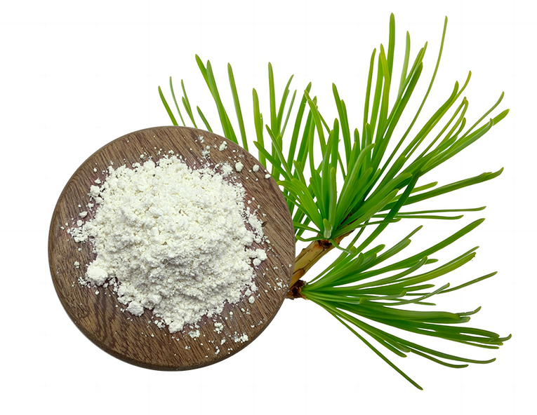
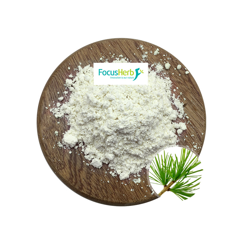
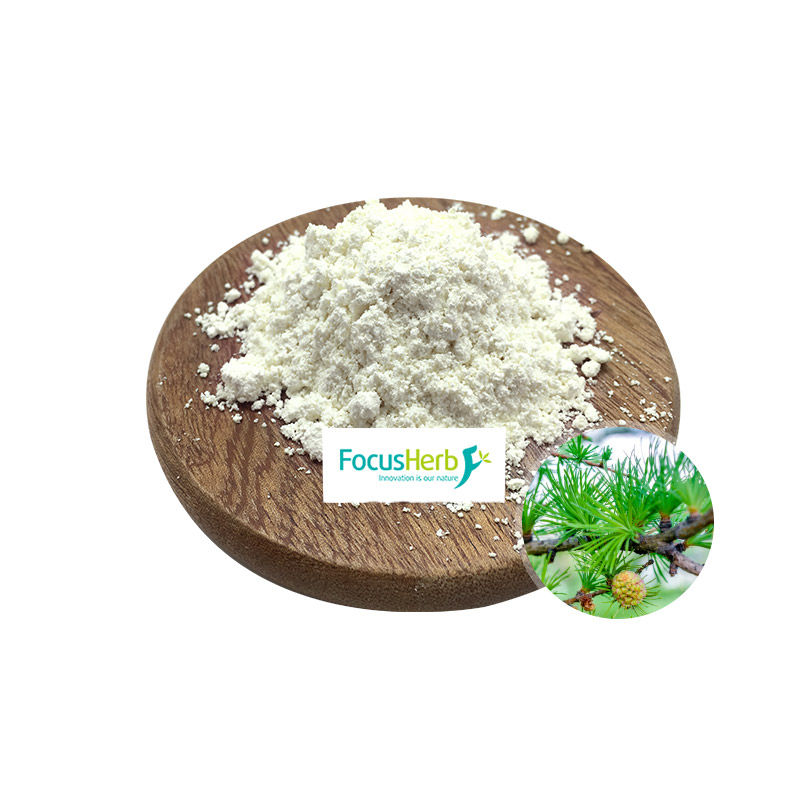
Cordyceps Militaris Extract (mainly derived from Cordyceps sinensis and its mycelium) is a natural product with multiple biological activities, and its efficacy covers multiple fields such as immune regulation, anti-tumor, and organ protection.

Immune regulation: a two-way balance mechanism to reconstruct the body’s defense system

Throughout life, the immune system is like a sturdy fortress, constantly guarding the body’s health. Cordyceps Militaris Extract, like a wise commander, can precisely and bidirectionally regulate this fortress, keeping the immune system in optimal condition in the face of various challenges.

Bidirectional Regulatory Effects on Immune Cells

The active ingredients in Cordyceps Militaris Extract, such as cordyceps polysaccharides and cordycepin, are key “weapons” for immune regulation. Like a group of well-trained soldiers, they precisely target every aspect of the immune system.

When the body is attacked by pathogens, these active ingredients quickly spring into action, activating T lymphocytes, natural killer cells (NK cells), and macrophages. T lymphocytes are like elite troops on the battlefield, capable of identifying and attacking pathogen-infected cells; NK cells are like special forces, rapidly eliminating tumor cells and virus-infected cells; and macrophages are like “scavengers” on the battlefield, responsible for engulfing and clearing pathogens and dead cells. The active ingredients in Cordyceps Militaris Extract enhance the phagocytic activity of these immune cells and the secretion of cytokines (such as interferon-γ and interleukin-2), strengthening their ability to eliminate pathogens and effectively defending against disease invasion.

In some inflammatory reactions or autoimmune diseases, the immune system becomes overactivated, like an army out of control, attacking its own tissues and causing tissue damage. In these situations, Cordyceps Militaris Extract can exert its regulatory effect, inhibiting the overexpression of the pro-inflammatory cytokine TNF-α, preventing tissue damage caused by hyperimmunity and restoring the immune system to a balanced state. This bidirectional regulatory effect enables Cordyceps Militaris Extract to precisely regulate the body under varying immune conditions, maintaining immune homeostasis.

Anti-tumor: A Multi-Pathway Synergistic Tumor Growth Inhibition Strategy

In humanity’s long battle against cancer, Cordyceps Militaris Extract has emerged as a shining star, bringing new hope to cancer treatment. Its anti-tumor mechanism of action is complex and sophisticated, involving multiple pathways and targets, demonstrating potent tumor growth inhibition.

Dual Mechanisms of Direct Killing and Immune-Mediated Cytotoxicity: Cordycepin, the key anti-tumor component in Cordyceps Militaris Extract, acts like a precise “scalpel,” directly targeting tumor cells and delivering a fatal blow. Studies have shown that cordycepin can inhibit tumor cell DNA/RNA synthesis, fundamentally blocking genetic information transmission and protein synthesis, thereby inhibiting tumor cell growth and division. It can also induce G2/M cell cycle arrest, effectively “pausing” tumor cell growth and preventing them from successfully entering the division phase, thereby suppressing tumor cell proliferation.
In in vitro experiments, cordycepin has demonstrated significant anti-proliferation effects on lung cancer, liver cancer, and breast cancer cell lines, with inhibition rates reaching 70%-85%. Taking lung cancer cells as an example, cordycepin can tightly bind to their DNA, preventing DNA replication and transcription, preventing normal metabolism and division, and ultimately leading to cell death. This direct cytotoxic effect provides an effective means for tumor treatment.

Immune Activation Effect: In addition to directly killing tumor cells,Cordyceps Militaris Extract can also activate the immune system, enhancing the body’s immune surveillance and killing capabilities against tumor cells. Like a powerful “commander,” it can activate the maturation of dendritic cells, turning them into “scouts” of the immune system. These cells are better able to identify and take up tumor antigens and present these antigens to T lymphocytes, thereby initiating a specific immune response.

Cordyceps Militaris Extract can also synergize with natural killer (NK) cells to specifically kill tumor cells. NK cells are the “special forces” of the immune system, capable of rapidly identifying and attacking tumor cells. Cordyceps Militaris Extract enhances NK cell activity, enabling them to more effectively kill tumor cells. In animal models, treatment with Cordyceps Militaris Extract has reduced tumor volume by 40%, demonstrating the importance of its immune-activating effect in anti-tumor treatment. While chemotherapy drugs kill tumor cells, they also damage the body’s immune system, leading to adverse reactions such as bone marrow suppression. Leukopenia is a common complication. Cordyceps Militaris Extract can alleviate chemotherapy-induced bone marrow suppression, reducing the incidence of leukopenia by 35%. It promotes the proliferation and differentiation of hematopoietic stem cells in the bone marrow, increasing white blood cell production, thereby boosting the body’s immunity and helping patients better tolerate chemotherapy.

Cardiovascular Protection: Multi-Dimensional Protection from Cellular Energy to Vascular Homeostasis

In the human body, the cardiovascular system is like a vast and complex transportation network. The heart is the core “pump” of this network, and blood vessels are the “highways” connecting various organs and tissues. Problems in the cardiovascular system are like a traffic network in chaos, severely impacting the health of the entire body. Cordyceps Militaris Extract demonstrates remarkable efficacy in cardiovascular protection, supporting the cardiovascular system from multiple perspectives and playing a vital role in maintaining cardiovascular health.

Bidirectional Regulation of Myocardial Function and Vascular Endothelium

Anti-Myocardial Ischemia and Hypoxia: In the cardiovascular system, the normal function of cardiomyocytes is crucial for the heart’s pumping ability. The adenosine component in Cordyceps Militaris Extract acts as the “energy guardian” of cardiomyocytes, significantly enhancing the efficiency of mitochondrial energy metabolism. Mitochondria are the cell’s “energy factories,” responsible for generating the energy needed for cellular activity. By regulating mitochondrial function, adenosine enables cardiomyocytes to utilize oxygen more efficiently, thereby reducing myocardial oxygen consumption. Studies have shown that Cordyceps Militaris Extract can enhance the heart’s tolerance to hypoxia by 25%-30%, meaning that the heart can maintain normal function longer under hypoxic conditions. Cordyceps Militaris Extract can also play a significant role in treating heart problems such as arrhythmias. For example, in rats with arrhythmias induced by aconitine, Cordyceps Militaris Extract was able to shorten the duration of the arrhythmia by 40%. Aconitine is a substance that can cause arrhythmias by interfering with the electrophysiological activity of cardiomyocytes, disrupting the heart’s rhythm. Cordyceps Militaris Extract effectively alleviated aconitine-induced arrhythmias by stabilizing the myocardial membrane potential and regulating ion channel function, restoring the heart’s rhythm.

Optimizing blood lipids and hemodynamics: Dyslipidemia is a major risk factor for cardiovascular disease. It increases blood viscosity and slows blood flow, thereby increasing the risk of atherosclerosis. The active ingredients in Cordyceps Militaris Extract can inhibit the activity of HMG-CoA reductase, a key enzyme in cholesterol synthesis. Inhibiting its activity can reduce cholesterol synthesis. Clinical research data shows that Cordyceps Militaris Extract can reduce serum total cholesterol (TC) by 18%-22% and triglycerides (TG) by 20%-25%, while also increasing high-density lipoprotein (HDL-C) levels. HDL, known as “good cholesterol,” transports cholesterol in the blood to the liver for metabolism, thereby lowering blood cholesterol levels. By lowering “bad cholesterol” and raising “good cholesterol,” Cordyceps Militaris Extract effectively regulates blood lipid levels and slows the progression of atherosclerosis.

Cordyceps Militaris Extract also improves hemodynamics, promoting smoother blood flow through blood vessels. By reducing blood viscosity and platelet aggregation, it reduces resistance to blood flow and improves vascular elasticity, thereby providing a healthier blood flow environment for the cardiovascular system.


Metabolic Regulation: Coordinated Optimization of the Energy Factory and Glucose and Lipid Homeostasis

In human life, the metabolic process is like a sophisticated factory, responsible for maintaining normal body function and energy balance. Cordyceps Militaris Extract plays a vital role in metabolic regulation, synergistically optimizing energy and glucose metabolism, providing the body with sufficient energy and maintaining glucose and lipid homeostasis, thereby promoting good health.

(I) Anti-Fatigue and Enhanced Mitochondrial Function

In modern society, people face increasing work and life pressures, and fatigue has become a common problem for many. Whether athletes after intense training and competition or workers engaged in long-term, high-intensity labor, they all yearn for effective methods to relieve fatigue and improve their endurance and recovery. The emergence of Cordyceps Militaris Extract offers new hope.

Cordyceps Militaris Extract acts as a magical energy regulator, comprehensively optimizing skeletal muscle mitochondria by activating the AMPK signaling pathway. The AMPK signaling pathway is a key intracellular energy regulation pathway. When cellular energy levels decrease, AMPK is activated, acting like a “switch” that triggers a series of metabolic reactions to increase energy production. Cordyceps Militaris Extract activates the AMPK signaling pathway, effectively injecting a powerful boost into this “switch,” enabling it to function more effectively.

Cordyceps Militaris Extract significantly increases the number of skeletal muscle mitochondria. These mitochondria act like miniature “energy factories,” providing ample energy for muscle movement. Mitochondrial oxidative phosphorylation efficiency is also significantly enhanced, meaning that mitochondria can more efficiently convert nutrients into ATP (adenosine triphosphate), the direct energy source for cellular activity. ATP production increases by 20%-25%, providing enhanced energy support for muscle movement.

Experimental data strongly supports the anti-fatigue effects of Cordyceps Militaris Extract. In a swimming experiment with mice, mice given Cordyceps Militaris Extract experienced a 35% increase in the time it took to fatigue, demonstrating that Cordyceps Militaris Extract significantly improves the mice’s exercise endurance, allowing them to maintain their performance for longer periods of time. After exercise, lactate levels in mice are a key indicator of fatigue; the accumulation of lactic acid can lead to increased muscle soreness and fatigue. Cordyceps Militaris Extract can accelerate the removal of lactic acid after exercise, acting like a diligent “cleaner,” quickly clearing lactic acid from muscles, significantly relieving muscle soreness and allowing the body to return to normal more quickly.

(II) Dual Regulation of Glucose and Lipid Metabolism

Glucose Homeostasis: With changing lifestyles and the advent of an aging society, the incidence of diabetes has increased annually, becoming a major public health issue threatening human health. For diabetic patients, controlling blood sugar levels is key to treatment, and Cordyceps Militaris Extract demonstrates unique advantages in this regard.

Cordyceps polysaccharides are the key component of Cordyceps Militaris Extract that regulates blood sugar. They act like a smart “blood sugar steward,” maintaining stable blood sugar levels through multiple mechanisms. In the intestines, cordyceps polysaccharides slow glucose absorption, acting like a “speed bump” for glucose absorption, allowing glucose to enter the bloodstream more slowly and preventing a sharp rise in blood sugar. Cordyceps polysaccharides also enhance insulin sensitivity. Insulin is a key hormone in blood sugar regulation, promoting glucose uptake and utilization by cells, thereby lowering blood sugar levels. Cordyceps polysaccharides enhance insulin sensitivity, acting like “wings” for insulin, enabling it to function more effectively and enabling cells to better absorb and utilize glucose, thereby lowering blood sugar levels.

The blood sugar-lowering effects of Cordyceps Militaris Extract have been fully demonstrated in a type 2 diabetes model. Glycated hemoglobin (HbA1c), a key indicator of average blood sugar levels over the past two to three months, decreased by 1.2% after treatment with Cordyceps Militaris Extract, demonstrating its long-term effectiveness in controlling blood sugar levels. Postprandial blood sugar fluctuations were also reduced by 30%, which is crucial for reducing the incidence of diabetic complications. A sharp rise in postprandial blood sugar can damage blood vessels and nerves, increasing the risk of diabetic complications. Cordyceps Militaris Extract effectively controls postprandial blood sugar fluctuations, providing a strong safeguard for the health of diabetic patients.

Lipid metabolism: In addition to blood sugar problems, dyslipidemia is also a common problem in metabolic diseases, closely related to obesity and cardiovascular disease. Cordyceps Militaris Extract also excels in regulating lipid metabolism, offering a new approach to addressing dyslipidemia.

In the liver, Cordyceps Militaris Extract inhibits fatty acid synthesis, effectively pausing the fatty acid synthesis production line, thereby reducing fatty acid synthesis and thus reducing fat accumulation. It also promotes β-oxidation in adipocytes, a key pathway for fat breakdown. By promoting β-oxidation, Cordyceps Militaris Extract acts like a powerful engine for fat breakdown, enabling adipocytes to more efficiently break down fat and provide energy.

The results were surprising. In experiments with mice induced by high-fat diet obesity, administration of Cordyceps Militaris Extract resulted in a 15%-20% weight loss, demonstrating that Cordyceps Militaris Extract can effectively reduce weight and improve obesity in obese mice. Visceral fat accumulation is a major risk factor for metabolic syndrome, and Cordyceps Militaris Extract significantly reduces this accumulation, acting like a “fat transporter,” removing fat from the viscera, reducing pressure on internal organs, and lowering the risk of metabolic syndrome.

Liver and Kidney Protection: Deep Regulation from Cell Repair to Function Maintenance

In the human body, the liver and kidneys are like two crucial “chemical plants” and “purifiers,” responsible for key tasks such as metabolism, detoxification, and excretion, playing an indispensable role in maintaining a stable internal environment. Problems with these two organs can trigger a series of health crises. Cordyceps Militaris Extract demonstrates remarkable efficacy in liver and kidney protection, deeply regulating the liver and kidneys from cell repair to functional maintenance, providing a strong safeguard for liver and kidney health.

(I) Liver Damage Repair and Fibrosis Inhibition

In daily life, the liver faces numerous challenges, such as chronic alcohol consumption, drug abuse, and viral infections. These factors can cause liver damage and even lead to liver fibrosis. Fibrosis is an excessive repair response to chronic damage, resulting in hardened liver tissue and a gradual decline in function. Cordyceps Militaris Extract acts as a “guardian” of the liver, effectively addressing these challenges.

The active ingredients in Cordyceps Militaris Extract regulate the Nrf2/ARE antioxidant pathway, establishing a powerful antioxidant defense for the liver. This line of defense plays a crucial role in the fight against chemical liver injury. When the liver is attacked by chemicals, it produces a large number of free radicals. These free radicals act like a swarm of troublemakers, causing lipid peroxidation damage to liver cell membranes, leading to structural and functional impairment. Cordyceps Militaris Extract can activate the Nrf2/ARE antioxidant pathway, injecting a powerful boost into the liver’s antioxidant system, enabling it to produce more antioxidant enzymes, such as superoxide dismutase (SOD) and glutathione peroxidase (GSH-Px). These antioxidant enzymes rapidly scavenge free radicals, reducing lipid peroxidation damage to liver cell membranes and thus protecting liver cell integrity.

Experimental data strongly demonstrate the protective effect of Cordyceps Militaris Extract. In a chemical liver injury model, administration of Cordyceps Militaris Extract reduced serum ALT and AST levels by 25%-35%. ALT and AST are important indicators of liver cell damage. Elevated serum levels indicate liver cell damage. Cordyceps Militaris Extract significantly reduces these two indicators, demonstrating its ability to effectively repair damaged liver cells and alleviate liver inflammation. Hepatic stellate cell activation is a key step in the development of liver fibrosis. When the liver is damaged, hepatic stellate cells are activated and transform into myofibroblasts, which synthesize and secrete collagen fibers in large quantities, leading to excessive collagen deposition in the liver and the development of liver fibrosis. Cordyceps polysaccharides can inhibit hepatic stellate cell activation, essentially pressing the “pause button” on the activation process. By modulating intracellular signaling pathways, they inhibit the proliferation and differentiation of hepatic stellate cells, reduce collagen synthesis, and promote collagen degradation, thereby maintaining normal collagen content in the liver.

The anti-fibrotic effects of Cordyceps Militaris Extract have been fully demonstrated in a mouse model of liver fibrosis. Treatment with Cordyceps Militaris Extract increased the rate of collagen degradation by 30%, demonstrating that Cordyceps Militaris Extract can effectively inhibit the development of liver fibrosis and promote liver tissue repair and regeneration. Cordyceps Militaris Extract has important adjunctive therapeutic value for patients with alcoholic liver disease, drug-induced liver injury, and chronic hepatitis. It can reduce the severity of liver damage, improve liver function, and slow disease progression, offering new hope for recovery.

(II) Protecting Kidney Function and Toxin Metabolism

The kidneys are crucial excretory organs in the human body, filtering waste and excess water from the blood and excreting them as urine. However, some nephrotoxic substances, such as chemotherapy drugs like cisplatin, as well as diseases like chronic nephritis, can cause severe damage to the kidneys, impairing their normal function. Cordyceps Militaris Extract plays an important role in kidney protection, mitigating the damage caused by nephrotoxic substances, promoting toxin metabolism, and maintaining normal kidney function.

Cisplatin is a widely used chemotherapy drug in clinical practice, but its nephrotoxicity limits its clinical application. After entering the body, cisplatin accumulates in the kidneys, damaging renal tubular cells and leading to impairments in renal reabsorption and excretion. Cordyceps Militaris Extract can mitigate the damage caused by cisplatin and other nephrotoxic substances to the renal tubules, acting like a protective layer for these cells. By modulating intracellular signaling pathways, it inhibits cisplatin-induced apoptosis and oxidative stress, protecting mitochondrial function in renal tubular cells and maintaining cellular energy metabolism. In animal studies, administration of Cordyceps Militaris Extract reduced serum creatinine (SCr) and blood urea nitrogen (BUN) levels by 20%-25%, and improved glomerular filtration rate (GFR). Serum creatinine and BUN are important indicators of renal function. Elevated serum levels indicate impaired renal excretion. Cordyceps Militaris Extract significantly reduced these levels, demonstrating its effectiveness in improving renal function and promoting toxin excretion.

Proteinuria is a common symptom in patients with chronic nephritis. It not only leads to protein loss but also further damages kidney tissue, accelerating the progression of kidney disease. Cordyceps Militaris Extract can reduce proteinuria, acting as a comprehensive cleansing and repair of the kidney’s “filter.” It modulates the expression of factors associated with renal interstitial fibrosis (TGF-β1 and CTGF), inhibits the onset and progression of renal interstitial fibrosis, and reduces extracellular matrix deposition, thereby improving glomerular filtration function and reducing proteinuria. Clinical research data shows that Cordyceps Militaris Extract can reduce 24-hour urine protein by 30%, a significant improvement for patients with chronic nephritis. It can slow the progression of kidney disease, protect kidney function, and improve patients’ quality of life. The mechanism of action of Cordyceps Militaris Extract in kidney protection is complex and sophisticated, protecting the kidneys from multiple angles and providing comprehensive support for kidney health.


Respiratory System: A Double Barrier of Airway Dilation and Lung Tissue Protection

In the human body, the respiratory system functions as a sophisticated gas exchange factory, responsible for inhaling oxygen and expelling carbon dioxide to maintain normal life. However, modern air pollution, smoking, and various respiratory diseases pose serious threats to respiratory health. Cordyceps Militaris Extract demonstrates remarkable efficacy in protecting the respiratory system. By dilating the bronchi and clearing mucus, it protects against pulmonary fibrosis and oxidative damage, creating a strong double barrier for the respiratory system, effectively maintaining respiratory health.

(I) Bronchodilation and Mucus Clearance

In the respiratory system, normal bronchial dilation and effective mucus clearance are crucial for maintaining airway patency. Components such as cordycepic acid and adenosine in Cordyceps Militaris Extract act as “guardians” of the bronchi, playing a vital role in bronchial dilation and mucus clearance.

During an attack of diseases such as asthma and chronic bronchitis, bronchial smooth muscle spasms, leading to airway narrowing, impaired gas exchange, and symptoms such as dyspnea. Cordycepic acid and adenosine precisely target bronchial smooth muscle, modulating intracellular signaling pathways to relax the muscle and expand the airway diameter by 15%-20%. This is like opening a wide door to a narrow airway, allowing air to flow in and out smoothly, effectively alleviating the patient’s dyspnea.

In the respiratory tract, mucus secretion and clearance are a dynamic equilibrium. When the airways are irritated, mucus secretion increases. If not cleared promptly, sputum accumulates, obstructing the airways and exacerbating dyspnea. Cordyceps Militaris Extract promotes the dilution of mucus in the airways, thinning thick sputum and making it easier to pass. It also increases the frequency of cilia, which act like “little brooms” in the airways, pushing sputum upwards, thereby effectively clearing it. Studies have shown that Cordyceps Militaris Extract can increase sputum clearance efficiency by 25%, which is crucial for maintaining airway patency.

For patients with COPD, coughing and expectoration are common symptoms, significantly impacting their quality of life. Clinical studies have shown that Cordyceps Militaris Extract improves cough and sputum symptoms in COPD patients by up to 70%. It effectively relieves cough symptoms by dilating the bronchi, reducing sputum accumulation, and alleviating airway irritation. By promoting sputum excretion, it reduces the burden of sputum production and allows for smoother breathing.

(II) Pulmonary Fibrosis and Protection against Oxidative Damage

Pulmonary fibrosis is a serious lung disease characterized by the accumulation of collagen fibers in lung tissue, leading to stiffening, decreased elasticity, and impaired gas exchange. Cordyceps Militaris Extract has demonstrated significant efficacy in protecting against pulmonary fibrosis, offering new hope for patients.

Abnormal proliferation of lung fibroblasts and excessive collagen deposition are key components of the development and progression of pulmonary fibrosis. Cordyceps Militaris Extract inhibits lung fibroblast proliferation, effectively pausing it and reducing collagen synthesis. It also promotes a reduction in collagen deposition and accelerates collagen degradation, thereby reducing hydroxyproline levels in lung tissue by 20%-30%. Hydroxyproline is a major component of collagen, and a decrease in its content indicates that the severity of pulmonary fibrosis has been effectively alleviated.

In modern society, people face increasingly severe air pollution. Pollutants such as PM2.5 and cigarette tar can cause severe oxidative damage to the lungs. Once these pollutants enter the lungs, they produce a large number of free radicals, such as superoxide anions. These free radicals act like “troublemakers” and attack alveolar epithelial cells, causing cell damage and death. The active ingredients in Cordyceps Militaris Extract possess potent antioxidant properties, scavenging free radicals in the lungs. This acts like a protective layer, reducing the damage caused by free radicals to the alveolar epithelial cells.

For those who are chronically exposed to polluted environments or smoke, their lungs are constantly exposed to pollutants and smog, making them prone to various lung problems. Cordyceps Militaris Extract can help maintain lung health, mitigate damage caused by pollutants and smog, enhance lung resistance, and prevent the onset of lung disease. Like a loyal “lung guardian,” it constantly protects lung health and provides strong support for people’s respiratory health.


Hematopoiesis and Endocrine System: Deeply Empowering from Cell Generation to Hormone Regulation

In the human body, the hematopoietic and endocrine systems function like two closely integrated “factories.” The hematopoietic system is responsible for producing various blood cells, providing the body with sufficient oxygen and nutrients to maintain normal physiological functions. The endocrine system, through the secretion of various hormones, regulates growth, development, metabolism, and reproduction, maintaining a stable internal environment. Cordyceps Militaris Extract demonstrates remarkable efficacy in regulating hematopoiesis and endocrine function. From cell generation to hormone regulation, it deeply empowers these two systems, providing strong support for overall health.

(I) Enhanced Hematopoietic Function

In the human bone marrow, hematopoietic stem cells are like a group of miraculous “seed cells.” They possess the ability to self-renew and differentiate into various blood cell types, making them crucial for maintaining the normal function of the hematopoietic system. Components such as adenosine and cordyceps polysaccharides in Cordyceps Militaris Extract act as growth catalysts for these “seed cells,” promoting the proliferation and differentiation of hematopoietic stem cells in the bone marrow and ensuring a continuous supply of blood cells. The efficacy of Cordyceps Militaris Extract has been fully demonstrated in models of anemia and post-chemoradiotherapy bone marrow suppression. It can increase red blood cell, white blood cell, and platelet counts by 15%-20%, effectively injecting a powerful boost into the body’s “blood cell factory,” enabling more efficient production of various blood cells. Red blood cells act as the body’s “oxygen transporters.” Their increased number improves oxygen transport capacity, alleviates symptoms of Qi and blood deficiency, and invigorates the body. White blood cells are the “guardians” of the immune system. Their increased number strengthens immunity and protects against pathogens. Platelets play a vital role in hemostasis and coagulation, and their increased number effectively prevents bleeding disorders.

For patients with aplastic anemia, their bone marrow hematopoietic function is severely impaired, like the “blood cell factory” paralyzed and unable to produce blood cells normally. Cordyceps Militaris Extract can modulate the hematopoietic microenvironment, providing a favorable growth environment for hematopoietic stem cells and promoting their proliferation and differentiation, thereby serving as an adjunct therapy for patients with aplastic anemia. Like a miraculous “repairman,” it helped the patient’s bone marrow hematopoietic function gradually return to normal, bringing new hope for recovery.

(II) Endocrine Regulation Effect

In the human endocrine system, the gonadal axis and the hypothalamic-pituitary-adrenal axis act as two important “hormone regulatory hubs.” They are responsible for regulating the secretion of sex hormones and stress hormones, respectively, and play a vital role in maintaining normal physiological functions and responding to various stressful situations. The active ingredients in Cordyceps Militaris Extract act like “intelligent controllers” for these “hormone regulatory hubs,” precisely regulating them to maintain balanced hormone secretion.

Cordyceps Militaris Extract has androgen-like effects, promoting the secretion of gonadal axis hormones, essentially accelerating the gonadal axis’s “hormone production line” and increasing the production of sex hormones. For male patients with hypoactive sexual dysfunction, problems with the gonadal axis lead to insufficient sex hormone secretion, resulting in decreased sexual function. Cordyceps Militaris Extractcan improve male sexual function by promoting the secretion of gonadal axis hormones, allowing them to regain confidence and vitality. In response to various stressful situations, the hypothalamic-pituitary-adrenal axis is activated, secreting stress hormones such as cortisol to help the body cope with stress. However, if the stress response is excessive or prolonged, it can lead to various health problems, such as anxiety and fatigue. Cordyceps Militaris Extract can regulate the hypothalamic-pituitary-adrenal axis, enhancing the adaptive secretion of cortisol in response to stress. This acts like an “intelligent regulator” within the hypothalamic-pituitary-adrenal axis’s “stress response system,” enabling it to regulate cortisol secretion based on the body’s actual conditions and prevent the damage caused by excessive stress.

Clinical studies have shown that Cordyceps Militaris Extract has some benefits for menopausal syndrome and chronic fatigue syndrome. In menopausal syndrome, ovarian dysfunction and imbalanced sex hormone secretion lead to a range of uncomfortable symptoms, such as hot flashes, night sweats, and mood swings. Cordyceps Militaris Extract can regulate sex hormone levels, alleviate symptoms, and improve the quality of life of menopausal patients. For patients with chronic fatigue syndrome, they experience chronic fatigue and a reduced ability to regulate stress. Cordyceps Militaris Extract can improve the body’s stress regulation by regulating the hypothalamic-pituitary-adrenal axis, alleviating fatigue symptoms and allowing patients to regain energy.


The Precision Health Value of Cordyceps Militaris Extract

With its diverse active ingredients, Cordyceps Militaris Extract establishes a comprehensive health intervention system, from immune regulation to organ protection. Its core advantage lies in its “multi-target synergy,” providing both a preventive barrier for healthy individuals and complementing medications in chronic disease management. Only by adhering to scientific dosage, suitable populations, and quality standards can its natural health potential be maximized, making it a smart choice for proactive health management.

Turkey Tail Mushroom Extact: A New Force in Medicinal Fungi with Six Core Benefits

Natural immune regulator: polysaccharide code that activates the body’s defense system

In the body’s defense system, Turkey Tail Mushroom Extact
acts like a mysterious polysaccharide key, unlocking the door to natural immune regulation and becoming a powerful aid in safeguarding health.

Immune Cell Activation Mechanism

The core components of Turkey Tail Mushroom Extact
—a complex of β-glucans and glycoproteins—have unique immunomodulatory properties. Once inside the body, they act like well-trained commanders, precisely identifying TLR-2/TLR-4 receptors on the surface of immune cells and rapidly activating the phagocytic activity of macrophages, increasing their effectiveness by 60%-80%, enabling them to more efficiently identify and phagocytose invading pathogens. Simultaneously, these stimuli stimulate T lymphocytes and NK cells to actively respond, accelerating their proliferation and differentiation, further strengthening the immune cell ranks. Serum interferon-γ (IFN-γ) levels are significantly increased by 35%. As a key immune regulator, IFN-γ activates more immune cells, enhancing their collaboration and building a multi-layered, comprehensive immune response network, comprehensively enhancing the body’s immune defenses.


New Options for Adjuvant Cancer Therapy: Multi-Pathway Inhibition of Tumor Development

In the protracted battle against cancer, Turkey Tail Mushroom Extact, with its unique multi-pathway mechanism of action, has become a rising star in the field of adjuvant cancer therapy, bringing new hope to cancer patients.

Dual Anti-Tumor Mechanisms

Direct Killing: The triterpenes in Turkey Tail Mushroom Extact
act as a sharp weapon directly against tumor cells. Studies have shown that these triterpenes can cleverly induce tumor cell cycle arrest at the G2/M phase, effectively pressing the “pause button” on tumor cell division. During this critical period, tumor cell DNA synthesis is strongly inhibited, preventing them from dividing and proliferating smoothly. In vitro experiments have shown that Turkey Tail Mushroom Extact can inhibit the proliferation of lung and gastric cancer cell lines by 75%-85%. In experiments with the lung cancer cell line A549, flow cytometry revealed that the addition of Coriolus versicolor extract significantly increased the proportion of cells in the G2/M phase, from 20% to over 50%. Cell proliferation was also significantly slowed. Within 72 hours, the cell number increased by only 1.5 times, compared to a 3-fold increase in the control group. This demonstrates its potent ability to directly kill tumor cells.

Immune-mediated: Coriolus versicolor extract also demonstrates excellent immune-mediated anti-tumor activity. Acting as an excellent immune “commander,” it activates dendritic cells, significantly enhancing their antigen-presenting capacity. This allows for more effective presentation of tumor antigens to T lymphocytes, initiating a specific immune response. In this process, Coriolus versicolor extract also synergizes with natural killer (NK) cells, enhancing their ability to recognize and kill tumor cells. In animal model experiments, administration of Coriolus versicolor extract to mice bearing tumors reduced tumor volume by 40%. Further testing by the experimenters found that the activity of NK cells in the mice increased by 80%, and the number of T lymphocytes infiltrating around the tumor tissue increased by 50%. This shows that Turkey Tail Mushroom Extact successfully stimulated the body’s own immune system to fight tumors through immune-mediated mechanisms.


Liver Protection Shield: Multi-Dimensional Protection from Cell Repair to Metabolic Regulation

The liver, the body’s “chemical factory,” undertakes crucial functions such as metabolism, detoxification, and immunity. Its health is directly linked to overall human health. Turkey Tail Mushroom Extact, with its unique bioactive components, plays a vital role in liver protection, serving as a powerful “shield” for maintaining liver health.

Mechanisms of Liver Damage Repair

The protective effects of Turkey Tail Mushroom Extact
on the liver are multi-dimensional and profound. At the cellular level, its core components, polysaccharides and triterpenoids, precisely regulate the Nrf2/ARE signaling pathway. When the liver is attacked by harmful substances such as alcohol, drugs, and viruses, Nrf2 is activated and translocated to the cell nucleus, where it binds to the ARE element and activates the gene expression of a series of antioxidant and detoxification enzymes, such as heme oxygenase-1 (HO-1) and glutathione S-transferase (GST). These enzymes effectively scavenge free radicals and oxidative stress products within the liver, enhancing the antioxidant capacity of liver cells, thereby improving the stability of liver cell membranes and protecting them from further damage. In a chemical liver injury model, treatment with Coriolus versicolor extract significantly reduced serum alanine aminotransferase (ALT) and aspartate aminotransferase (AST) levels by 25%-35%. ALT and AST are important enzymes within hepatocytes. When hepatocytes are damaged, they are released into the blood. Therefore, elevated serum ALT and AST levels are key markers of liver damage. Coriolus versicolor extract can reduce these two enzymes, indicating that it can effectively mitigate the severity of hepatocyte damage and promote hepatocyte repair.

The polysaccharides in Coriolus versicolor extract also promote the secretion of hepatocyte growth factor (HGF). HGF is a cytokine crucial for liver regeneration and repair. It stimulates the proliferation and differentiation of hepatocytes, accelerating the replacement of damaged hepatocytes. In a liver fibrosis model, Coriolus versicolor extract significantly accelerated the degradation of collagen fibers in liver fibrosis, increasing the rate of liver tissue repair by 30%. Liver fibrosis is a pathological condition caused by long-term liver damage. If not promptly addressed, it can progress to cirrhosis or even liver cancer. Turkey Tail Mushroom Extact enhances the liver’s self-repair ability by promoting the secretion of HGF, effectively delaying the progression of liver fibrosis.

Metabolic Syndrome Regulator: Dual Optimization of Blood Lipids and Blood Sugar

In today’s society, the incidence of metabolic syndrome is rising year by year, posing a serious threat to human health. Metabolic syndrome is a complex set of metabolic disorders, primarily consisting of hyperglycemia, hyperlipidemia, and hypertension. These interrelated symptoms collectively increase the risk of chronic diseases such as cardiovascular disease and diabetes. Coriolus versicolor extract, as a natural bioactive substance, demonstrates unique advantages in regulating metabolic syndrome, providing new insights and approaches for the prevention and treatment of metabolic-related diseases.

(I) Lipid Metabolism Regulation

Coriolus versicolor acid in Coriolus versicolor extract is a key component in regulating lipid metabolism. Studies have shown that coriolus versicolor acid can specifically inhibit the activity of HMG-CoA reductase, a key rate-limiting enzyme in cholesterol synthesis. In a high-fat diet-induced mouse model, administration of Coriolus versicolor extract reduced serum total cholesterol (TC) levels by 18% and low-density lipoprotein cholesterol (LDL-C) levels by 22%. This is because versicolor acid binds tightly to the active site of HMG-CoA reductase, blocking the enzyme’s catalytic function and inhibiting cholesterol synthesis, thereby effectively lowering blood cholesterol levels.

Coriolus versicolor extract can also activate the AMPK signaling pathway, a key energy sensing and metabolic regulation pathway within cells. When intracellular energy levels decrease, AMPK is activated, initiating a series of catabolic processes to generate energy. In fat metabolism, activated AMPK promotes the β-oxidation of fatty acids, converting them into acetyl-CoA, which enters the tricarboxylic acid cycle for energy production. Experimental data show that triglyceride (TG) levels in cells and animal models treated with Coriolus versicolor extract decreased by 25%. This process not only reduces fat accumulation in the body but also improves energy utilization efficiency, which is of great significance for improving lipid metabolism disorders.

(II) Maintaining Blood Glucose Homeostasis

The polysaccharides in Coriolus versicolor extract play a vital role in maintaining blood glucose homeostasis. On the one hand, Coriolus versicolor polysaccharides can form a viscous substance in the intestines, slowing carbohydrate digestion and glucose absorption. In an in vitro simulated intestinal digestion experiment, the addition of Coriolus versicolor polysaccharides significantly slowed the rate of glucose release, reducing peak concentrations by 30%. This suggests that Coriolus versicolor polysaccharides can effectively prevent a sharp rise in blood sugar after a meal, alleviating the burden on pancreatic islet cells.

On the other hand, Coriolus versicolor polysaccharides can improve insulin sensitivity and enhance cellular responses to insulin. In a mouse model of type 2 diabetes, eight weeks of continuous administration of Coriolus versicolor extract reduced the mice’s glycated hemoglobin (HbA1c) levels by 1.2%, and postprandial blood sugar fluctuations by 30%. HbA1c is an important indicator of long-term blood sugar control, and its reduction suggests that Coriolus versicolor extract can effectively improve blood sugar control in diabetic mice.

When Coriolus versicolor extract is used in combination with antidiabetic drugs such as metformin, careful monitoring of blood sugar levels is necessary. Since both drugs have blood sugar-lowering effects, combined use may enhance the blood sugar-lowering effect and increase the risk of hypoglycemia. Therefore, during combined medication, patients should pay close attention to changes in their blood sugar, monitor blood sugar levels regularly, and adjust drug dosages in a timely manner according to blood sugar conditions to ensure that blood sugar is controlled within a safe and stable range.


Dual Anti-Inflammation and Antioxidation Pathways: A Double Shield Against Chronic Diseases

In the prevention and treatment of chronic diseases, inflammation and oxidative stress are two “invisible killers” that constantly threaten human health. Coriolus versicolor extract, with its unique biological activity, demonstrates remarkable efficacy in anti-inflammatory and antioxidant activities, building a solid dual shield against chronic diseases.

Oxidative Stress Regulation

The triterpenes and polysaccharides in Coriolus versicolor extract are key components for scavenging free radicals and inhibiting oxidative stress. Studies have shown that the triterpenes in Coriolus versicolor extract have a scavenging capacity 5-8 times that of vitamin C for DPPH radicals, hydroxyl radicals, and superoxide anion radicals. In an oxidative stress model, Coriolus versicolor extract reduced malondialdehyde (MDA) levels by 40%. MDA is the end product of lipid peroxidation, and its level directly reflects the degree of oxidative stress in the body. Coriolus versicolor extract can significantly reduce MDA levels, demonstrating its ability to effectively inhibit lipid peroxidation and reduce free radical damage to cells. Turkey Tail Mushroom Extact can also reduce the production of inflammatory factors by inhibiting the NF-κB signaling pathway. NF-κB is a key inflammatory transcription factor that plays a central regulatory role in the inflammatory response. When the body is stimulated by external stimuli, NF-κB is activated and translocates to the cell nucleus, initiating the expression of a series of inflammation-related genes, leading to the secretion of inflammatory factors such as interleukin-6 (IL-6) and tumor necrosis factor-α (TNF-α). Turkey Tail Mushroom Extact can inhibit the nuclear translocation and transcriptional activity of NF-κB, thereby reducing the secretion of inflammatory factors such as IL-6 and TNF-α by 35%-50%.

In a rheumatoid arthritis model, Turkey Tail Mushroom Extact significantly reduced joint swelling and inflammation, and decreased the levels of IL-6 and TNF-α in joint fluid. In the treatment of chronic bronchitis, Turkey Tail Mushroom Extact can improve patients’ symptoms such as cough and sputum, reduce the infiltration of airway inflammatory cells, inhibit the release of inflammatory factors, effectively relieve inflammatory responses, and improve patients’ quality of life.

A Reconstructor of the Intestinal Microbiome: The Synergistic Effect of Mucosal Protection and Microbial Balance

As the body’s largest digestive and immune organ, the balance of its microbiome is crucial to overall health. Turkey Tail Mushroom Extact plays a crucial role in maintaining a balanced intestinal microbiome, acting like a precise regulator, safeguarding intestinal health by strengthening the intestinal barrier and regulating the microbiome.

(I) Intestinal Barrier Strengthening

The strengthening effect of Turkey Tail Mushroom Extact on the intestinal barrier is one of its key mechanisms for maintaining intestinal health. At the cellular level, Yunzhi polysaccharides promote the secretion of mucin MUC2 by goblet cells, creating a more robust “protective coating” for the intestinal mucosa. Mucin MUC2 is a major component of the intestinal mucus layer, forming a thick mucus barrier on the surface of the intestinal mucosa, effectively preventing pathogens, toxins, and other harmful substances from directly contacting intestinal epithelial cells, thereby reducing the risk of intestinal infections.

Yunzhi polysaccharides also significantly increase the expression of tight junction proteins (ZO-1 and Occludin) by up to 60%. Tight junction proteins are key components of the tight junctions between intestinal epithelial cells. Like tightly connected “rivets,” they tightly connect intestinal epithelial cells together, forming a tight barrier and effectively reducing intestinal permeability. In a leaky gut model, administration of Coriolus versicolor extract significantly reduced intestinal permeability and significantly improved endotoxemia by 45%. Endotoxemia is a series of pathological reactions caused by a damaged intestinal barrier, allowing endotoxins in the intestine to enter the bloodstream. Coriolus versicolor extract can effectively improve endotoxemia, fully demonstrating its remarkable efficacy in strengthening the intestinal barrier.

(II) Microbiome Modulating Effects

Coriolus versicolor extract has a significant modulating effect on the intestinal microbiota, earning it the title of “master of intestinal microbiome balance.” In in vitro culture experiments, the addition of Coriolus versicolor extract increased the abundance of beneficial bacteria such as Bifidobacterium and Lactobacillus by 80%. These beneficial bacteria are crucial components of the intestinal microbiome. They inhibit the growth and adhesion of pathogens by producing substances such as short-chain fatty acids and bacteriocins, thereby maintaining a balanced intestinal microbiome. Turkey Tail Mushroom Extact can also significantly increase the diversity index of the intestinal flora by 20%. Microbial diversity is a key indicator of intestinal microbial health. Higher microbial diversity indicates greater stability and adaptability of the intestinal microbiome, enabling it to better respond to changes in the external environment.

In a clinical study of patients with functional constipation, after four weeks of Turkey Tail Mushroom Extact use, the bowel movement frequency normalization rate reached 72%. This is because Turkey Tail Mushroom Extact regulates the intestinal flora, promotes the growth of beneficial bacteria, and produces more short-chain fatty acids, which stimulates intestinal motility and improves constipation symptoms. Experimental data showed a 35% increase in short-chain fatty acid levels in the patients’ intestines, further confirming the mechanism by which Turkey Tail Mushroom Extact improves intestinal function by regulating the intestinal flora.

The Precise Health Benefits of Turkey Tail Mushroom Extact

With its rich and diverse active ingredients, Turkey Tail Mushroom Extact provides a comprehensive, multi-dimensional health protection system. From a natural immunomodulator that activates the immune system to a new adjuvant therapy option for tumors, inhibiting tumor progression through multiple pathways; from a liver shield that protects liver health to a metabolic regulator that regulates metabolic syndrome; from dual anti-inflammatory and antioxidant pathways that combat chronic diseases to a gut microbiome remodeler, Turkey Tail Mushroom Extact provides powerful support for human health on multiple levels.

Both for the general public pursuing a healthy lifestyle and for chronic disease patients facing various health challenges, Turkey Tail Mushroom Extact holds significant value. For healthy individuals, it serves as a strong barrier against disease, enhancing the body’s resistance and preventing the onset of various illnesses. For patients with chronic diseases, it not only serves as an effective supplement to medication, synergizing with medications, mitigating adverse drug reactions, and improving treatment outcomes, but also plays a vital role in the recovery process, promoting physical recovery.

When using Turkey Tail Mushroom Extact, strict adherence to scientific dosage and quality standards is crucial. Only by ensuring its safety and effectiveness can the full health benefits of Turkey Tail Mushroom Extact be realized. In the future, with the continuous deepening of research and continuous advancement of technology, the modern application of Turkey Tail Mushroom Extact, an ancient medicinal fungus, will continue to expand, becoming a smart choice for people to actively manage their health.

Oleanolic acid: a comprehensive analysis of the research and application of pentacyclic triterpenoid natural active ingredients

Oleanolic acid (OA) is a typical pentacyclic triterpenoid compound, occupying a unique position within the vast family of phytochemicals. Its molecular formula is C₃₀H₄₈O₃, with a molecular weight of 456.71. This precise molecular structure is like the foundation of a solid “chemical edifice.” Its chemical structure, based on the oleanane core, resembles the main framework of a building, while the hydroxyl group at position 3 and the carboxyl group at position 28 are key components within this framework, acting as the key active groups that determine its activity. The presence of these groups endows OA with unique chemical reactivity and physiological functions.

Physically, oleanolic acid appears as white needle-shaped crystals or powder. This pure appearance resembles a pure gift from nature. However, it is poorly soluble in water, with an extremely low solubility of less than 0.1 mg per milliliter. This is like dropping sand into water; it’s difficult to make it completely dissolve. However, it is readily soluble in organic solvents such as methanol and ethanol, blending readily with these solvents, like a wanderer finding a familiar home. This characteristic is crucial for its extraction and formulation.

Its melting point is between 283 and 288°C. This high melting point indicates its high stability, similar to a determined individual who maintains their steadfastness under normal conditions. However, it also has its weaknesses: it must be stored away from light, as light can damage its structure and affect its stability, much like delicate flowers needing to avoid direct sunlight. Under alkaline conditions, oleanolic acid can form water-soluble salts. This transformation acts as a unique “coat” that allows it to move freely in water, greatly expanding its applications in pharmaceuticals and biomedicine.

Oleanolic acid is found in over 2,000 plant species, making it a valuable resource in the botanical world. Among its numerous sources, Ligustrum lucidum fruit, olive leaves, and Sophora japonica seeds stand out as key sources. The fruit of Ligustrum lucidum contains a rich oleanolic acid content, as high as 6%-12%, making it a treasure trove. Olive leaves, while containing relatively low levels, still contain 1%-2%, making them a significant resource. Sophora japonica seeds, with a concentration between 2% and 5%, also contribute significantly to the production of oleanolic acid. These plants are not only important sources of oleanolic acid but also possess both medicinal and edible properties, making them truly versatile.

Among medicinal plants, the whole herb of Gentianaceae and the tuberous roots of Cucurbitaceae (such as the genus Rhizoma Corydalis) are traditional raw materials for extraction. The whole herb of Gentianaceae acts as a natural “pharmacy,” providing a rich resource for extracting oleanolic acid. The tuberous roots of the genus Rhizoma Corydalis have also proven their value as a source of oleanolic acid through long-standing medicinal practice. They play a vital role in traditional medicine, bringing hope for health to countless patients. In the realm of medicine and food, while apple and pear peels, as well as olive leaves, contain only trace amounts of OA, these seemingly insignificant amounts are driving the application of oleanolic acid in functional foods. Imagine savoring apples and pears while lurking within their peels, these ingredients possess special benefits, silently contributing to our health. Oleanolic acid, found in olive leaves, is also emerging as a promising candidate in the development of functional foods, offering new options for those pursuing a healthier lifestyle.

Multi-target-driven bioactive network

Liver protection: from injury repair to fibrosis reversal

In the field of liver protection, oleanolic acid is a shining star. With its unique triple mechanism of “antioxidant, anti-inflammatory, and pro-regenerative” activities, it provides a strong defense for liver health and plays a key role in the treatment of various liver diseases.
Oxidative stress is like an invisible killer of liver health, generating large amounts of reactive oxygen species (ROS). These ROS attack liver cells, leading to lipid peroxidation, like a raging fire within liver cells, causing severe damage. Oleanolic acid, like a heroic “firefighter,” activates the Nrf2/ARE pathway, effectively pressing the “start button” on the intracellular antioxidant defense system, significantly increasing the activities of the antioxidant enzymes superoxide dismutase (SOD) and glutathione peroxidase (GSH-Px). These antioxidant enzymes, like well-trained “firefighters,” rapidly clear ROS from liver cells and reduce oxidative damage. Oleanolic acid also effectively reduces malondialdehyde (MDA) levels, a product of lipid peroxidation. Its reduction means that “fire damage” within liver cells is effectively controlled, significantly alleviating oxidative stress damage to liver cells.

Liver fibrosis is a dangerous stage in the progression of liver disease. It is like the continuous proliferation of “scar tissue” within the liver, seriously affecting normal liver function and even potentially progressing to cirrhosis. Oleanolic acid has demonstrated strong efficacy in combating liver fibrosis by downregulating the TGF-β1/Smad3 signaling pathway, effectively severing the “fuse” that triggers the development of liver fibrosis. Through this action, oleanolic acid reduces the synthesis of extracellular matrix such as collagen and inhibits the activation and proliferation of hepatic stellate cells, thereby effectively suppressing liver fibrosis. In an animal model of carbon tetrachloride-induced liver fibrosis, oleanolic acid inhibited liver fibrosis by as much as 40%-50%, demonstrating its significant efficacy in treating liver fibrosis. Promoting liver cell regeneration is another important mechanism by which oleanolic acid protects the liver. When the liver is damaged, the regenerative capacity of liver cells is crucial for restoring liver function. Oleanolic acid acts like a “cell regeneration commander,” stimulating DNA synthesis in liver cells, providing ample “raw materials” for their division and proliferation. In related experiments, rats treated with oleanolic acid showed a significant increase in nuclear division in residual liver tissue, with regeneration rates increasing by 30% compared to the control group. This indicates that oleanolic acid significantly promotes liver cell regeneration, accelerating liver tissue repair and functional recovery.

Anti-Inflammation and Immune Regulation: Multi-Pathway Inhibition and Bidirectional Regulation

The inflammatory response plays a crucial role in the development and progression of many diseases. It is like a “double-edged sword.” Moderate inflammation is a self-protective mechanism of the body, but excessive inflammation can cause severe damage to tissues and organs, much like an uncontrolled fire can burn down a house. Oleanolic acid exhibits excellent anti-inflammatory properties, primarily by blocking the inflammatory cascade. Inhibiting the NF-κB pathway is one of its key anti-inflammatory mechanisms. NF-κB is a crucial transcription factor that acts as the “commander-in-chief” of the inflammatory response. Once activated, it triggers the release of a series of pro-inflammatory cytokines, such as tumor necrosis factor-α (TNF-α) and interleukin-6 (IL-6), triggering a strong inflammatory response. Oleanolic acid can inhibit NF-κB activation, effectively placing a “tight ring” around the “commander-in-chief,” preventing it from issuing pro-inflammatory commands. This effectively reduces the release of pro-inflammatory cytokines and alleviates the inflammatory response. In experimental arthritis models, oleanolic acid inhibited joint swelling by up to 50%, demonstrating its potent anti-inflammatory properties.

Immune regulation is a crucial physiological process for maintaining overall health. The immune system acts as the body’s “defense force,” requiring a balanced balance to effectively defend against invading pathogens and prevent the development of autoimmune diseases. Oleanolic acid exerts a bidirectional regulatory effect in immune regulation, demonstrating its unique “intelligence.” On the one hand, it enhances the phagocytic function of macrophages, the “phagocytic pioneers” of the immune system. Oleanolic acid can increase the phagocytic index of macrophages by 25%, enabling them to more effectively engulf pathogens and foreign matter, thereby strengthening the body’s immune defenses. On the other hand, oleanolic acid can inhibit the overactivation of T cells. T cells play a key regulatory role in the immune system, but overactivated T cells can attack self-tissues and trigger autoimmune diseases. Oleanolic acid acts like a “brake” on overactive T cells, maintaining their activity at a moderate level, thereby maintaining the balance of the immune system. In a model of autoimmune liver injury, oleanolic acid effectively ameliorated immune damage to the liver and protected liver function through this dual regulatory effect.

Anti-tumor Potential: Targeted Apoptosis Induction and Cycle Arrest

In the field of anti-tumor therapy, oleanolic acid has demonstrated remarkable potential. Like a precise “anti-cancer weapon,” it exhibits broad-spectrum inhibitory activity against a variety of tumor cells, offering new hope for cancer treatment.

Inducing tumor cell apoptosis is one of the key mechanisms of oleanolic acid’s anti-tumor effects. Apoptosis is a programmed cell death process, like a cellular “self-destruction program.” Under normal circumstances, apoptosis helps the body eliminate damaged or abnormal cells and maintain a healthy balance. However, tumor cells often evade apoptosis, allowing them to continue to proliferate and spread. Oleanolic acid can disrupt this “evasion mechanism” of tumor cells by upregulating the expression of pro-apoptotic proteins such as Bax and Cleaved-Caspase-3, acting as a “self-destruction switch” for tumor cells. It also downregulates the expression of anti-apoptotic proteins such as Bcl-2, acting as a “survival shield” for tumor cells, thereby inducing apoptosis. In studies of lung cancer A-549 cells and liver cancer HepG2 cells, oleanolic acid demonstrated significant pro-apoptotic effects, with half-maximal inhibitory concentrations (IC₅₀) ranging from 20-40μM, indicating that oleanolic acid can effectively induce apoptosis in tumor cells at relatively low concentrations.

In addition to inducing apoptosis, oleanolic acid can also inhibit tumor cell proliferation by arresting the cell cycle. The cell cycle acts like a “clock” for cell growth and division. While the normal cell cycle is strictly regulated, tumor cells often experience disruptions in their cycle, leading to uncontrolled proliferation. Oleanolic acid can arrest tumor cells at the G2/M phase, essentially pausing the cell cycle “clock” at this stage, preventing them from successfully entering the division phase and thereby inhibiting their proliferation. In an S180 sarcoma xenograft model, a 200mg/kg dose of oleanolic acid resulted in a 45% tumor inhibition rate, strongly demonstrating its inhibitory effect on tumor growth in vivo.


From Traditional Processes to Green Innovation

Traditional Methods: Bottlenecks of Solvent Extraction and Alkali Extraction and Acid Precipitation

Traditional oleanolic acid extraction methods primarily include solvent extraction and alkaline extraction and acid precipitation. These methods have played an important role in the history of oleanolic acid extraction, laying the foundation for subsequent research and application. However, they also face some significant bottlenecks.
Solvent reflux is a common solvent extraction method, using 70%-95% ethanol as the solvent. This choice is primarily based on the solubility of oleanolic acid in organic solvents such as ethanol. In practice, the material-to-liquid ratio is typically controlled between 1:15-20, much like preparing a specific “solution.” The optimal ratio ensures optimal extraction. Extraction typically takes 8-12 hours. This long extraction time not only consumes a significant amount of energy, like a continuously running machine, consuming significant amounts of electricity, but also results in low production efficiency, making it unsuitable for large-scale production. The yield of this method is relatively low, reaching only 3%-5%. Furthermore, during the extraction process, the lack of solvent selectivity can easily introduce impurities such as fatty acids and chlorophyll. These impurities, like sand mixed into the “treasure,” present significant challenges for subsequent separation and purification.

Alkali extraction and acid precipitation are another traditional extraction method. They exploit the fact that oleanolic acid forms water-soluble salts under alkaline conditions and precipitates under acidic conditions. The process begins by dissolving the oleanolic acid in the plant material with a sodium hydroxide solution. This step is like placing the “treasure” into a special “dissolution tank,” allowing it to “swim” freely in the solution. Then, hydrochloric acid is added to adjust the pH to 2-3, causing the oleanolic acid to precipitate, essentially allowing the “treasure” to “reappear” from the solution. This method offers certain advantages in removing water-soluble impurities and can provide a preliminary cleansing of the extract. However, strict pH control is required during the process, as excessively high or low pH values ​​can damage the oleanolic acid structure and affect its quality. Furthermore, under acidic conditions, the hydroxyl groups of oleanolic acid may undergo oxidation, which, like a layer of rust on a treasure, reduces the quality of the product. Therefore, this method is generally only suitable for preliminary impurity removal and cannot meet the demand for high-purity oleanolic acid.

Modern Technology: A Strategy for Improving Efficiency and Purity

With the continuous advancement of science and technology, modern extraction and purification technologies have brought new hope to the preparation of oleanolic acid. These technologies have demonstrated significant advantages in improving extraction efficiency and product purity, providing strong support for the large-scale production and widespread application of oleanolic acid.

Ultrasound-assisted extraction (UAE) is a modern technology that utilizes the unique effects of ultrasound to enhance the extraction process. Regarding process parameters, the ultrasonic power is generally set at 300-500W. This power range acts as an optimal “energy level” for the ultrasound, effectively promoting the dissolution of oleanolic acid. The extraction temperature is controlled at 50-60°C, which is neither too high to destroy the oleanolic acid structure nor too low to affect the extraction effect. Requiring only 30 minutes, the extraction process is significantly shortened compared to traditional solvent reflux methods, effectively accelerating the extraction process. The optimized material-to-liquid ratio of 1:10 allows for more complete solvent contact with the raw material, improving extraction efficiency. Advantageously, the yield is increased by 20%-30% compared to traditional methods, while impurity levels are reduced by 15%. This significant improvement makes ultrasound-assisted extraction highly promising for industrial-scale primary extraction, providing higher-quality raw materials for subsequent purification steps.

Supercritical CO₂ extraction (SFE-CO₂) is an advanced technology that utilizes supercritical CO₂ as an extraction solvent. Key requirements include the use of 5%-10% ethanol as an entrainer, which acts as a guide, helping the CO₂ dissolve oleanolic acid. Under controlled pressures of 25-30 MPa and temperatures of 40-50°C, CO₂ exhibits unique solubility properties, resulting in strong selectivity for oleanolic acid. This technology offers significant advantages, achieving oleanolic acid purity exceeding 90% with no residual solvent, resulting in a highly pure product particularly suitable for the preparation of pharmaceutical-grade raw materials. However, the high cost associated with large equipment investments and demanding operating conditions has limited its large-scale application.
Macroporous resin purification is a commonly used purification technique. Regarding resin selection, D101/AB-8 polar resins demonstrate excellent performance, acting as “impurity filters” that effectively adsorb impurities and retain oleanolic acid. Elution is typically performed with 50%-70% ethanol, which optimally elutes oleanolic acid from the resin. After purification with macroporous resin, the purity of oleanolic acid can be increased from 30% to over 98%, with a recovery rate of 85%. This remarkable purification effect makes macroporous resin purification a core technology for industrial purification and plays a vital role in the production process of oleanolic acid.


Diverse Application Areas: From Clinical Treatment to Functional Products

Pharmaceutical Field: Adjunctive Treatment for Liver Disease and Synergistic Anti-tumor Effects

Clinical Applications in Liver Disease: Oleanolic acid has demonstrated significant clinical value in the treatment of liver disease, becoming an important adjunctive therapy for liver disease. Oleanolic acid tablets (20 mg per tablet, taken three times daily) are a common treatment option for patients with acute and chronic hepatitis. Clinical studies have shown that this regimen can achieve a 60%-70% normalization rate for alanine aminotransferase (ALT), effectively lowering ALT levels and alleviating liver inflammation. When oleanolic acid is combined with inosine, its enzyme-lowering effect is significantly enhanced, increasing by 30% compared to oleanolic acid alone. This is like two “therapeutic partners” working together to safeguard liver health.

Oleanolic acid also plays an important role in preventing hepatic encephalopathy. Hepatic encephalopathy (HE), a complication that can occur when liver disease reaches its advanced stages, can lead to cognitive decline and severely impact patients’ quality of life. Oleanolic acid can reduce brain homogenate tyrosine levels, effectively cutting off the “raw material supply” for the production of false neurotransmitters. This inhibits the production of false neurotransmitters, improves cognitive function, and provides a new approach for the prevention and treatment of HE.

2. Anti-tumor Adjuvant: In anti-tumor treatment, the combination of oleanolic acid and chemotherapy drugs offers new hope for cancer patients. Many tumor cells develop resistance to chemotherapy drugs, akin to donning a “resistance armor” that significantly reduces the effectiveness of chemotherapy. Oleanolic acid, by inhibiting the NF-κB pathway, effectively reverses this resistance and enhances the efficacy of chemotherapy. Oleanolic acid can also reduce the renal toxicity of the chemotherapy drug cisplatin. In an animal study, mice treated with a combination of cisplatin and oleanolic acid showed significant reductions in oxidative stress markers in their kidney tissue, demonstrating that oleanolic acid can mitigate cisplatin-induced oxidative damage to the kidneys and protect renal function. This allows patients receiving chemotherapy to improve treatment efficacy while reducing the side effects of chemotherapy drugs, thereby enhancing their quality of life.

Cosmetics and Personal Care: Anti-Aging and Barrier Repair

Anti-Aging: In the cosmetics and personal care sector, oleanolic acid, due to its excellent antioxidant and anti-inflammatory properties, has become a key active ingredient in many high-end skincare products, providing strong support for anti-aging and barrier repair. With aging, skin gradually loses elasticity, developing wrinkles and other signs of aging. Oleanolic acid acts as a “guardian of youth” for the skin, promoting collagen synthesis in the dermis and increasing type I collagen content by 20%. This injects new “elasticity” into the skin, making it firmer and more supple. It also improves wrinkle depth, reducing it by 15%-20%, restoring a youthful glow. Chanel’s Blue Essence uses oleanolic acid as a core ingredient, leveraging its powerful anti-aging properties to deliver an exceptional skincare experience.

Problem Skin Care: Oleanolic acid also demonstrates excellent results for problematic skin conditions such as acne and seborrheic dermatitis. These problematic skin conditions are often accompanied by excessive sebum secretion and inflammation, causing numerous challenges for patients. Oleanolic acid inhibits 5α-reductase activity by 30%, acting like a “control valve” on the sebum secretion switch, reducing sebum production and alleviating symptoms at the source. Furthermore, it reduces inflammation in acne and seborrheic dermatitis by up to 60%, effectively alleviating inflammation and promoting skin repair and recovery. (III) Health Supplements and Functional Foods: A New Direction in Metabolic Regulation
Lowering Blood Sugar/Lipids: Oleanolic acid is gradually emerging in the health supplement and functional food sectors, offering new options for health management. With rising living standards, the incidence of metabolic diseases such as diabetes and hyperlipidemia is increasing, posing a serious threat to people’s health. Oleanolic acid can regulate the AMPK pathway, activating an intracellular “energy metabolism switch,” promoting glucose transport and increasing cellular glucose uptake and utilization, thereby lowering blood sugar levels. Clinical studies have shown that after eight weeks of continuous use of health supplements containing oleanolic acid, subjects’ triglycerides decreased by 12%. This result demonstrates that oleanolic acid is also effective in regulating blood lipids. It is particularly suitable for people with prediabetes, helping them improve their metabolic status and prevent further disease progression.
Immune Enhancement: Oleanolic acid also has immune-boosting effects, increasing lymphocyte proliferation by 25%, effectively injecting powerful “fighting power” into the immune system’s “soldiers,” enhancing the body’s immunity. As a natural immunomodulator, oleanolic acid is widely added to dietary supplements to help boost immunity and prevent disease. During seasonal changes and when immunity weakens, dietary supplements containing oleanolic acid can provide additional protection and promote healthier living.


Industry Status and Future Trends: Challenges and Innovation Paths

Market Landscape and Key Players

Oleanolic acid is gradually emerging on the global market stage, demonstrating its unique commercial appeal. By 2024, the Chinese oleanolic acid market had reached 130 million yuan, a figure that, like a shining star, traces a brilliant trajectory across the market sky. Even more exciting, the market size is expected to continue to rise through 2031, with a projected compound annual growth rate (CAGR) exceeding 8% between 2025 and 2031. This unstoppable growth trend, like a high-speed train, is injecting powerful momentum into the development of the oleanolic acid industry.

From a regional perspective, the Asia-Pacific region is undoubtedly the “giant” in the oleanolic acid market, accounting for 66% of the global market share, standing tall at the top of the global market like a towering mountain. This dominant position is not only reflected in production. The Asia-Pacific region’s abundant botanical resources, such as Ligustrum lucidum and olive leaves, provide unique conditions for oleanolic acid extraction, like a vast treasure trove bestowed by nature. In the consumer sector, the region’s vast population and growing health needs have also led to continued strong demand for oleanolic acid. People’s pursuit of health, like a yearning for light, has laid a solid foundation for the oleanolic acid market’s prosperity. The European and American markets also hold significant positions in the global oleanolic acid market, accounting for approximately 19% and 12% of the market share, respectively. Like two shining stars, they radiate a unique brilliance in the global market.

In this fiercely competitive market, a few core manufacturers have emerged as industry leaders. Shaanxi Fuheng Biotechnology is one such company. The company specializes in extracting oleanolic acid from olive leaves. Its 3,000-acre olive plantation in Longnan, Gansu, functions as a green “raw material factory,” ensuring quality and stability from the source. The company strictly adheres to GMP production controls and can process up to 600 tons of raw materials annually. Leveraging advanced imported testing equipment and a professional technical team, they ensure high product quality, including low pesticide and solvent residues and high purity, earning them a strong reputation in the market.

Sabinsa is a global enterprise with a broad business scope encompassing the R&D, production, and sales of oleanolic acid. Leveraging its strong technical capabilities and extensive market channels, the company has secured a significant position in the global oleanolic acid market, like a giant ship sailing through the seas of commerce, reaching the world.

Furuitang Pharmaceutical has excelled in the development of oleanolic acid formulations, and its oleanolic acid formulations have enjoyed high market recognition. The company continuously increases its R&D investment, striving to develop more effective and safe oleanolic acid formulations to provide patients with better treatment options, like a diligent craftsman meticulously crafting each product. These three leading companies hold a combined market share of 46%, forming the “first tier” of the market. Their competition and cooperation in technological innovation, product quality, and market expansion are driving the development of the oleanolic acid industry, like three horses pulling the industry forward.

Technical Bottlenecks and Breakthroughs

Although oleanolic acid has demonstrated tremendous application potential in various fields, it currently faces several technical bottlenecks. These bottlenecks act as stumbling blocks, hindering the further development of the oleanolic acid industry.

Water solubility is a major challenge facing oleanolic acid. Due to its poor water solubility, its oral bioavailability is extremely low, typically less than 10%, placing a heavy shackle on its application. To overcome this obstacle, researchers are actively exploring new technologies and approaches. Nanoliposome technology is one such approach. Nanoliposomes act as tiny “transport vehicles” that can encapsulate oleanolic acid, improving its solubility and stability in water. By encapsulating oleanolic acid in nanoliposomes, its bioavailability can be increased by 3-5 times. This significant improvement offers new hope for the development of oral formulations for oleanolic acid.

Cyclodextrin inclusion technology is also an effective approach to addressing water solubility issues. β-cyclodextrin can form an inclusion complex with oleanolic acid, encapsulating the hydrophobic portion of oleanolic acid within its cavity while exposing the hydrophilic groups. This significantly improves oleanolic acid’s water solubility and dispersibility. Oleanolic acid-β-cyclodextrin inclusion complexes prepared by coprecipitation have a 10-15-fold increase in water solubility compared to oleanolic acid monomers. They also effectively reduce the decomposition and loss of oleanolic acid during storage and use, extending its duration of action.

Raw material standardization is also a pressing issue in the development of the oleanolic acid industry. Currently, the cultivation of Ligustrum lucidum fruit and olive leaves, the main raw materials for oleanolic acid, is fragmented and lacks standardized management. This results in large fluctuations in oleanolic acid content, generally ranging from 6% to 12%. Furthermore, the levels of pesticide residues and other harmful substances are difficult to control, with some raw materials containing pesticide residues exceeding 0.1 ppm. This not only affects oleanolic acid extraction efficiency and product quality, but also poses a potential threat to consumer health.

To address this issue, establishing GAP (Good Agricultural Practice) cultivation bases for Ligustrum lucidum fruit and olive leaves is crucial. Within GAP cultivation bases, every process, from seed selection and cultivation environment control to fertilization and pest and disease control, is managed in strict accordance with standardized operating procedures. Precise soil testing and fertilization regulation ensure sufficient nutrients for plant growth. Green pest control technologies, such as biological and physical pest control, effectively control pests and diseases and reduce pesticide use, thereby ensuring the stability and consistency of oleanolic acid content in the raw materials. Meanwhile, pesticide residue levels are kept to extremely low levels, ensuring product safety and consistent quality. (III) Frontier Research Directions

In cutting-edge research, oleanolic acid presents a broad potential for development, with a series of innovative research directions creating new opportunities for its application expansion.

Targeted drug design is a key area of ​​current oleanolic acid research. Based on the structure of oleanolic acid, researchers have developed highly selective NF-κB inhibitors through chemical modification. For example, by introducing a Cy3.5-labeled derivative into the structure of oleanolic acid, a novel oleanolic acid derivative was successfully developed. This derivative not only specifically inhibits the NF-κB pathway but also can be used for tumor imaging. In tumor cells, the NF-κB pathway is often overactivated, leading to tumor proliferation, invasion, and metastasis. This novel oleanolic acid derivative acts like a precise “scalpel,” specifically inhibiting the NF-κB pathway and blocking tumor cell growth signals. Simultaneously, leveraging the fluorescence properties of Cy3.5, it enables precise imaging of tumor cells, providing a new approach for early diagnosis and treatment of tumors.

Developing new indications is also a hot topic in oleanolic acid research. In addition to its application in liver disease treatment, researchers are exploring the potential of oleanolic acid in treating atherosclerosis and diabetic nephropathy. Regarding atherosclerosis, oleanolic acid can inhibit the oxidation of OX-LDL (oxidized low-density lipoprotein), reducing its damage to vascular endothelial cells, thereby delaying the progression of atherosclerosis. In an animal study, mice treated with oleanolic acid showed a significant reduction in the area of ​​lipid plaques in their vascular walls, demonstrating its significant anti-atherosclerotic effects.

Regarding diabetic nephropathy, oleanolic acid can protect renal tubular epithelial cells, inhibiting apoptosis and inflammation, thereby improving renal function. Diabetic nephropathy is a common complication of diabetes, severely impacting patients’ quality of life and prognosis. This activity of oleanolic acid provides new insights into the treatment of diabetic nephropathy and is expected to advance oleanolic acid from a traditional liver disease drug to a metabolic syndrome treatment, bringing health benefits to more patients. As a prime example of a natural medicine and functional ingredient, oleanolic acid, through its multi-target pharmacological activity and the integration of green extraction technologies, is driving its in-depth application in liver disease treatment, anti-aging, metabolic management, and other fields. With innovative delivery systems and in-depth research into its mechanisms, this natural ingredient is expected to evolve from a “liver disease adjuvant medication” to a core ingredient in “multi-scenario health solutions,” demonstrating its vast industrial value and scientific potential.

Thymoquinone: From Natural Active Ingredient to Innovative Applications

Thymoquinone (CAS No. 490-91-5, molecular weight: 164.20), the primary active ingredient in the seeds of the Ranunculaceae plant Nigella sativa, holds a unique position in traditional medicine. Known as “Si Ya Dan” in Uyghur, it is not only a traditional medicinal herb but also a common spice in Xinjiang. Both the Chinese Pharmacopoeia and the Uyghur Pharmacopoeia document its benefits, including diuretic, blood circulation, and lactation-promoting properties. Chemically, thymoquinone possesses a unique quinone structure, which confers numerous biological activities and is key to the medicinal value of Nigella sativa seeds. Research has shown that certain groups within its chemical structure can interact with various targets within the body, resulting in a range of physiological activities, including antioxidant and anti-inflammatory properties.

Thymoquinone exhibits remarkable physiological activity. Through a complex series of biological processes, it modulates oxidative stress, inflammatory responses, and cell signaling pathways. In regulating oxidative stress, it acts as a potent antioxidant, scavenging excess free radicals in the body. The accumulation of free radicals in the body can cause oxidative damage to cells and tissues, leading to a variety of diseases. Thymoquinone’s antioxidant properties can effectively mitigate this damage. In regulating inflammatory responses, it can inhibit the activation of inflammatory pathways such as NF-κB. NF-κB is a key transcription factor that plays a central role in inflammatory responses. Inhibiting its activation can reduce the release of inflammatory mediators, thereby alleviating the inflammatory response.

Thymoquinone also has extensive medicinal applications. Preclinical studies have demonstrated its potential therapeutic benefits for a variety of conditions, including diabetes, cardiovascular disease, and cancer. In the treatment of diabetes, it can promote the regeneration of pancreatic beta cells and increase insulin secretion, effectively lowering blood sugar levels. Researchers at the Indian Council of Medical Research have found that black cumin seeds (the main active ingredient of which is thymoquinone) can gradually partially regenerate pancreatic beta cells, increase decreased serum insulin concentrations, and reduce elevated blood sugar. It is comparable in improving glucose tolerance to metformin, with few significant adverse effects and minimal toxicity. In the treatment of cardiovascular disease, thymoquinone protects the cardiovascular system through multiple pathways, including lowering blood lipids, inhibiting platelet aggregation, and alleviating oxidative stress and inflammation. Studies using animal models have shown that six weeks of low-dose black seed (containing thymoquinone) administration to diabetic animals resulted in decreased levels of total cholesterol, LDL (“bad”) cholesterol, and glucose, while increasing HDL (“good”) cholesterol. In cancer treatment, thymoquinone can induce apoptosis in tumor cells and inhibit their proliferation and metastasis. Studies have shown that it exerts anti-cancer effects by modulating related signaling pathways, such as inhibiting the NF-κB signaling pathway. It can also induce apoptosis in human colon cancer cells by activating the expression of p21 through the p53-independent pathway, arresting the cell cycle at the S phase. Furthermore, thymoquinone possesses multiple medicinal properties, including immunomodulatory, antibacterial, and antiviral properties, demonstrating significant research and application potential in the medical field.

Thymoquinone Extraction Technology: From Traditional Methods to Process Innovation

Limitations and Challenges of Traditional Extraction Methods

Traditional thymoquinone extraction methods primarily include solvent extraction, steam distillation, and supercritical CO₂ extraction. However, each method faces its own challenges in practical application.
In solvent extraction, the structural characteristics of Nigella sativa seeds present a major obstacle. The thick seed walls and the abundance of macromolecules such as starch and cellulose within them act as a fortified defense, making it difficult for standard organic solvents to penetrate and effectively disrupt the cell walls, significantly impacting extraction efficiency. This is like the attackers struggling to break through a fortified enemy stronghold in a siege, hindering their progress. Furthermore, Nigella sativa seeds contain a large amount of fatty acids. When using traditional solvent extraction, these fatty acids can be incorporated into the extract, leading to an increase in impurities. These impurities not only complicate separation but also interfere with the separation of thymoquinone, making it difficult to achieve high thymoquinone purity. This is like searching for a precious gem amidst a pile of debris: the greater the debris, the greater the difficulty. The natural content of thymoquinone in Nigella sativa seeds is approximately 5.35%, but due to the limitations of solvent extraction, the actual yield of thymoquinone is often low.

Steam distillation also has significant drawbacks. Because the active ingredients in Nigella sativa seed volatile oil are susceptible to sublimation and photolysis, these active ingredients are like volatile spirits that easily disappear under high temperatures and light during the traditional steam distillation process, resulting in a significant loss of active ingredients. This is similar to cooking a delicious dish: if the heat and cooking time are not properly controlled, the nutrients in the ingredients will be destroyed.

Although supercritical CO₂ extraction offers a relatively high yield and can extract relatively high amounts of thymoquinone, the equipment, maintenance, and upkeep are extremely expensive. It’s like owning a luxury sports car: despite its excellent performance, the purchase and maintenance costs are prohibitive for many. The high cost makes this method difficult to apply in large-scale production, limiting the industrial development of thymoquinone.

Innovative Extraction Process and Equipment Optimization

To overcome the shortcomings of traditional extraction methods, researchers have implemented a series of innovations and optimizations in the extraction process and equipment. From pretreatment technology and distillation process control to device integration innovation, each step embodies technological wisdom.

In terms of pretreatment technology, low-temperature soaking and crushing, as well as controlling the material-liquid ratio, are two key steps. During low-temperature soaking and crushing, the black cumin seeds are soaked in deionized water at 4-8°C for 16-20 hours. This process allows the seeds to slowly absorb moisture in a gentle environment, softening them. The seeds are then crushed and passed through a 40-60 mesh sieve to achieve an appropriate particle size range. Next, they are ultrasonically treated in an ice bath at 0±2°C for 20-30 minutes. The ice bath acts as a cryogenic shield, and the ultrasound emulsifies the cell structure, effectively disrupting the cell walls and facilitating the release of ingredients such as thymoquinone. Regarding the material-liquid ratio, ultrasonically crushed black cumin seeds are mixed with a material-liquid ratio of 1:8 to 1:12. Choosing this ratio is crucial, just like mixing a delicious drink: the ratio of water to raw materials directly impacts the drink’s taste and quality. A suitable material-liquid ratio balances solvent usage with extraction efficiency, avoiding wasteful use of excessive solvent and compromised extraction results with insufficient solvent.

During the distillation process, staged distillation, impurity treatment, and double condensation and reflux techniques effectively enhance the extraction quality of thymoquinone. Staged heating and boiling gradually transform the solution into a mixed oil-water vapor, like a slow simmer, allowing the essence to be slowly released. Furthermore, integrated physical defoaming and desulfurization equipment reduces foam interference and sulfide contamination during the distillation process. Foam, like playful bubbles, can disrupt the distillation process, while sulfides can contaminate the extract. These devices effectively address these issues. Double condensation and reflux further enhance the purity of thymoquinone. The first stage, condensation, collects the oil-water mixture, acting as a preliminary screening process. The second stage, secondary condensation, promotes stratification, acting as a finer screening process, further separating the oil and water. Real-time monitoring of the volatile oil content during this process acts like a precise dashboard for the extraction process, providing timely insight into the progress of the extraction. Repeated distillation is performed until the yield stabilizes. This method increases the purity of thymoquinone in Nigella sativa seed extract to over 8%.

Innovative device integration has been developed, encompassing distillation, buffering, condensation, reversing flow, and reflux systems. This integrated system functions like an efficient factory, with each system working in tandem. The slow-flow device and oil-level meter act like sophisticated controllers, enabling precise process control. The slow-flow device allows the oil-water mixture to fall slowly into the oil-level meter, avoiding shock and fluctuations, ensuring more accurate measurements. The oil-level meter separates the oil-water mixture and measures the oil level, providing crucial data support for the entire extraction process. Through such device integration innovation, environmentally friendly and efficient extraction is achieved, which not only improves the extraction efficiency but also reduces the impact on the environment, providing strong guarantees for the large-scale production of thymoquinone.


Multiple Biological Activities and Mechanisms of Action: From Molecular Targets to Disease Intervention

Metabolic Disease Regulation

Antidiabetes: Thymoquinone has demonstrated remarkable efficacy in diabetes treatment research. It regulates blood sugar levels through multiple mechanisms, with promoting pancreatic β-cell regeneration being a key pathway. Experiments conducted by the Indian Council of Medical Research have shown that black cumin seeds (the main active ingredient of which is thymoquinone) can gradually partially regenerate pancreatic β-cells and significantly increase serum insulin concentrations, effectively injecting new vitality into the body’s blood sugar regulation system. Pancreatic β-cells are important cells that secrete insulin, a key hormone in regulating blood sugar. When pancreatic β-cells are damaged or dysfunctional, insulin secretion decreases, leading to elevated blood sugar levels. Thymoquinone promotes β-cell regeneration, increasing insulin secretion and thus lowering blood sugar levels.

Thymoquinone also exerts antidiabetic effects by inhibiting hepatic gluconeogenesis and intestinal glucose absorption. Hepatic gluconeogenesis is the process by which the liver converts non-sugar substances into glucose. In diabetes, gluconeogenesis is often overactive, leading to further increases in blood sugar levels. Thymoquinone acts as a precise regulator, inhibiting this process and reducing glucose production. It also reduces intestinal glucose absorption, acting as a barrier to glucose entry into the body and reducing glucose uptake. Thymoquinone is as effective as the commonly used diabetes drug metformin in improving glucose tolerance, but with few of metformin’s common gastrointestinal side effects, offering a safer and gentler treatment option for diabetics.

Lipid regulation and cardiovascular protection: Dyslipidemia is a major risk factor for cardiovascular disease, and thymoquinone has excellent lipid-regulating properties. It can lower low-density lipoprotein (LDL) cholesterol and triglyceride levels, acting like a waste removal agent in the blood vessels, reducing the accumulation of these harmful substances in the vessel walls. It also increases high-density lipoprotein (HDL) cholesterol, known as “good cholesterol.” It transports cholesterol from blood vessel walls to the liver for metabolism, thereby reducing the risk of atherosclerosis and protecting cardiovascular health.

Thymoquinone has even more profound effects on cardiovascular health. Studies have shown that it can inhibit the production of inflammatory factors in cardiomyocytes. When the myocardium is damaged or diseased, a large number of inflammatory factors are produced, which further aggravate myocardial damage and affect cardiac function. Thymoquinone acts as an anti-inflammatory, inhibiting the production of these inflammatory factors and alleviating the damage caused by inflammation to the myocardium. In a cisplatin-induced myocardial injury model, thymoquinone significantly reduced pathological changes such as congestion, edema, and nuclear pyknosis in myocardial fibers, and increased the expression of the anti-apoptotic protein B-cell lymphoma-2, thereby protecting cardiomyocytes and improving cardiac function. This suggests that thymoquinone has great potential in the prevention and treatment of cardiovascular disease, providing new insights and approaches for its treatment.

Immunity and Inflammation Regulation

Inhibition of Anti-inflammatory Pathways: Inflammation is the body’s defensive response to various stimuli, but excessive inflammation can lead to tissue damage and disease. Thymoquinone has a unique mechanism of action in its anti-inflammatory capacity, primarily by blocking the cyclooxygenase (COX) and 5-lipoxygenase (5-LO) pathways involved in arachidonic acid metabolism. Arachidonic acid metabolism in the body produces inflammatory mediators such as prostaglandins and leukotrienes, which play a key role in inflammatory responses. COX and 5-LO are key enzymes in arachidonic acid metabolism. Thymoquinone inhibits the activity of these two enzymes, effectively cutting off the source of inflammatory mediator production, thereby reducing the production of prostaglandins and leukotrienes and achieving anti-inflammatory effects.

Thymoquinone has demonstrated promising therapeutic effects in studies of inflammatory diseases such as rheumatoid arthritis. A comparative study found that an ethanol extract of Nigella sativa (the main active ingredient of which is thymoquinone) and diclofenac sodium (a commonly used anti-inflammatory drug) both significantly reduced inflammatory responses in the paws of albino rats. Although the anti-inflammatory effect of the ethanol extract of black cumin is slightly less potent than that of diclofenac sodium, its efficacy lasts longer. This suggests that thymoquinone has unique advantages in treating inflammatory diseases, potentially offering a longer-lasting, gentler treatment option.

Bidirectional Immune Regulation: The immune system is a crucial line of defense for the body against disease, and thymoquinone exerts a bidirectional regulatory effect on the immune system, helping to maintain a balanced immune system. In a diabetic rat model, studies have shown that thymoquinone can enhance lymphocyte activity and increase levels of cytokines such as tumor necrosis factor-α (TNF-α) and interleukin-4 (IL-4). Lymphocytes are an important component of the immune system and play a key role in immune responses. Cytokines such as TNF-α and IL-4 are crucial signaling molecules for communication between immune cells, and changes in their levels influence the intensity and direction of immune responses. Thymoquinone’s enhancement of these indicators suggests that it can strengthen immune system function and enhance the body’s resistance.

Thymoquinone also enhances the phagocytic function of macrophages. Macrophages are crucial cells in the immune system, capable of engulfing and clearing foreign matter such as pathogens and senescent cells. Thymoquinone acts like a powerful energy injection into macrophages, enhancing their phagocytic function and enabling them to better perform their immune defenses. In another aspect of immune regulation, thymoquinone can inhibit overactivation of T helper cells and balance the Th1/Th2 immune response. T helper cells play a crucial regulatory role in the immune response. Th1 and Th2 are two distinct subtypes of T helper cells, secreting different cytokines and mediating distinct immune responses. An imbalance in the Th1/Th2 immune response can lead to immune-related diseases. Thymoquinone can regulate this balance, maintaining normal immune function and preventing and treating immune-related diseases.

Anti-tumor and Cell Protection

Promoting apoptosis and cell cycle arrest: Cancer is a serious threat to human health, and thymoquinone has demonstrated significant anti-tumor efficacy. It can induce apoptosis in tumor cells through multiple mechanisms, with the p53-independent pathway being a key pathway. In human colon cancer cells, thymoquinone can activate the p53-independent pathway to express p21, a cyclin-dependent kinase inhibitor. Increased expression of p21 arrests the cell cycle in the S phase, effectively pausing cell growth and division and preventing tumor cell proliferation. Tumor cells require DNA replication during the S phase, and cell cycle arrest in the S phase leads to blocked DNA replication, thereby inducing apoptosis.

In studies of lung cancer cells, thymoquinone has also demonstrated potent anticancer activity. It activates the mitogen-activated protein kinase (MAPK) pathway, which plays a crucial role in regulating cell growth, differentiation, and apoptosis. Activation of the MAPK pathway triggers a series of intracellular signaling events, ultimately leading to apoptosis in lung cancer cells. Researchers have found that thymoquinone significantly inhibits the viability of A-549 lung cancer cells, and this inhibitory effect is concentration-dependent, with increasing thymoquinone concentration leading to a stronger inhibitory effect on cell viability. This suggests that thymoquinone has potential application value in the treatment of lung cancer and provides a new direction for drug development.

Neuroprotection and Liver Protection: The nervous system and liver are crucial to the normal physiological function of the human body, and thymoquinone plays an important role in protecting these two systems. Regarding neuroprotection, thymoquinone can scavenge free radicals in the brain. The accumulation of free radicals in the brain can cause oxidative damage to nerve cells, leading to cognitive impairment and neurodegenerative diseases. Thymoquinone acts as a powerful antioxidant, neutralizing these free radicals and reducing their damage to nerve cells, thereby improving memory impairment and protecting the normal function of the nervous system.

Thymoquinone has multiple benefits in terms of liver protection. It can enhance the activity of glutathione peroxidase (GSH-Px) in the liver. GSH-Px is an important antioxidant enzyme that catalyzes glutathione to participate in antioxidant reactions, scavenging peroxides in the body and protecting liver cells from oxidative damage. When the liver is damaged by drugs, alcohol, or other substances, GSH-Px activity decreases. Thymoquinone can enhance its activity, accelerating the repair process of drug-induced liver damage. Thymoquinone also shows potential in the prevention and treatment of fatty liver disease. It can regulate liver lipid metabolism, reduce fat accumulation in the liver, prevent the progression of fatty liver disease, and protect normal liver function.

Skin and Hair Health Maintenance

Anti-Aging and Repair: The skin is the largest organ in the human body, and its health and appearance directly impact people’s quality of life. Thymoquinone has excellent performance in maintaining skin health, possessing both anti-aging and repairing effects. In a UV-induced skin damage model, studies have shown that thymoquinone can inhibit apoptosis in skin fibroblasts. UV rays are a major factor in skin aging and damage, inducing apoptosis in skin fibroblasts, leading to a loss of skin elasticity and radiance. Thymoquinone acts as a guardian of skin cells, inhibiting this apoptotic process and protecting the vitality of skin fibroblasts.

Thymoquinone also reduces collagen degradation. Collagen is a key structural protein in the skin, providing it with elasticity and firmness. With aging and environmental influences, collagen gradually degrades, leading to sagging and wrinkling. Thymoquinone can inhibit collagen degradation, maintaining collagen levels in the skin, thereby delaying aging and maintaining elasticity and radiance. Thymoquinone also has promising results in the treatment of inflammatory skin conditions. A study conducted by Iranian researchers found that black cumin seed oil (the main active ingredient of which is thymoquinone) was as effective as betamethasone cream in improving quality of life and reducing the severity of hand eczema. Furthermore, thymoquinone does not have the common side effects of steroid medications like betamethasone, such as swelling of the face or hands, soreness or tingling in the mouth or throat, chest tightness, difficulty breathing, skin color changes, freckles, easy bruising, and muscle weakness, offering a safer, more natural option for the treatment of inflammatory skin conditions.

Promoting Hair Growth: Hair loss is a common problem for many people, and thymoquinone has demonstrated unique efficacy in promoting hair growth. Hair follicle growth is a complex process regulated by multiple signaling pathways. The core issues of hair loss are often related to prolonged telogen phase, shortened anagen phase, increased follicular cell apoptosis, and local inflammation/oxidative stress damage. Thymoquinone’s effects primarily focus on ameliorating these pathological conditions.

It activates the Wnt/β-catenin pathway, a key pathway that promotes the transition of hair follicles from telogen to anagen. Underactivation of this pathway leads to prolonged dormancy in hair follicles, resulting in hair thinning and loss. Thymoquinone activates this pathway by upregulating the expression of key proteins in the pathway, such as β-catenin and LEF1, prompting telogen follicles to awaken and enter the anagen phase. Furthermore, thymoquinone inhibits the release of proteins involved in catagen, such as TGF-β1, delaying the onset of catagen, thereby prolonging hair growth and increasing hair length and density. Thymoquinone also plays an important role in anti-inflammatory and antioxidant properties. Local inflammation and oxidative stress are major factors that damage the hair follicle microenvironment and lead to hair follicle atrophy. Thymoquinone can inhibit the activity of local inflammatory factors on the scalp, such as TNF-α, IL-1β, and NF-κB, reducing inflammatory damage to hair follicles and preventing follicle atrophy caused by chronic inflammation. It also enhances the antioxidant capacity of the tissue surrounding the hair follicles, such as by increasing levels of glutathione (GSH) and superoxide dismutase (SOD), scavenging free radicals and protecting the activity of hair follicle stem cells, thus providing a stable microenvironment for hair regeneration.

For androgenic alopecia, thymoquinone also inhibits 5α-reductase, reducing the production of dihydrotestosterone (DHT). DHT is a key factor in androgenic alopecia. It binds to androgen receptors in hair follicle cells, causing hair follicle miniaturization and ultimately leading to hair loss. By reducing DHT production, thymoquinone mitigates its adverse effects on hair follicles and alleviates hair loss symptoms. Thymoquinone can also regulate the scalp microenvironment through antibacterial and anti-inflammatory effects, inhibit dandruff and scalp dryness, create good conditions for healthy hair growth, and have a comprehensive and significant effect in improving hair loss problems.


Diverse Application Areas: From Pharmaceutical R&D to Daily Care

Pharmaceutical Field: Disease Prevention and Treatment

Preclinical Research: Thymoquinone has demonstrated significant potential in the pharmaceutical field, particularly in preclinical studies targeting a variety of diseases. In research on hepatitis C, scientists have discovered that thymoquinone can exert its effects by inhibiting the E2F1 transcription factor. The E2F1 transcription factor plays a key role in cell cycle regulation and DNA damage repair. In hepatitis C virus-infected cells, abnormal expression of E2F1 promotes viral replication and abnormal cell proliferation. Thymoquinone acts like a precise “molecular scissor,” specifically inhibiting the E2F1 transcription factor, thereby blocking the viral replication pathway and providing a new target and approach for the treatment of hepatitis C.

In research on HR-positive breast cancer, thymoquinone targets the MUC4 protein. MUC4 is a transmembrane glycoprotein that plays a crucial role in the proliferation, migration, and invasion of breast cancer cells. Research has shown that thymoquinone can inhibit the expression of the MUC4 protein, thereby suppressing the growth and metastasis of breast cancer cells. This discovery provides a new approach for the treatment of HR-positive breast cancer and may lead to the development of more effective therapeutic drugs.

For non-small cell lung cancer, thymoquinone demonstrates therapeutic potential by activating PTEN phosphatase. PTEN phosphatase is an important tumor suppressor that inhibits tumor cell growth and proliferation by regulating intracellular signaling pathways. In non-small cell lung cancer, PTEN phosphatase activity is often suppressed, leading to uncontrolled tumor cell growth. Thymoquinone can activate PTEN phosphatase and restore its inhibitory effect on tumor cells, offering new hope for the treatment of non-small cell lung cancer. These preclinical study results demonstrate that thymoquinone has unique potential for targeted therapy in the field of cancer treatment and provide a solid theoretical foundation for future drug development.

Formulation development: Thymoquinone has achieved some important results in formulation development. Currently, thymoquinone powders with purities of 5%-20% are available on the market and serve as a key raw material for the preparation of various pharmaceuticals. Taking antidiabetic medications as an example, thymoquinone can be used in combination with the traditional antidiabetic drug metformin. Metformin is a commonly used antidiabetic drug in clinical practice. It primarily lowers blood sugar by inhibiting hepatic glucose output and increasing insulin sensitivity. Thymoquinone, on the other hand, regulates blood sugar by promoting pancreatic β-cell regeneration, inhibiting hepatic gluconeogenesis, and inhibiting intestinal glucose absorption. The combination of the two can produce a synergistic effect, significantly enhancing antidiabetic efficacy.

In a clinical trial, researchers divided patients into two groups: one treated with metformin alone, while the other received thymoquinone in combination with metformin. After a period of treatment, the combined treatment group demonstrated significantly better blood sugar control than the metformin group alone, with significant reductions in glycated hemoglobin levels and improvement in insulin resistance. Furthermore, the addition of thymoquinone can reduce the metformin dosage, thereby reducing potential gastrointestinal side effects of metformin, such as nausea, vomiting, and diarrhea, and improving patient compliance. In addition to antidiabetic drugs, thymoquinone can also be used to prepare anti-inflammatory and immunomodulatory drugs. During inflammatory reactions, thymoquinone can inhibit the release of inflammatory mediators such as interleukin-1β (IL-1β) and tumor necrosis factor-α (TNF-α), thereby reducing inflammation. In terms of immunomodulation, it can modulate the activity of T helper cells, balance the Th1/Th2 immune response, and enhance the body’s immunity. These properties make thymoquinone valuable in the preparation of anti-inflammatory and immunomodulatory drugs, providing new treatment options for related diseases.

Health Products and Functional Foods

Metabolic Management: With increasing awareness of health, the market for health products and functional foods is booming, and thymoquinone has demonstrated outstanding performance in this area, particularly in metabolic management. Based on thymoquinone’s blood sugar-lowering and lipid-regulating properties, a series of dietary supplements have been developed. These supplements can effectively help people manage metabolic health and prevent and improve metabolic-related diseases. In a clinical trial targeting obese and prediabetic individuals, researchers had participants take a daily dietary supplement containing thymoquinone for eight weeks. Results showed a significant reduction in both body mass index (BMI) and waist circumference, as well as a marked improvement in insulin resistance. Insulin resistance is a major risk factor for diabetes and cardiovascular disease. Thymoquinone can modulate insulin signaling pathways and increase insulin sensitivity, thereby reducing insulin resistance and lowering the risk of both diabetes and cardiovascular disease. These clinical findings demonstrate that dietary supplements containing thymoquinone have significant benefits in metabolic management, offering a safe and effective approach to health management for individuals with obesity and prediabetes.

Holistic Health Maintenance: In addition to its metabolic management applications, thymoquinone can also be added to various functional foods as a natural antioxidant to support overall health. In modern life, people face a variety of stressors and environmental pollution, which can increase the production of free radicals in the body, triggering oxidative stress and damaging health. Thymoquinone possesses powerful antioxidant properties, scavenging free radicals in the body and reducing oxidative stress damage to cells and tissues, thereby alleviating fatigue and boosting immunity.

For individuals at high risk of diabetes, whose risk is elevated due to genetics, lifestyle, and other factors, thymoquinone can reduce their risk by regulating blood sugar and improving insulin resistance. For those experiencing sub-health conditions, who may not experience obvious symptoms but experience fatigue and weakened immunity, thymoquinone can help alleviate fatigue, boost immunity, and restore their health. Functional foods such as energy bars and juices infused with thymoquinone are widely popular among consumers for their delicious taste and health benefits. These functional foods offer a convenient and delicious way to consume thymoquinone, satisfying the dual pursuit of health and deliciousness.

Daily Chemicals and Personal Care Products

Skin Care: Thymoquinone has also demonstrated its unique appeal in daily chemical and personal care products, particularly in skin care, where its application is growing. Due to its excellent anti-inflammatory and antioxidant properties, thymoquinone is widely added to various skincare products, such as eczema creams and anti-aging creams, to provide comprehensive care for skin health.

Adding thymoquinone to eczema cream formulas can effectively alleviate eczema symptoms. Eczema is a common inflammatory skin disease characterized by symptoms such as itching, redness, swelling, and oozing. Thymoquinone’s anti-inflammatory properties can reduce skin inflammation and alleviate itching and redness. It also promotes skin cell repair and regeneration, accelerating the healing process of eczema. In a clinical trial of eczema patients, researchers compared an eczema cream containing thymoquinone with traditional eczema treatments. The results showed that patients using the thymoquinone cream experienced significantly greater improvement in eczema symptoms than those using traditional treatments, with faster skin repair and a lower recurrence rate.

Thymoquinone also plays a key role in anti-aging creams. As we age, our skin gradually develops wrinkles and sagging, a sign of aging. This is primarily due to the gradual reduction of collagen and elastin fibers in the skin, as well as free radical damage to skin cells. Thymoquinone’s antioxidant properties can scavenge free radicals in the skin, reducing this damage, thereby delaying skin aging. It also promotes collagen synthesis, increasing skin elasticity and firmness, and reducing the formation of wrinkles. Thymoquinone is a core ingredient in some high-end anti-aging creams and has gained popularity among consumers. To enhance the efficacy of thymoquinone in skincare products, researchers have also adopted transdermal absorption technology. This technology allows thymoquinone to more effectively penetrate the skin barrier and reach deeper into the skin, thereby enhancing its skincare benefits. Through transdermal technologies such as microneedling and liposomes, the penetration rate of thymoquinone’s active ingredients has been significantly improved, providing more effective skin care.

Haircare products: Hair loss is a problem that plagues many people, and the use of thymoquinone in haircare products offers new hope for addressing this issue. It’s incorporated into hair care products such as anti-hair loss shampoos and hair growth serums, promoting hair growth and alleviating hair loss through various mechanisms.

Adding thymoquinone to anti-hair loss shampoo can regulate scalp oil secretion. Excessive oil production can clog hair follicles, hindering hair growth. Thymoquinone regulates the secretory function of the sebaceous glands, maintaining normal oil production and creating a favorable environment for hair growth. It also has antibacterial and anti-inflammatory properties, inhibiting the growth of bacteria and fungi on the scalp, preventing scalp inflammation, and reducing hair loss caused by inflammation.

The thymoquinone in hair growth serums primarily works by promoting the proliferation and differentiation of hair follicle cells. Hair follicles are the foundation of hair growth, and the proliferation and differentiation of hair follicle cells directly influences hair growth. Thymoquinone activates signaling pathways within hair follicle cells, promoting their proliferation and differentiation, making them healthier and ultimately promoting hair growth. In a clinical trial, subjects who used a hair growth serum containing thymoquinone experienced a significant increase in hair density and a marked improvement in hair loss symptoms after eight weeks of use. This demonstrates that thymoquinone has a significant effect in promoting hair growth, bringing hope to those suffering from hair loss.

Research Status and Future Outlook: Challenges and Innovative Directions

Current Research Progress

In-depth Exploration of the Mechanism of Action: In-depth research into its mechanism of action has revealed complex and subtle regulatory effects. Thymoquinone can precisely regulate key signaling pathways such as Nrf2 and AMPK, thereby playing a role in complex conditions such as metabolic syndrome and neurodegenerative diseases. Taking metabolic syndrome as an example, the Nrf2 signaling pathway plays a central role in the cellular anti-oxidative stress response, while the AMPK signaling pathway is involved in regulating cellular energy and lipid metabolism. Thymoquinone can activate the Nrf2 signaling pathway, increasing the expression of downstream antioxidant enzymes such as heme oxygenase-1 (HO-1) and superoxide dismutase (SOD). These antioxidant enzymes act as intracellular “cleaning guards,” scavenging excess free radicals and alleviating oxidative stress damage, thereby improving oxidative stress in patients with metabolic syndrome. Thymoquinone can also regulate the AMPK signaling pathway, inhibiting fatty acid synthase (FAS) activity and reducing fatty acid synthesis while promoting β-oxidation of fatty acids and increasing energy expenditure. This, in turn, regulates lipid metabolism and lowers blood lipid levels, making it of great significance for the treatment of metabolic syndrome.

In neurodegenerative disease research, researchers have found through cell-based experiments and animal models that thymoquinone can regulate the Nrf2 signaling pathway, reducing oxidative stress damage in neurons and inhibiting neuroinflammatory responses, thereby protecting neurons and slowing the progression of neurodegenerative diseases. It can also regulate other related signaling pathways, such as inhibiting the activation of the NF-κB signaling pathway, reducing the release of inflammatory factors, and alleviating neuroinflammatory damage to neurons. Through in-depth research on these signaling pathways, scientists have gradually identified the specific targets of thymoquinone in metabolic syndrome and neurodegenerative diseases, providing a solid theoretical foundation for the development of precision medicine based on thymoquinone. Dosage Form Optimization: To overcome the poor water solubility and low bioavailability of thymoquinone, researchers have conducted extensive research on dosage form optimization, developing novel delivery systems such as nanoliposomes and microcapsules. Nanoliposomes are nanoscale particles composed of lipid materials such as phospholipids. They encapsulate thymoquinone, forming a stable nanostructure. This structure exhibits excellent biocompatibility and targeting properties, effectively enhancing the water solubility of thymoquinone. The phospholipid bilayer of nanoliposomes resembles the structure of cell membranes, making it easier to penetrate cell membranes and deliver thymoquinone into cells, thereby improving its bioavailability. Researchers have found that when thymoquinone is formulated into nanoliposomes, its absorption rate and distribution within the body are significantly improved, enhancing its efficacy.
Microencapsulation technology uses natural or synthetic polymers to encapsulate thymoquinone into tiny capsules. These polymers act as a protective shell, shielding thymoquinone from the damaging effects of the gastrointestinal environment and reducing its degradation in the GI tract. Microcapsules can also achieve controlled release of thymoquinone, slowly releasing the drug according to the body’s needs and prolonging its duration of action. When preparing microcapsules, different polymer materials and preparation processes can be selected to adjust the release characteristics of the microcapsules. By using enteric-coated materials as the microcapsule wall material, the microcapsules remain stable in the acidic environment of the stomach while releasing thymoquinone in the intestinal tract, improving the efficacy and safety of the drug. The development of these novel delivery systems provides more effective means for the clinical application of thymoquinone and is expected to further expand its application in the medical field.

Challenges in Industrial Application

Raw Material Standardization: Raw material standardization is a key issue in industrial applications. The cultivation process of black cumin seeds, the primary raw material for thymoquinone extraction, lacks standardization, resulting in significant variability in thymoquinone content between different production locations and batches, generally fluctuating between 5% and 8%. This instability directly affects the quality and efficacy of the thymoquinone extract. Black cumin seeds may also contain impurities such as fatty acids and heavy metals. These impurities not only affect the purity of thymoquinone but also pose potential risks to human health. To address these issues, comprehensive black cumin seed cultivation standards are needed, with strict controls across every aspect, including soil selection, planting density, fertilization management, and pest and disease control, to ensure consistent quality. During the planting process, appropriate soil should be selected to ensure fertility and air permeability, and planting density should be appropriately controlled to avoid overcrowding that can lead to poor growth. Scientific fertilization, reducing the use of chemical fertilizers and increasing the use of organic fertilizers, is crucial to improve the quality of black cumin seeds.

After harvesting, advanced testing technologies are required to rigorously test the thymoquinone content and impurities in the black cumin seeds to ensure the consistent quality of the extract. Techniques such as high-performance liquid chromatography (HPLC) can be used to accurately measure thymoquinone content, while atomic absorption spectroscopy (AAS) can be used to detect impurities such as heavy metals. Only through rigorous testing and screening can the black cumin seeds used to extract thymoquinone meet quality standards, providing a reliable raw material guarantee for the industrial production of thymoquinone.

Cost and Scalability: While thymoquinone extraction processes are undergoing continuous improvement, they still face challenges of high costs and difficulty in scalable production. While processes such as supercritical extraction and ultrasound-assisted extraction can improve thymoquinone extraction yields, these processes require significant equipment investment and high energy consumption, resulting in high production costs. Supercritical extraction equipment requires a high-pressure environment and specialized solvents, resulting in significant equipment purchase and maintenance costs, making it unaffordable for many companies. To reduce costs and promote industrial production, further optimization of extraction process parameters is necessary. In supercritical extraction, parameters such as pressure, temperature, and time can be adjusted to optimize extraction conditions, improve extraction efficiency, reduce solvent usage, and thus reduce costs. New extraction technologies, such as enzyme-assisted extraction and microwave-assisted extraction, can also be explored, potentially offering even higher extraction efficiency and lower costs.

For large-scale production, a comprehensive production process and quality control system must be established to improve production efficiency and product quality. The production workshop layout should be rationally planned, and advanced automated equipment should be employed to achieve a continuous and automated production process, reducing the errors and costs associated with manual operations. A strict quality control system should also be established, with rigorous monitoring of every step, from raw material procurement and production to product testing, to ensure that products meet quality standards. Only by continuously optimizing processes and improving production systems can low-cost, large-scale production of thymoquinone be achieved, promoting its widespread application in various fields.

Future Research Directions

Combination therapy: Combination therapy is a key area of ​​future thymoquinone research. In the field of cancer treatment, drug resistance in tumor cells is a pressing issue. The combination of thymoquinone and chemotherapy drugs has the potential to exert synergistic effects, enhancing the efficacy of chemotherapy drugs and reducing the development of tumor resistance. In the treatment of breast cancer, researchers have found that thymoquinone can enhance the killing effect of chemotherapy drugs on tumor cells by modulating tumor cell signaling pathways. It can inhibit the expression of drug resistance proteins in tumor cells, making them more sensitive to chemotherapy drugs, thereby improving the effectiveness of chemotherapy. Thymoquinone can also alleviate the side effects of chemotherapy drugs, such as suppressing chemotherapy-induced bone marrow suppression and gastrointestinal reactions, thereby improving patients’ quality of life.

Developing thymoquinone-based compound preparations also holds great promise for the prevention and treatment of diabetic complications. Diabetic patients often develop multiple complications, such as diabetic nephropathy and diabetic neuropathy, which severely impact their quality of life and health. Thymoquinone possesses multiple antioxidant, anti-inflammatory, and metabolic regulatory properties. Combining it with other medications can prevent and treat diabetic complications from multiple perspectives. Combining it with angiotensin-converting enzyme inhibitors (ACEIs) can better protect renal function and reduce proteinuria in patients with diabetic nephropathy. Combining it with medications such as methylcobalamin can improve nerve function and alleviate symptoms such as pain and numbness in patients with diabetic neuropathy. The development of compound preparations can achieve synergistic effects among multiple drugs, providing more effective treatments for diabetic complications.

Functional Expansion: In addition to existing research and applications, expanding the functional capabilities of thymoquinone is also a key focus of future research. In terms of anti-addiction properties, research has shown that thymoquinone may have the potential to aid in opioid withdrawal. Opioid addiction is a serious social problem, causing immense suffering for patients and their families. Thymoquinone can alleviate symptoms of opioid withdrawal, such as anxiety, depression, and pain, by modulating relevant neurological signaling pathways, helping patients better navigate the withdrawal process. Studies have shown that thymoquinone can regulate brain levels of neurotransmitters such as dopamine and gamma-aminobutyric acid, which play a crucial role in opioid addiction and withdrawal. By regulating these neurotransmitters, thymoquinone can alleviate withdrawal symptoms and reduce opioid dependence.

Thymoquinone also demonstrates unique benefits in protecting the reproductive system. In traditional medicine, black cumin seeds have been used to promote lactation and menstruation. Modern research suggests that thymoquinone may promote lactation and menstruation by regulating reproductive hormone secretion and improving blood circulation to the reproductive organs. In lactating women, thymoquinone can promote breast cell proliferation and differentiation, increasing milk production. In women with menstrual irregularities, thymoquinone can regulate the endocrine system, improving menstrual cycles and menstrual volume. In-depth research into the mechanisms of action of thymoquinone in protecting the reproductive system could lead to the development of novel reproductive health products, providing better protection for women’s reproductive health.

As a star ingredient in natural medicine, thymoquinone’s extraction technology innovations and bioactivity exploration offer promising opportunities for the development of new drugs and functional products. With in-depth research on its mechanisms and advancement of industrial transformation, it is expected to play a greater role in the treatment of metabolic diseases, cancer, skin health, and other areas, becoming a bridge between traditional medicine and modern technology.

Crocin: A natural active ingredient from molecular structure to diverse applications

Natural crocin is primarily found within the delicate stigmas of the Crocus sativus plant (Iridaceae) and the dried fruits of the Gardenia jasminoides plant (Rubiaceae). Crocin content in saffron is considerable, accounting for over 10% of its dry weight, making it a high-quality source of crocin. However, saffron is extremely picky about its growing environment, requiring specific climate and soil conditions. Furthermore, its cultivation is cumbersome, requiring significant labor, material, and time. These factors have resulted in a scarce and expensive saffron resource. Extracting crocin from saffron is prohibitively expensive, making it difficult to meet the demands of large-scale industrial production.

In contrast, Gardenia jasminoides, a widely distributed and abundant plant, has gradually become the primary raw material for industrial crocin extraction. Gardenia jasminoides is widely cultivated in many regions of my country, including Hunan, Fujian, Zhejiang, Jiangxi, and Sichuan. Although the crocin content in its fruit is relatively low compared to saffron, at approximately 0.5%, scientists have successfully optimized the extraction process, enabling efficient crocin extraction from gardenia jasminoides and achieving large-scale production. For example, advanced extraction techniques, such as ultrasound-assisted extraction and microwave-assisted extraction, not only increase crocin yields but also reduce environmental impact, making crocin extraction from gardenia jasminoides more feasible and cost-effective in industrial production.

Technological Breakthroughs: Crocin Extraction Process and Technology Optimization

Traditional Extraction Methods and Their Advantages and Disadvantages

Traditional extraction methods have long dominated the development of crocin extraction technology, laying the foundation for the subsequent development of more advanced technologies. Solvent extraction is one of the most commonly used, classic methods. In practice, organic solvents such as methanol and ethanol are typically used as extraction media. Taking the extraction of crocin from gardenia fruit as an example, the fruit is first crushed to increase its contact area with the solvent. The crushed gardenia powder is then immersed in an organic solvent. Heating or ultrasound-assisted methods are then used to promote the release of crocin from plant cells. Heating increases molecular motility and accelerates the dissolution of crocin, while ultrasound, through its cavitation effect, disrupts plant cell walls, facilitating crocin dissolution. The greatest advantage of this method is its relatively simple operation, minimal equipment requirements, and low cost, making it widely used in early crocin extraction. However, its disadvantages are also significant. The extraction efficiency is not ideal, typically only 30%-50%, meaning that a large amount of crocin is retained in the raw material, resulting in a waste of resources. Furthermore, organic solvent residues may remain during the extraction process. If not completely removed, these residual solvents can adversely affect subsequent applications of crocin. For example, in the food and pharmaceutical industries, residual solvents can pose a health risk, necessitating cumbersome subsequent purification processes, which significantly increases production costs and cycle time. Another traditional extraction method is enzymatic hydrolysis. Enzymatic hydrolysis primarily utilizes enzymes such as cellulase and pectinase, which act like “gentle scissors” to gently break down the polysaccharide structure in plant cell walls, releasing crocin. Under suitable conditions, these enzymes can specifically target specific areas of the cell wall, releasing the crocin intact from the cell without damaging its structure. The advantages of enzymatic hydrolysis include high extraction yields of over 65%, and the mild conditions of the entire extraction process, which do not damage the heat-sensitive crocin. This is crucial for maintaining the bioactivity of crocin, making it particularly suitable for pharmaceutical applications where high bioactivity is crucial. However, enzymatic hydrolysis also faces a significant limitation: the high cost of enzyme preparations. The complex production and purification process of enzymes results in high costs, with enzyme preparations accounting for up to 40% of the total production cost. This places significant economic pressure on large-scale enzymatic hydrolysis for crocin extraction, limiting its widespread adoption in industrial production.

Modern High-Efficiency Extraction Technology

With the continuous advancement of science and technology, modern high-efficiency extraction technologies have emerged to overcome the drawbacks of traditional extraction methods, bringing new changes to crocin extraction. Ultrasound-assisted extraction is a representative example. It cleverly utilizes the cavitation effect of ultrasound. When ultrasound propagates through a liquid, it generates countless tiny bubbles. These bubbles, upon instantaneous collapse, generate extremely high temperatures and pressures, acting like miniature bombs. This rapidly and effectively accelerates cell disruption and releases crocin from cells. Research has shown that at a temperature of 40°C, with an ethanol concentration of 60% and a material-to-liquid ratio of 1:40, an extraction yield of 72% can be achieved in just 30 minutes. Compared to traditional solvent extraction methods, this significant advantage, with a 40% increase in efficiency, has made ultrasound-assisted extraction highly popular in industrial production. Furthermore, the equipment used in this method is highly versatile and doesn’t require specialized equipment. This further reduces production costs and allows companies to quickly adopt this new technology without major equipment upgrades, making it the preferred method for industrial crocin extraction.

Microwave-assisted extraction is also a highly promising modern extraction technology. It leverages the thermal effect of microwaves to achieve efficient extraction. Microwaves cause water molecules within plant cells to vibrate rapidly, generating heat and rapidly raising the temperature. This rapid heating process rapidly disrupts the cell structure, releasing the crocin within the cells. It also promotes mass transfer of crocin in the solvent, significantly improving extraction efficiency. In practice, microwave-assisted extraction can shorten extraction time to as little as 20 minutes, with an extraction yield as high as 75%, far exceeding traditional extraction methods. However, microwave-assisted extraction requires careful control of microwave power. Excessive microwave power can generate excessive heat, potentially disrupting the crocin structure and reducing its biological activity and efficacy. Therefore, precise control of microwave power and duration is required, requiring high operator skill and increasing operational complexity. Given these characteristics, microwave-assisted extraction is more suitable for small-batch preparation in a laboratory setting for scientific research and sample analysis. It can quickly obtain high-purity crocin samples, facilitating further research.

Process Optimization and Quality Control

To further improve the extraction efficiency and quality of crocin, process optimization and quality control are key steps. Through extensive experimental research, researchers employed a combination of single-factor and orthogonal experiments to meticulously explore and optimize various extraction parameters. For example, in single-factor experiments, the effects of factors such as ethanol volume fraction, material-liquid ratio, ultrasonication time, and ultrasonication temperature on the extraction yield were examined, and the approximate optimal range for each factor was determined. Based on this, a four-factor, three-level orthogonal experiment was designed. Through a comprehensive and systematic analysis of the extraction yield under different factor combinations, the optimal extraction parameters were ultimately determined: 60% ethanol volume fraction, 40°C ultrasonication temperature, and 30 minutes ultrasonication time. Under these optimized conditions, efficient crocin extraction was achieved. For quality control, high-performance liquid chromatography (HPLC) is used to analyze the purity of the extracted crocin. HPLC technology offers advantages such as high separation efficiency, rapid analysis speed, and high sensitivity, enabling accurate separation and detection of crocin and its impurities. HPLC testing allows for real-time monitoring of purity changes during the extraction process, ensuring consistent extract quality. Through optimized processes and rigorous quality control, extract purity exceeding 98% can be achieved. This high-purity crocin fully meets the standards for pharmaceutical-grade raw materials, providing a solid foundation for its widespread use in the pharmaceutical field. This ensures safer and more effective use of crocin in pharmaceutical production, resulting in better therapeutic outcomes for patients.

Biological Activity: Pharmacological Efficacy and Mechanism of Action of Crocin

Core Pharmacological Actions

At the forefront of life science research, crocin has demonstrated remarkable biological activity, playing a vital role in multiple key areas, including antioxidant, anti-inflammatory, metabolic regulation, cardiovascular protection, and anti-tumor activities, bringing new hope and possibilities to human health.
Crocin is a powerful free radical scavenger, with an IC50 for DPPH radicals as low as 12.5μg/mL, a value that directly demonstrates its remarkable antioxidant capacity. Like a precise “molecular guardian,” crocin rapidly captures and neutralizes free radicals generated in the body, reducing oxidative damage to cells and tissues. Crocin also excels in regulating inflammatory responses, primarily by inhibiting the NF-κB signaling pathway, effectively reducing the release of inflammatory factors such as TNF-α and IL-6. In animal models of arthritis, crocin significantly reduced joint swelling, decreased inflammatory cell infiltration, and significantly improved joint function. In a colitis model, intestinal inflammation symptoms were alleviated and mucosal damage was reduced. By inhibiting inflammatory responses, crocin provides a potential therapeutic strategy for these inflammatory diseases.

From the perspectives of metabolic regulation and cardiovascular protection, crocin has a positive effect on lipid metabolism. Clinical studies have shown that it can significantly lower serum total cholesterol (TC) and triglyceride (TG) levels while increasing HDL-C (high-density lipoprotein cholesterol). HDL-C acts as a “scavenger” in the blood vessels, transporting excess cholesterol back to the liver for metabolism, thereby lowering blood cholesterol levels and reducing the risk of atherosclerosis. Crocin activates the PPAR-γ receptor, modulating the expression of genes related to lipid metabolism and promoting fatty acid oxidation and metabolism, thereby improving lipid metabolism disorders. Furthermore, crocin inhibits platelet aggregation by interfering with platelet interactions and preventing platelet thrombosis. This is crucial for preventing thrombotic diseases such as myocardial infarction and stroke, providing a strong defense against cardiovascular disease.

Crocin’s anti-tumor activity has also attracted widespread attention from researchers. Extensive in vitro data demonstrates its significant inhibitory effects on various tumor cell types. For example, crocin exhibits IC50 values ​​of 25μM and 30μM against lung cancer A549 cells and liver cancer HepG2 cells, respectively. This suggests that crocin can effectively inhibit tumor cell growth at relatively low concentrations. Further research has revealed its anti-tumor mechanism, which primarily involves inducing tumor cell apoptosis and inhibiting angiogenesis. In terms of inducing apoptosis, crocin can activate intracellular apoptotic signaling pathways, prompting tumor cells to undergo programmed cell death. In terms of inhibiting angiogenesis, it can suppress the expression and activity of factors such as vascular endothelial growth factor (VEGF), blocking the tumor’s blood supply and thereby “starving” the tumor cells. More importantly, crocin exhibits extremely low toxicity to normal cells, making it highly targeted in cancer treatment. It can precisely target tumor cells while minimizing damage to normal tissues and cells, offering a highly promising natural drug option for targeted cancer therapy.

Regulation of the Neural and Metabolic Systems

Crocin also demonstrates remarkable performance in regulating the neural and metabolic systems, bringing new hope to the treatment of neurological and metabolic diseases.

In the nervous system, crocin has the unique ability to cross the blood-brain barrier, enabling it to act directly on the brain, exerting both neuroprotective and neuromodulatory effects. Studies have found that crocin can effectively inhibit the aggregation of β-amyloid protein, a key pathological hallmark of Alzheimer’s disease. These aggregates form plaques in the brain, disrupting connections between neurons and leading to neuronal death, which in turn causes cognitive impairment and memory loss. By inhibiting β-amyloid aggregation and reducing plaque formation, crocin improves neurotransmission and cognitive function. In experiments with Alzheimer’s disease mice, crocin treatment significantly improved learning and memory abilities by up to 35%. This exciting result offers new avenues for Alzheimer’s disease treatment. Furthermore, crocin can regulate glutamate metabolism. Glutamate is a key excitatory neurotransmitter in the brain, but imbalances in its metabolism can lead to neurotoxicity and psychiatric symptoms such as anxiety and depression. By regulating glutamate uptake, release, and metabolism, crocin maintains glutamate homeostasis, thereby alleviating anxiety and depression, offering important therapeutic benefits for neurodegenerative diseases. In terms of metabolic systems, crocin is closely related to energy and material metabolism. It can regulate intracellular energy metabolism pathways, enhance mitochondrial function, and improve cellular energy production efficiency. Mitochondria are the cell’s “energy factories,” responsible for producing ATP, the energy required for cellular activity. Crocin can increase the number and activity of mitochondria and promote the expression of enzymes involved in the mitochondrial respiratory chain, thereby increasing ATP production and providing sufficient energy for cells. In terms of material metabolism, crocin is involved in regulating sugar and fat metabolism. It can increase insulin sensitivity, promote glucose uptake and utilization, and lower blood sugar levels. Furthermore, by regulating the expression of genes related to fat metabolism, it promotes fat breakdown and oxidation, reduces fat accumulation in the body, and helps maintain a healthy weight and metabolic state. It has potential application value in the prevention and treatment of metabolic diseases such as diabetes and obesity.

Diverse Applications: Exploring Cross-Disciplinary Value from Pharmaceuticals to Daily Chemicals

Pharmaceuticals: From Clinical Practice to Formulation Development

Crocin’s remarkable biological activity lays a solid foundation for its extensive application in the pharmaceutical field. From clinical research to formulation development, each step holds immense medical value and potential. Researchers have conducted in-depth clinical studies on the therapeutic effects of crocin on cardiovascular and inflammatory diseases. In a clinical study of patients with hyperlipidemia, crocin enteric-coated tablets were administered as adjuvant therapy. Over time, observation showed significant reductions in serum total cholesterol (TC) and triglyceride (TG) levels, and increases in high-density lipoprotein cholesterol (HDL-C), effectively improving the patients’ lipid metabolism. Furthermore, a clinical study of arthritis patients using a topical crocin formulation demonstrated significant relief of joint swelling and pain, along with significant improvements in joint function. These clinical research findings provide strong practical support for crocin’s pharmaceutical applications.

Based on the positive results of clinical research, significant progress has been made in the formulation development of crocin. In the oral preparation field, we have successfully developed enteric-coated crocin tablets, with dosages ranging from 1 to 3 mg per tablet, primarily for the adjunctive treatment of hyperlipidemia. The recommended dose is three times daily, after meals, which facilitates absorption and utilization, maximizing its lipid-regulating effects. However, due to crocin’s antiplatelet properties, drug interactions should be carefully considered during use. Combination with anticoagulants such as warfarin should be avoided, as this may enhance the anticoagulant effect, increasing the risk of bleeding and threatening the patient’s health.

We have also achieved remarkable results in the field of topical preparations, developing a 0.5% crocin ointment that demonstrates significant efficacy in treating traumatic injuries and joint swelling and pain. When applied to the injured area, the ointment promotes local blood circulation, rapidly alleviating pain and swelling. Research data indicates that within 48 hours of application, the crocin ointment can reduce swelling by up to 60%, effectively alleviating pain and accelerating wound healing. The crocin gel developed for inflammatory acne has demonstrated unique benefits. It effectively inhibits the growth of Propionibacterium acnes, reduces inflammation, and improves inflammatory acne symptoms. Clinical trials have demonstrated an 82% efficacy rate, bringing relief to patients suffering from acne and providing a safe and effective natural remedy for skin diseases.

Functional Food and Cosmetic Applications

Due to its exceptional stability and bioactivity, crocin demonstrates unique application value in functional foods and cosmetics, injecting new vitality into product innovation and quality improvement in these industries.

As a natural orange pigment, crocin offers numerous advantages in the food additive sector. Its lightfastness and pH stability are excellent, with a color retention rate of 90% after 24 hours of exposure to light at 60°C. This means that crocin maintains its color during food processing and storage, even under the influence of light and temperature fluctuations, and resists fading. It maintains excellent stability within a pH range of 4-8, adapting to the acidic and alkaline environments of a wide range of foods. Due to these properties, crocin is widely used in coloring products such as baked goods and beverages. In baked goods, it can impart an attractive orange hue to bread and cakes, enhancing their visual appeal and stimulating consumer purchase. In beverages, whether fruit juices, carbonated drinks, or functional beverages, crocin imparts a unique color and enhances the perceived quality. More importantly, crocin is more than just a pigment; it also imparts antioxidant properties to foods, helping to extend their shelf life while providing certain health benefits to consumers. However, when using crocin as a food additive, the dosage must be strictly controlled; the recommended dosage is ≤50mg/kg to ensure food safety and quality stability.

In cosmetics, crocin, as an active ingredient, plays a key role in anti-aging and eye care products. Anti-aging creams containing crocin (at a concentration of 0.1%-0.3%) can reduce melanin production by inhibiting tyrosinase activity. Tyrosinase is a key enzyme in melanin synthesis, and crocin can inhibit it by up to 45%, effectively fading dark spots and promoting fairer, more even-toned skin. Long-term use of this cream can improve skin texture and tone, slow aging, and maintain a youthful appearance. In eye creams, crocin can improve microcirculation around the eyes and alleviate dark circles. Because the skin around the eyes is delicate and poor blood circulation can easily lead to dark circles, crocin boosts circulation, increasing oxygen and nutrient supply, and reducing blood congestion, thereby alleviating the symptoms of dark circles. Clinical trials have shown that after four weeks of continuous use, visible reductions in dark circles are observed, resulting in brighter, more radiant eye area and an overall enhanced facial appearance and appearance.

Expansion into the Industrial and Feed Industries

Crocin’s application has also expanded into the industrial and feed sectors, bringing new development opportunities and innovative ideas to these industries. In the feed sector, research has found that adding 50-100 ppm of crocin to feed can positively impact the performance of laying hens. On the one hand, it can increase egg production by approximately 5%, which translates into higher economic benefits and production efficiency for the laying hen industry. Furthermore, crocin can significantly darken the color of egg yolks, reaching a Roche chroma of ≥12. This brightens the yolk color, meeting consumer demand for high-quality eggs and improving their market competitiveness. Furthermore, the addition of crocin may also have a positive impact on the health of laying hens, enhancing their immunity and reducing the incidence of disease, further ensuring the stable development of the laying hen industry.

In the textile industry, crocin, as a natural dye, shows promising application prospects. It has a high affinity for cotton fibers and achieves dye fastness levels of 4 or higher, meaning that dyed fabrics retain their vibrant color through daily wear and washing. Moreover, as a natural dye, saffron meets the standards of environmentally friendly dyes. Compared with traditional chemical dyes, it does not contain harmful chemicals, causes less pollution to the environment during production and use, does not cause damage to the ecosystem, and does not pose potential hazards to human health. Therefore, it has attracted the attention and favor of more and more textile companies, and has promoted the development of the textile industry in a green and environmentally friendly direction.


Safety Boundaries: Crocin Usage Guidelines and Risk Warnings

Indicators and Contraindications

Although crocin has many significant benefits, it is not suitable for everyone. Special attention should be paid to the appropriate population and contraindications when using it to ensure its safe and effective use. Pregnant and breastfeeding women are specifically prohibited from using it. This is because crocin is a mild irritant and may stimulate uterine contractions. For pregnant women, these contractions can cause serious consequences such as miscarriage, posing a significant threat to the health of the fetus. For breastfeeding women, crocin may be passed through breast milk to the baby, affecting its normal development. Therefore, for the safety of both mother and child, its use should be strictly prohibited.

Patients with bleeding disorders, such as gastric ulcers, whose gastric mucosa may be damaged and bleeding, and those with thrombocytopenia, whose platelet count is low and coagulation function is impaired, should use crocin with caution. Because crocin has antiplatelet effects, it can further inhibit platelet function, exacerbating bleeding tendencies, worsening the condition, and potentially leading to serious internal bleeding and other dangerous conditions. Therefore, careful evaluation and strict medical guidance are essential before use. People with allergies should also exercise caution. Due to individual differences, those with allergies are at a higher risk of developing an allergic reaction to crocin. Allergic reactions may include itching, rash, shortness of breath, and even life-threatening anaphylactic shock in severe cases. To determine if they are allergic to crocin, it is recommended that such individuals undergo a patch test before use. This involves applying a small amount of crocin preparation to the skin and observing for 24-48 hours. If a positive reaction such as redness, swelling, or itching occurs, discontinue use immediately to avoid an allergic reaction. Research data shows that the positive reaction rate to crocin in people with allergies is approximately 0.8%. While this rate is relatively low, the consequences of an allergic reaction can be serious, so it should not be taken lightly.

Dosage Control and Drug Interactions

When using crocin, strict dosage control is crucial. At the same time, close attention should be paid to potential interactions with other medications to ensure safety and effectiveness. Extensive scientific research and clinical practice have confirmed that the maximum safe daily dose of crocin for adults is 10 mg. Exceeding this dose may trigger a range of adverse reactions, most commonly gastrointestinal reactions such as nausea, vomiting, and diarrhea. These symptoms can cause discomfort and affect health.

Regarding drug interactions, particular caution is warranted when using crocin in combination with antiplatelet drugs such as aspirin and clopidogrel. These drugs inherently inhibit platelet aggregation, and combined use with crocin may result in additive antiplatelet effects, significantly increasing the risk of bleeding. To monitor changes in coagulation function, the International Normalized Ratio (INR) should be regularly monitored during combined use, maintaining an INR value between 2 and 3. This range minimizes the risk of bleeding while ensuring the effectiveness of antiplatelet therapy. For example, during clinical treatment, doctors regularly perform blood tests and monitor INR values ​​based on the patient’s specific condition. If the INR value is found to be outside the normal range, the medication dosage will be adjusted or the combination medication will be discontinued promptly to ensure the patient’s medication safety.

Quality Standards and Storage Requirements

To ensure the quality and stability of crocin, strict adherence to quality standards and proper storage requirements are key. Regarding quality standards, pharmaceutical-grade crocin must meet the stringent standards of the 2025 edition of the Chinese Pharmacopoeia. Purity is a key indicator of crocin quality, requiring a purity of ≥98%. High-purity crocin guarantees the reliability and stability of its efficacy. Heavy metal content is also a parameter that requires strict control, with a limit of ≤10 ppm to prevent potential harm to the human body. There is also a clear limit on the total colony count, requiring ≤1000 CFU/g, to ensure that crocin is free of microbial contamination and product safety.

Regarding storage, crocin is sensitive to environmental conditions and requires proper storage. First, it is crucial to strictly protect crocin from light, as UV rays and other components in light can accelerate the decomposition of crocin, reducing its active ingredients and thus its efficacy. Therefore, crocin should be stored in light-proof containers, such as brown glass bottles, to avoid direct sunlight. Second, it must be sealed to prevent chemical reactions between crocin and oxygen and moisture in the air, which could affect its quality. It is generally recommended to store crocin in a cool, dry place, with a temperature of ≤25°C and a humidity of ≤60%. Under these conditions, crocin can maintain its quality and activity for up to two years, ensuring its continued use. Improper storage conditions, such as excessive temperature and humidity, can cause crocin to deteriorate, change its color, reduce its efficacy, and even produce harmful substances. Therefore, strict storage requirements must be adhered to.

As a star ingredient with both natural properties and multiple biological activities, innovations in extraction technology and the expansion of its application scenarios are driving the transition from traditional herbal medicine to modern precision medicine. In the future, with breakthroughs in synthetic biology technology and in-depth research on its mechanism of action, saffron is expected to unleash greater potential in personalized medicine, functional foods and other fields, becoming an important bridge connecting nature and health.

Dihydroquercetin: Six benefits for the human body

Dihydroquercetin is a hydrogenated derivative of quercetin, a natural flavonoid found in plants such as pine trees, grape seeds, and onions. It possesses antioxidant, anti-inflammatory, and metabolic regulatory properties and is currently being researched for use in food, health supplements, and pharmaceuticals.

Antioxidant defense: the core mechanism of free radical scavenging

In vivo, the production and clearance of free radicals are in a dynamic equilibrium. Once this equilibrium is disrupted, excessive free radicals trigger oxidative stress, damaging cells and tissues, and ultimately leading to a variety of diseases. Dihydroquercetin, as a potent antioxidant, plays a key role in maintaining the body’s redox balance. Its antioxidant mechanism is primarily based on the free radical-snatching ability of the phenolic hydroxyl group and its unique cyclic antioxidant activity.

Free Radical-Snatching Function of the Phenolic Hydroxyl Group

Dihydroquercetin contains five phenolic hydroxyl groups in its molecular structure, which gives it a powerful free radical-snatching ability. From a chemical perspective, free radicals possess unpaired electrons and are extremely reactive. They aggressively steal electrons from other molecules within cells, attacking biomolecules such as DNA, proteins, and lipids, and causing various oxidative damage. The phenolic hydroxyl groups in dihydroquercetin act like “electron donors,” actively donating hydrogen atoms. When free radicals attack, the hydrogen atoms in the phenolic hydroxyl groups bind to the free radicals’ unpaired electrons, stabilizing them and preventing them from further attacking other substances within the cell, thus interrupting the chain reaction initiated by the free radicals.

For example, superoxide anion radicals are a common free radical produced during cellular respiration. If accumulated in large quantities, they attack lipids in the mitochondrial membrane, affecting mitochondrial function and disrupting cellular energy metabolism. Dihydroquercetin reacts rapidly with superoxide anion radicals, donating hydrogen atoms to convert them into relatively stable hydrogen peroxide, thus preventing further damage to mitochondria. Another example is the hydroxyl radical, a highly oxidizing free radical that can directly damage DNA structure and cause serious consequences such as gene mutations. Dihydroquercetin can also bind to hydroxyl radicals through its phenolic hydroxyl groups, effectively reducing the risk of hydroxyl radicals damaging DNA, acting like a protective shield for vital substances within the cell.

Continuous Cycling Antioxidant Activity

Unlike most antioxidants that are consumed once, dihydroquercetin possesses unique cyclic antioxidant properties. When the phenolic hydroxyl group of dihydroquercetin donates hydrogen atoms to combine with free radicals, it forms phenoloxyl radicals. While phenoloxyl radicals are generally unstable and may continue to trigger new oxidative reactions, the phenoloxyl radicals formed by dihydroquercetin have a unique structure and are highly stable. This stability stems from the electron conjugation effect within the molecule, which allows the unpaired electrons of the phenoloxyl radical to be delocalized within the entire molecular structure, thereby reducing the free radical’s activity.

In the intracellular aqueous environment, water molecules can interact with phenoloxyl radicals. The hydrogen atoms in the water molecules bind to the phenoloxyl radicals, allowing the phenoloxyl radicals to regain their hydrogen atoms and return to the original dihydroquercetin molecule. This process is known as “regenerative” antioxidant activity. It’s like a soldier who, despite expending energy (donating hydrogen atoms) in battle, is able to quickly replenish his energy (regaining hydrogen atoms) and continue fighting. For example, when responding to chronic oxidative stress, antioxidants like vitamin C are depleted after scavenging free radicals. However, dihydroquercetin can be continuously regenerated, providing a sustained antioxidant effect. Studies have shown that in models of chronic inflammation, dihydroquercetin’s sustained antioxidant capacity can effectively reduce oxidative damage in inflamed tissues, promote the resolution of inflammation, and provide more lasting protection.


Cardiovascular System Protection: Dual Protection from Blood Vessels to the Myocardium

The cardiovascular system, the body’s “lifeline,” is directly linked to its quality and length of life. Dihydroquercetin, with its unique biological activity, plays a key role in cardiovascular system protection, from maintaining vascular elasticity to safeguarding the health of myocardial cells, providing a multi-dimensional defense for the cardiovascular system.

Maintaining Vascular Elasticity and Improving Microcirculation

In the cardiovascular system, endothelial cells serve as a crucial barrier between blood and the vessel wall. Their normal function is crucial for maintaining vascular health. However, in modern life, unhealthy lifestyles such as a long-term high-salt and high-fat diet, lack of exercise, smoking, and environmental pollution can damage endothelial cells and trigger an inflammatory response. Once the inflammatory response is activated, monocytes adhere to the endothelium, gradually forming atherosclerotic plaques. These thicken and harden the vessel wall, reducing its elasticity and thus impairing normal blood flow. Dihydroquercetin can inhibit inflammatory signaling pathways, such as the nuclear factor-κB (NF-κB) pathway, reducing the release of inflammatory factors such as interleukin-1β (IL-1β), interleukin-6 (IL-6), and tumor necrosis factor-α (TNF-α). This can inhibit the inflammatory response in vascular endothelial cells, prevent monocyte adhesion, and delay the formation of atherosclerotic plaques. A study in an animal model of hyperlipidemia found that dihydroquercetin treatment significantly reduced the expression of inflammatory factors in vascular endothelial cells, significantly reduced monocyte adhesion to the endothelium, and significantly reduced the area of ​​atherosclerotic plaques.

Dihydroquercetin also has the effect of dilating vascular smooth muscle. It acts on vascular smooth muscle cells, activating a series of intracellular signaling pathways, leading to vascular smooth muscle relaxation. Specifically, dihydroquercetin can promote the synthesis and release of nitric oxide (NO) in vascular endothelial cells. NO is a potent vasodilator that rapidly diffuses into vascular smooth muscle cells, activating guanylate cyclase and increasing intracellular cyclic guanosine monophosphate (cGMP) levels, leading to vascular smooth muscle relaxation. This vasodilatory effect effectively reduces vascular resistance, increases coronary blood flow, and ensures adequate blood supply to the heart. Clinical studies have also shown that long-term dihydroquercetin consumption significantly increases endothelial nitric oxide (NO) production, enhances vasodilatory function, and effectively controls blood pressure, offering significant preventive value for cardiovascular diseases such as hypertension and arteriosclerosis.

Myocardial Cell Protection and Energy Metabolism Optimization

Myocardial ischemia-reperfusion injury is a common and serious problem in cardiovascular diseases, particularly in patients with coronary artery disease. When a coronary artery is blocked, myocardial cells are damaged by ischemia. Restoration of blood perfusion triggers a complex series of pathophysiological reactions, leading to further damage and even apoptosis of myocardial cells, severely impairing cardiac function and increasing the risk of sudden cardiac death. Dihydroquercetin demonstrates remarkable protective properties against myocardial ischemia-reperfusion injury. From the perspective of the apoptosis pathway, dihydroquercetin can regulate the expression of apoptosis-related proteins within cells. It upregulates the expression of the anti-apoptotic protein Bcl-2, which acts like a “lifeguard” within the cell, inhibiting apoptosis. Simultaneously, dihydroquercetin downregulates the expression of the pro-apoptotic protein Bax, reducing factors that induce apoptosis, thereby inhibiting the apoptotic pathway in cardiomyocytes and enabling their survival in the harsh environment of ischemia-reperfusion injury.

Mitochondria are the “energy factories” of cells. For cardiomyocytes, which have high energy demands, proper mitochondrial function is particularly crucial. During myocardial ischemia-reperfusion, mitochondria are vulnerable to damage, resulting in a breakdown of their structural integrity, disrupted energy metabolism, and an inability to provide sufficient ATP for cardiomyocytes. Dihydroquercetin can protect mitochondrial structural integrity in multiple ways. Dihydroquercetin has antioxidant properties, scavenging free radicals around mitochondria and reducing oxidative damage to mitochondrial membranes. Furthermore, dihydroquercetin regulates mitochondrial membrane potential, maintaining normal mitochondrial function and ensuring a stable supply of ATP.

A large number of animal studies have provided strong evidence for dihydroquercetin’s cardioprotective effects. In animal models of myocardial infarction, treatment with dihydroquercetin significantly reduced infarct size and improved myocardial contractility. This suggests that dihydroquercetin not only reduces cardiomyocyte death but also promotes functional recovery. This makes it of great significance as an adjunctive therapy for patients with coronary artery disease, offering new strategies and hope for reducing the risk of sudden cardiac death.


Metabolic Regulation: Balancing Blood Glucose and Lipids

In modern life, the incidence of metabolic diseases such as diabetes and hyperlipidemia is increasing, posing a serious threat to human health. Dihydroquercetin, a natural compound with multiple biological activities, demonstrates unique effects in regulating blood glucose and lipids, offering new insights and hope for the prevention and treatment of metabolic diseases.

Improving Insulin Sensitivity and Optimizing Glucose Metabolism

Under normal physiological conditions, insulin is a key hormone that regulates blood glucose levels. It acts like a “key,” unlocking the “door” for cellular glucose uptake, allowing glucose from the blood to enter cells and provide energy for their vital activities. However, in patients with diabetes, especially type 2 diabetes, various factors can reduce cellular sensitivity to insulin, weakening the ability of this “key” to unlock the door. Even when sufficient insulin is secreted, cells are unable to properly uptake glucose, leading to elevated blood glucose and the development of insulin resistance.

Dihydroquercetin has a significant effect in improving insulin resistance and optimizing glucose metabolism. From a cellular signaling perspective, dihydroquercetin can activate the 5′-AMP-activated protein kinase (AMPK) signaling pathway. AMPK is a key regulator of intracellular energy metabolism. When intracellular energy levels decrease, AMPK becomes activated, acting as an intracellular “energy police,” regulating cellular metabolic processes through a series of reactions, increasing energy production and reducing energy expenditure. Dihydroquercetin’s activation of AMPK promotes glucose uptake by skeletal muscle and adipocytes. In skeletal muscle cells, activated AMPK causes glucose transporter 4 (GLUT4) to move from storage vesicles within the cell to the cell membrane surface. GLUT4 acts as a “carrier” for glucose entry into the cell. Increased GLUT4 levels enable more efficient glucose transport from the blood into skeletal muscle cells, thereby lowering blood glucose levels.

In the liver, dihydroquercetin inhibits the activity of key gluconeogenic enzymes, reducing hepatic glucose output and thereby lowering fasting blood glucose levels. Clinical studies have also provided strong evidence for dihydroquercetin’s hypoglycemic effects. In a clinical trial of patients with type 2 diabetes, daily supplementation with 50-100mg of dihydroquercetin resulted in a significant decrease in their glycated hemoglobin levels after a period of intervention. Glycated hemoglobin is an important indicator of average blood sugar levels over the past two to three months. This reduction in levels suggests that dihydroquercetin can effectively control blood sugar levels over the long term, improving the condition of diabetic patients and providing a new natural intervention for diabetes management.

Lipid Metabolism Regulation and Cholesterol Homeostasis

Dyslipidemia refers to abnormally elevated or decreased levels of lipid components in the blood, such as cholesterol, triglycerides, and lipoproteins. It is a significant risk factor for cardiovascular diseases such as atherosclerosis and coronary heart disease. Cholesterol synthesis, transport, and metabolism play key roles in lipid metabolism. Dihydroquercetin can regulate lipid metabolism at multiple levels to maintain cholesterol homeostasis.

Dihydroquercetin can inhibit the activity of 3-hydroxy-3-methylglutaryl coenzyme A (HMG-CoA) reductase. HMG-CoA reductase is the key rate-limiting enzyme in cholesterol synthesis, acting like the master switch in the cholesterol synthesis production line. When dihydroquercetin inhibits HMG-CoA reductase activity, the cholesterol synthesis production line is partially shut down, reducing cholesterol synthesis in the liver and lowering the source of cholesterol in the blood.

Dihydroquercetin can also promote the expression of low-density lipoprotein (LDL) receptors. LDL receptors act as “LDL receptors” on the cell surface, recognizing and binding to LDL in the blood and transporting it into the cell for metabolic breakdown. When dihydroquercetin promotes LDL receptor expression, the number of “LDL receptors” on the cell surface increases, enabling more efficient uptake of LDL in the blood, accelerating the clearance of “bad cholesterol” (LDL-C) from the blood, and reducing LDL-C levels.

Notably, dihydroquercetin’s antioxidant properties also play a crucial role in regulating lipid metabolism. In the blood, LDL is easily oxidized and modified, forming oxidized LDL (ox-LDL). Ox-LDL is highly cytotoxic and is taken up in large quantities by macrophages, leading to their transformation into foam cells. Foam cells accumulate within the blood vessel walls, gradually forming atherosclerotic plaques. Dihydroquercetin can prevent the oxidative modification of LDL, reduce the formation of ox-LDL, inhibit the initiating factors of atherosclerosis at the source, and protect the cardiovascular system from damage caused by lipid peroxidation products. Numerous animal and cell studies have confirmed these lipid-lowering effects of dihydroquercetin, providing a solid theoretical basis for its use in the prevention and treatment of hyperlipidemia.

Liver Protection: From Cell Regeneration to Fibrosis Intervention

As the largest organ in the human body, the liver undertakes numerous important physiological functions, including metabolism, detoxification, and immune regulation. However, many factors in modern life, such as alcohol consumption, drug abuse, environmental pollution, and poor dietary habits, pose significant challenges to the liver, leading to an increasing incidence of various liver diseases. Dihydroquercetin, with its unique biological activity, has demonstrated significant efficacy in liver protection, from promoting hepatocyte regeneration to inhibiting the progression of liver fibrosis, providing comprehensive protection for liver health.

Regulating the Balance between Hepatocyte Proliferation and Apoptosis

When the liver is damaged, the balance between hepatocyte proliferation and apoptosis is crucial for liver repair and functional recovery. Under normal circumstances, hepatocytes maintain a relatively stable state. However, when the liver is exposed to external factors such as alcohol, drugs, and viral infections, the normal physiological functions of hepatocytes are disrupted, and some hepatocytes undergo apoptosis or necrosis. At this time, the liver needs to activate the hepatocyte proliferation mechanism to replenish the damaged hepatocytes and maintain normal liver structure and function. Dihydroquercetin plays a key role in regulating the balance between hepatocyte proliferation and apoptosis. It activates the cell cycle protein Cyclin D1, pushing hepatocytes from the G1 phase into the S phase, promoting DNA replication and cell division. The cell cycle is a crucial process in cellular life. The G1 phase is the stage of cell growth and preparation for DNA replication, while the S phase is a critical period for DNA replication. Activating Cyclin D1 by dihydroquercetin acts like a supercharged “cell cycle engine” in hepatocytes, accelerating their progression into the DNA replication phase and thereby promoting hepatocyte proliferation.

Concurrently, dihydroquercetin downregulates the pro-apoptotic protein Bax, reducing programmed cell death in hepatocytes. Bax is a pro-apoptotic protein. When activated, it increases mitochondrial membrane permeability, releasing apoptotic factors such as cytochrome C, which in turn activates apoptotic signaling pathways, leading to hepatocyte apoptosis. By downregulating Bax expression, dihydroquercetin acts like a “lock” on hepatocyte apoptosis, reducing apoptosis and creating favorable conditions for hepatocyte regeneration.

Extensive animal studies have provided strong evidence for this effect of dihydroquercetin. In a model of alcoholic liver injury, dihydroquercetin treatment increased the expression of the hepatocyte proliferation marker PCNA by 40%. PCNA is a protein closely associated with DNA synthesis, and its elevated expression indicates enhanced hepatocyte proliferation. This experimental result strongly demonstrates that dihydroquercetin can significantly accelerate liver tissue repair and promote the recovery of damaged livers.

Anti-fibrosis and Inflammatory Microenvironment Modulation

Liver fibrosis is an essential stage in the progression of various chronic liver diseases to cirrhosis. Its primary characteristic is the excessive accumulation of extracellular matrix (ECM) in the liver, leading to structural destruction and functional impairment of liver tissue. Hepatic stellate cell activation plays a central role in the progression of liver fibrosis. When the liver is subjected to sustained injury, hepatic stellate cells become activated, transitioning from a quiescent state to an active state where they proliferate, synthesize, and secrete ECM.

Dihydroquercetin targets hepatic stellate cell activation by inhibiting the transforming growth factor β (TGF-β) signaling pathway. TGF-β is a key cytokine in liver fibrosis, activating hepatic stellate cells and promoting the synthesis and secretion of ECM. By inhibiting the TGF-β signaling pathway, dihydroquercetin acts like a “fuse” for hepatic stellate cell activation, reducing the deposition of ECM components such as type I collagen, thereby halting the fibrotic process.

The liver’s inflammatory microenvironment is also a key factor influencing liver health. Kupffer cells, macrophages within the liver, play a key role in the inflammatory response. When the liver is damaged, Kupffer cells become activated, releasing pro-inflammatory cytokines such as TNF-α and IL-6. These cytokines further exacerbate the liver’s inflammatory response, damaging liver cells. They also stimulate the activation of hepatic stellate cells, accelerating the progression of liver fibrosis.

Dihydroquercetin alleviates liver inflammation by inhibiting the release of pro-inflammatory cytokines such as TNF-α and IL-6 by Kupffer cells. This action, like pouring water on the “flame” of liver inflammation, reduces the damage to liver cells and creates a favorable microenvironment for their repair and regeneration. This dual protective effect of dihydroquercetin is crucial for patients with chronic hepatitis and fatty liver disease, delaying progression to cirrhosis and improving patients’ quality of life and survival rates.

Immunomodulation: Anti-inflammatory, Antimicrobial, and Immune Response Enhancement

Immunomodulation plays a crucial role in the human immune system, acting like a well-trained army, constantly protecting the body from pathogens. Dihydroquercetin, with its unique biological activity, plays a multifaceted role in immunomodulation, from precisely inhibiting inflammatory signaling pathways to exhibiting broad-spectrum antimicrobial activity and enhancing immune cell responses, providing comprehensive support for the human immune system.

Precision Inhibition of Inflammatory Signaling Pathways

Under normal circumstances, the body’s inflammatory response is a self-protective mechanism. When the body is invaded by pathogens or subjected to physical or chemical damage, the immune system rapidly initiates an inflammatory response to eliminate the pathogens and repair damaged tissues. However, when the inflammatory response becomes uncontrolled, the overproduction of pro-inflammatory cytokines can lead to an exaggerated inflammatory response, causing damage to the body and triggering various chronic inflammatory diseases, such as rheumatoid arthritis and inflammatory bowel disease. Dihydroquercetin plays a key role in regulating inflammatory responses, precisely inhibiting the nuclear factor κB (NF-κB) and mitogen-activated protein kinase (MAPK) pathways. NF-κB is a transcription factor ubiquitously present in cells. During inflammatory responses, it acts like a “commander.” Once activated, it translocates from the cytoplasm to the nucleus, initiating the transcription of a series of proinflammatory cytokine genes, leading to the massive release of proinflammatory cytokines such as interleukin-6 (IL-6) and tumor necrosis factor-α (TNF-α). Dihydroquercetin inhibits NF-κB activation, preventing its translocation from the cytoplasm to the nucleus. This deactivates the “signal source” for proinflammatory cytokine release and reduces the production of inflammatory mediators.
The MAPK pathway is also a key pathway for inflammatory signaling, encompassing multiple branches such as the extracellular signal-regulated kinase (ERK), c-Jun N-terminal kinase (JNK), and p38 MAPK. When cells are stimulated by inflammation, the MAPK pathway is activated. Through a series of phosphorylation cascades, inflammatory signals are transmitted to the cell nucleus, promoting the expression of inflammation-related genes. Dihydroquercetin can inhibit the activity of key kinases in the MAPK pathway, blocking the transmission of inflammatory signals and reducing the synthesis and release of proinflammatory cytokines.
Clinical studies have provided strong evidence for the anti-inflammatory effects of dihydroquercetin. In a clinical trial of patients with rheumatoid arthritis, daily dihydroquercetin supplementation significantly reduced joint swelling and pain, and significantly decreased blood levels of proinflammatory cytokines such as IL-6 and TNF-α. This suggests that dihydroquercetin’s anti-inflammatory effects are comparable to those of nonsteroidal anti-inflammatory drugs (NSAIDs), but without the common gastrointestinal side effects of NSAIDs, offering a safer and more effective natural option for the treatment of chronic inflammatory diseases.

Broad-Spectrum Antimicrobial Effects and Immune Cell Activation

In our daily lives, we are constantly threatened by various pathogens, including bacteria, viruses, and fungi. Dihydroquercetin exhibits broad-spectrum antimicrobial activity, providing an important line of defense for our health. In vitro studies have demonstrated significant inhibitory effects against a variety of common pathogens, including Staphylococcus aureus, Escherichia coli, and Candida albicans.

In terms of its mechanism of action, dihydroquercetin exerts its antimicrobial activity primarily by disrupting the integrity of pathogen cell membranes and interfering with DNA synthesis. For Staphylococcus aureus, dihydroquercetin interacts with the phospholipid molecules on the cell membrane, disrupting its structure and increasing its permeability, leading to the outflow of intracellular substances and ultimately causing bacterial death. In studies on Escherichia coli, dihydroquercetin was found to inhibit the activity of DNA gyrase, an essential enzyme in bacterial DNA replication. Inhibiting its activity prevents bacterial DNA replication from proceeding normally, thereby inhibiting bacterial growth and reproduction.

In addition to its direct antibacterial effects, dihydroquercetin can also enhance the body’s immune response by promoting T lymphocyte proliferation and natural killer (NK) cell activity, thereby improving the body’s ability to eliminate pathogens. T lymphocytes are the core cells of cellular immunity. Upon recognizing pathogens, they become activated, rapidly proliferating and differentiating into effector T cells. These effector T cells can directly attack and eliminate pathogen-infected cells. Dihydroquercetin can provide a favorable environment for T lymphocyte proliferation, promoting their division and differentiation, and enhancing cellular immunity.
NK cells are a key component of the body’s innate immunity, capable of directly killing virus-infected and tumor cells without prior antigen exposure. Dihydroquercetin can activate NK cells, enhancing their activity and releasing more cytotoxic substances, such as perforin and granzymes. These substances can puncture the cell membranes of infected cells, leading to cell death and effectively clearing virus-infected cells from the body. This immunomodulatory effect of dihydroquercetin has potential therapeutic value in viral diseases such as influenza and herpes, helping patients boost their immunity and better fight viral infections.


Trace element synergy: A golden partnership for enhanced performance

In the complex and delicate human body, various nutrients do not exist in isolation; rather, they collaborate and influence each other to maintain normal physiological functions. Dihydroquercetin and various trace elements exhibit a close synergistic relationship. This synergistic effect not only enhances dihydroquercetin’s biological activity but also promotes its absorption and utilization, bringing comprehensive benefits to human health.

Synergistic Antioxidant Network of Magnesium, Zinc, and Iron

In the human body’s antioxidant defense system, trace elements such as magnesium, zinc, and iron form a highly effective synergistic antioxidant network with dihydroquercetin. Magnesium ions, as a crucial cofactor for superoxide dismutase (SOD), play a key role in the antioxidant process. SOD is a core member of the human antioxidant enzyme system, catalyzing the conversion of superoxide anion radicals into hydrogen peroxide, thereby effectively scavenging superoxide anion radicals from the body and reducing their damage to cells. When dihydroquercetin works synergistically with magnesium ions, it significantly activates SOD activity, injecting a powerful boost into this “antioxidant machine,” enabling it to more efficiently scavenge superoxide anion free radicals. Studies have shown that the combined action of dihydroquercetin and magnesium ions can increase SOD activity by 30%-50%, significantly enhancing the body’s ability to scavenge superoxide anion free radicals and reducing cellular damage caused by oxidative stress.

Zinc ions also play a crucial role in this synergistic antioxidant network. They enhance dihydroquercetin’s ability to bind to cell membranes, allowing it to more easily penetrate the cell interior and expand its free radical scavenging range. The cell membrane is a crucial barrier and a primary target of free radical attack. When dihydroquercetin binds tightly to the cell membrane, it forms an “antioxidant defense line” on the cell membrane surface, promptly scavenging invading free radicals and protecting the integrity and function of the cell membrane. Zinc ions also regulate the composition and activity of many enzymes, including some involved in antioxidant activity, such as glutathione peroxidase (GSH-Px), further enhancing the body’s antioxidant capacity.

Iron ions play a vital role in human physiological processes. However, free iron ions can generate hydroxyl radicals through the Fenton reaction. Hydroxyl radicals are highly oxidizing free radicals that are extremely destructive to cells. Dihydroquercetin has the ability to chelate free iron, forming a stable complex with it. This reduces its participation in the Fenton reaction and reduces the production of hydroxyl radicals. In this process, dihydroquercetin acts as an “iron steward,” properly “safekeeping” free iron ions and preventing them from triggering harmful oxidative reactions, thus protecting cells from hydroxyl radical damage.

This multi-element collaborative “antioxidant matrix” significantly enhances the antioxidant efficacy of individual ingredients. Studies have found that when dihydroquercetin works synergistically with trace elements such as magnesium, zinc, and iron, its antioxidant capacity is 3-5 times greater than when dihydroquercetin is used alone. This synergistic antioxidant network can more effectively protect human cells from external oxidative stressors such as environmental pollution and ultraviolet radiation, reducing the risk of oxidative damage and preventing the onset of various chronic diseases.

Optimizing Nutrient Absorption and Bioavailability

During the human nutrient absorption process, trace elements and dihydroquercetin interact closely, significantly optimizing nutrient absorption and bioavailability and providing more adequate nutritional support.

Trace elements play a crucial role in promoting dihydroquercetin’s absorption across the gastrointestinal mucosa. Dihydroquercetin is both fat-soluble and water-soluble, requiring specialized transport mechanisms for its absorption. Trace elements such as zinc, iron, and magnesium can bind to dihydroquercetin, forming complexes. These complexes can utilize the trace element’s own transporters to more smoothly cross the gastrointestinal mucosa and enter the bloodstream. For example, zinc ions can form stable complexes with dihydroquercetin, leveraging zinc transporters on the surface of intestinal epithelial cells to carry dihydroquercetin into the cells, thereby promoting its absorption. Studies have shown that the synergistic effect of trace elements can increase the bioavailability of dihydroquercetin by over 60%, significantly enhancing its efficacy in the body.

Dihydroquercetin also promotes the absorption of trace elements. It modulates the intestinal microenvironment, affecting the solubility and presence of trace elements, making them more easily absorbed. Within the intestine, dihydroquercetin binds to iron ions, forming a more readily absorbed complex, increasing iron solubility and promoting its absorption. For populations at risk of trace element deficiencies, such as vegetarians, pregnant women, and the elderly, the synergistic effect of dihydroquercetin and trace elements can better meet their trace element needs and prevent the development of diseases associated with trace element deficiencies.

In terms of nutritional support for the nervous system, zinc ions are particularly important in assisting dihydroquercetin in crossing the blood-brain barrier. The blood-brain barrier is a crucial barrier protecting the central nervous system, preventing many harmful substances from entering the brain while also restricting the entry of some nutrients. Zinc ions bind to dihydroquercetin, altering its molecular structure and physicochemical properties, making it easier to cross the blood-brain barrier. Once in the brain, dihydroquercetin exerts its antioxidant and anti-inflammatory effects, scavenging free radicals in the brain, inhibiting neuroinflammatory responses, and enhancing antioxidant protection of the central nervous system. This provides new avenues and hope for the prevention of neurodegenerative diseases such as Alzheimer’s and Parkinson’s. Supplementing with dihydroquercetin and trace elements such as zinc may help maintain brain health and slow the onset and progression of neurodegenerative diseases.


Conclusion: Scientific Applications and Future Prospects of Dihydroquercetin

Dihydroquercetin, a natural compound with a wide range of biological activities, demonstrates remarkable efficacy in antioxidant, metabolic, organ, and immune regulation through its unique molecular structure and multi-target mechanisms, providing comprehensive support for human health. From free radical scavenging at the molecular level to regulating signaling pathways at the cellular level, and ultimately to maintaining overall organ function, dihydroquercetin’s effects permeate multiple aspects of human physiology, and its importance is self-evident.

In 2021, the National Health Commission of China approved dihydroquercetin (purity ≥95%) as a new food ingredient. This significant move opens new avenues for the widespread application of dihydroquercetin in the food and health sectors. In the functional food sector, dihydroquercetin, with its outstanding antioxidant and health-promoting properties, is expected to become a core ingredient in a new generation of functional foods, providing consumers with healthier and more functional food options. For example, adding dihydroquercetin to beverages, fermented milk, cocoa products, and other foods not only enhances their nutritional value but also imparts unique health properties, satisfying consumer demand for healthy foods.

Dihydroquercetin also holds promising application prospects in the pharmaceutical and healthcare sectors. With increasing public awareness of health, the demand for natural, safe, and effective health products is also growing. As a natural bioactive substance, dihydroquercetin has low toxicity and side effects, meeting modern consumers’ pursuit of health products. In the future, it may be developed into various health supplements, such as capsules, tablets, and oral solutions, to provide strong support for daily health maintenance. Furthermore, in drug development, dihydroquercetin’s multi-target mechanism of action makes it a potential lead compound for treating a variety of diseases, providing new insights and directions for new drug development. Looking ahead, dihydroquercetin research still has many promising avenues for further exploration. In the field of cancer prevention, although studies have shown that dihydroquercetin has inhibitory effects on various cancer cells, further research is needed to understand its specific mechanisms of action and clarify its targets during tumor development, progression, and metastasis, thereby providing a more solid theoretical foundation for cancer prevention and treatment. In the field of neuroprotection, with the aging population, the incidence of neurodegenerative diseases such as Alzheimer’s disease and Parkinson’s disease is increasing year by year, placing a heavy burden on society and families. Dihydroquercetin has shown promising potential in protecting the nervous system. Future research could focus on its protective mechanisms for nerve cells, its regulation of neurotransmitters, and its inhibitory effects on neuroinflammation, exploring its application in the prevention and treatment of neurodegenerative diseases.

With the continuous advancement of science and technology, precision medicine has become a key trend in medical development. As a natural active ingredient, dihydroquercetin has the potential to be deeply integrated with precision medicine. By analyzing multi-omics data, including individual genetics and proteomics, we can precisely determine the effects and optimal dosage of dihydroquercetin for different individuals, enabling personalized health management and disease treatment. This not only enhances the efficacy of dihydroquercetin but also maximizes its health-promoting effects, providing safer and more effective solutions for human health.

Dihydroquercetin, as a highly promising natural compound, has demonstrated significant value in multiple fields. We believe that with continued in-depth research and expanded applications, dihydroquercetin will play an even more important role in human health, bringing greater benefits to people’s health.

Lions Mane Mushroom powder: the natural key to protecting gastrointestinal health

Lions Mane Mushroom powder is more than just a name; it contains numerous core active ingredients that are beneficial for gastrointestinal health, including polysaccharides, peptides, terpenoids, and various amino acids. These ingredients complement each other to safeguard gastrointestinal health. Hericium erinaceus polysaccharides comprise a significant proportion, exceeding 30%, and play a key role in protecting the gastrointestinal lining.

At a molecular level, Hericium erinaceus polysaccharides regulate blood flow to the gastric mucosa, ensuring a constant and adequate blood supply and maintaining a healthy physiological state. They also inhibit the expression of inflammatory factors such as TNF-α and IL-6. These inflammatory factors, TNF-α and IL-6, are released in large quantities during gastrointestinal inflammation, triggering a series of inflammatory reactions and damaging the gastrointestinal mucosa. Hericium erinaceus polysaccharides act like firefighters, effectively curbing the spread of these inflammatory factors and reducing the damage to the gastrointestinal mucosa, thereby building a natural protective barrier. Not to be outdone, peptides and terpenoids work synergistically with polysaccharides to activate the proliferation signaling pathways of gastric mucosal cells. This is like injecting a “vitalizer” into gastric mucosal cells, enabling damaged cells to rapidly proliferate and repair, accelerating the healing of damaged tissue and keeping the gastrointestinal mucosa healthy and intact.

Modern pharmacological research has provided a solid theoretical basis for Lions Mane Mushroom powder’s ability to protect gastrointestinal health, revealing its miraculous multi-dimensional mechanisms of action in regulating gastrointestinal function. In terms of regulating gastric acid secretion, Lions Mane Mushroom powder exhibits unique “bidirectional” regulatory capabilities. For those with excessive stomach acid who often suffer from heartburn and acid reflux, the sterols in Lions Mane Mushroom powder can precisely inhibit the activity of H+/K+-ATPase. H+/K+-ATPase acts like a “switch” for gastric acid secretion; inhibiting its activity is like closing the “valve” that prevents excessive gastric acid secretion, effectively reducing irritation and corrosion of the gastric mucosa by gastric acid. Furthermore, for those with weak digestive function and insufficient gastric acid secretion, Lions Mane Mushroom powder can promote the secretion of pepsin. Pepsin is a crucial aid in protein digestion. Increased secretion of pepsin allows for more complete digestion of food in the stomach, reducing the digestive burden on the gastrointestinal tract.

Hickey erinaceus powder also excels in regulating intestinal flora. The dietary fiber and oligosaccharides it contains act as a “special treat” for probiotics. These probiotics, such as bifidobacteria and lactic acid bacteria, thrive under the nourishment of dietary fiber and oligosaccharides. Increased probiotic numbers not only improve intestinal flora imbalances but also inhibit the growth of harmful bacteria. Harmful bacteria produce various metabolites in the intestines, which often irritate the gastrointestinal mucosa and cause discomfort. When probiotics dominate, they reduce the disruption caused by harmful bacteria and the irritation of their metabolites on the gastrointestinal mucosa, maintaining a balanced intestinal microbiome and maintaining healthy digestive and absorptive functions.

Four Gastrointestinal Protective Benefits of Lions Mane Mushroom powder

Building a “Protective Shield” for the Gastric Mucosa

The gastric mucosa is like the “wall” of the stomach; once damaged, various gastrointestinal problems are prone to set in. Lions Mane Mushroom powder is an expert in protecting the gastric mucosa. Its macromolecular polysaccharides bind tightly to gastric mucosal glycoproteins, forming a viscoelastic protective layer. This protective layer acts like armor, effectively defending the gastric mucosa from physical and chemical factors such as alcohol, Helicobacter pylori, and NSAIDs.
Clinical studies have also provided strong evidence for the protective effects of Lions Mane Mushroom powder on the gastric mucosa. Studies have shown that long-term consumption of Lions Mane Mushroom powder increases gastric mucosal prostaglandin E2 (PGE2) levels by 20%-30%. PGE2 is a key factor in maintaining the integrity of the gastric mucosa. It increases blood flow to the gastric mucosa and promotes the secretion of mucus and bicarbonate. It acts as a “nutrient” and “protective material” for the gastric mucosa, keeping it in good condition and enhancing its ability to resist external damage.

Relieving Inflammation and Symptoms

For patients with chronic gastritis and gastric ulcers, inflammation and various discomfort symptoms often cause them unbearable suffering. The anti-inflammatory effects of Lions Mane Mushroom powder are like “extinguishing a fire” in the inflamed gastrointestinal tract. It can inhibit the adhesion of Helicobacter pylori to the gastric mucosa, reducing the root cause of inflammation. Furthermore, Lions Mane Mushroom powder can downregulate the activity of inflammatory mediators such as cyclooxygenase-2 (COX-2). COX-2 plays a key role in the inflammatory response. Its downregulation can effectively reduce inflammation and alleviate symptoms such as redness, swelling, and pain in the gastrointestinal mucosa.

User survey data indicates that Lions Mane Mushroom powder is very effective. After four weeks of continuous consumption, 82% of consumers experienced a significant decrease in the frequency of stomach pain and bloating, and a 65% improvement in acid reflux symptoms. This means that many people struggling with gastrointestinal discomfort have significantly improved their quality of life with the help of Lions Mane Mushroom powder, no longer suffering from frequent pain and discomfort.

Optimizing Digestion and Absorption Efficiency

For people with functional dyspepsia, inadequate digestion and absorption of food not only affects nutrient intake but also causes discomfort such as bloating and pain. The α-glucosidase inhibitor contained in Lions Mane Mushroom powder acts as a “regulator” of the digestive process, slowing the digestion of carbohydrates. After we eat, it allows carbohydrates to be broken down and absorbed more slowly, preventing a sudden spike in blood sugar and reducing the digestive burden on the gastrointestinal tract. Furthermore, Lions Mane Mushroom powder promotes pancreatic juice secretion and intestinal motility. Pancreatic juice contains various digestive enzymes that help break down proteins, fats, and carbohydrates. Increased intestinal motility allows food to move more smoothly through the intestines, improving digestion and absorption efficiency. Experiments have also confirmed this efficacy of Lions Mane Mushroom powder. The gastric emptying time in the Lions Mane Mushroom powder-treated group was 15%-20% shorter than in the control group. This suggests that consuming Lions Mane Mushroom powder allows the stomach to empty food more quickly, allowing the gastrointestinal tract more time and space for subsequent digestion and absorption, allowing the nutrients in food to be better utilized by the body.

Strengthening the Gastrointestinal Immune Barrier

The intestine is a vital immune organ in the human body, and local immunity in the gastrointestinal mucosa is crucial for preventing diseases such as infectious gastroenteritis. Hericium erinaceus polysaccharides play a key role in strengthening the gastrointestinal immune barrier by activating dendritic cells in the intestinal immune system. Dendritic cells act as “scouts” of the immune system. Once activated, they can better identify foreign pathogens and transmit information to other immune cells, initiating an immune response. Furthermore, Hericium erinaceus polysaccharides can promote the secretion of IgA antibodies. IgA antibodies act as the “guardians” of the gastrointestinal mucosa, attaching to the surface of the mucosa to prevent pathogen invasion, neutralize toxins, and effectively enhance local immunity.

Immune function testing data shows that people who consumed Lions Mane Mushroom powder for three consecutive months experienced an average 40% increase in intestinal sIgA levels. Increased sIgA levels indicate enhanced immune defenses in the gastrointestinal mucosa, significantly improving resistance to diseases like infectious gastroenteritis. This makes our gastrointestinal tract more resilient to pathogens and reduces the risk of illness.


Scientific Combinations and Time Management Plans

Nutritional Compatibility with Multiple Consumption Methods

Hickey Mouse Powder can be consumed in a variety of ways, each with unique nutritional benefits to meet the needs of different individuals.

For those seeking basic care and maintaining gastrointestinal health, warm water or milk is a simple and effective method. Take 5-10g of Lions Mane Mushroom powder and gently mix it with 40-50°C warm water or milk. This water temperature is crucial, ensuring that the Lions Mane Mushroom powder dissolves fully without destroying its active ingredients. In the morning, on an empty stomach, the gastrointestinal tract is like a sponge, highly absorptive. Taking Lions Mane Mushroom powder at this time allows for rapid absorption of its polysaccharides and other nutrients, laying a solid foundation for gastrointestinal health. Taking Lions Mane Mushroom powder one hour before bedtime can silently protect the gastrointestinal tract during sleep, aiding its self-repair during the night. If Lions Mane Mushroom powder alone is bland, you can also mix it with honey in a 1:1 ratio. Honey not only adds sweetness and improves the taste, but also contains a variety of vitamins and minerals. When combined with Lions Mane Mushroom powder, it can enhance mucosal repair. However, diabetics should be aware that honey contains a high amount of sugar and is not suitable for you. Instead, consider using low-fat milk powder to maintain nutritional value without affecting blood sugar stability.

For those seeking to improve gastrointestinal health through dietary therapy, a comprehensive meal plan is a good option. Adding Lions Mane Mushroom powder to porridge is a thoughtful way to nourish the stomach. Millet porridge and pumpkin porridge are common stomach-nourishing porridges. Add Lions Mane Mushroom powder five minutes before serving, using 8g of powder per 100g of rice. The starch in the porridge acts as a gentle “protective shield,” slowing the erosion of gastric acid on the gastric mucosa and nourishing it in a relatively mild environment. This method is particularly suitable for those with chronic gastritis as a breakfast to start the day healthy.

Soups are also a good companion to Lions Mane Mushroom powder. Add the powder to chicken or bone broth 10 minutes before removing it from the heat, using 10g of powder for every 500ml of broth. The animal protein in the broth and the Hericium erinaceus peptides work together like two tacitly aligned teams, promoting gastric mucosal cell regeneration. For those recovering from ulcers, a bowl of this soup can provide ample ammunition for gastric mucosal repair, helping it heal faster.

Noodle lovers are also in luck: Lions Mane Mushroom powder can be easily incorporated into pastries. When making steamed buns or noodles, add Lions Mane Mushroom powder at a ratio of 5% to 8% by weight of flour. The active ingredients in Lions Mane Mushroom powder remain stable and undegraded even after high-temperature cooking. The dietary fiber in pasta, combined with gluten, acts as an acid-base neutralizer, neutralizing excess stomach acid and relieving esophageal irritation. For patients with reflux esophagitis, this method of consumption not only satisfies their cravings but also helps maintain their gastrointestinal health through daily diet, killing two birds with one stone.

Individualized Consumption Timing Strategies

The timing of consuming Lions Mane Mushroom powder is also crucial and should be tailored to individual physical conditions. For healthy individuals, consuming Lions Mane Mushroom powder on an empty stomach (30 minutes before a meal) is a wise choice. During this time, the gastrointestinal tract is free from food interference, and digestive juices are actively secreted, allowing them to fully absorb the polysaccharides and other nutrients in Lions Mane Mushroom powder and maximize their utilization. This is like injecting a “source of vitality” into the gastrointestinal tract, keeping it in good condition throughout the day.

However, for those with sensitive stomachs, such as those with atrophic gastritis or hypomotility, the situation is different. Their gastrointestinal mucosa is relatively fragile, and their digestive function is weak. If Lions Mane Mushroom powder is consumed on an empty stomach, the powder may directly irritate the mucosa, causing discomfort. Therefore, these individuals should consume Lions Mane Mushroom powder one hour after a meal. One hour after a meal, the food in the stomach has been primarily digested, forming a “buffer layer.” Taking Lions Mane Mushroom powder at this time can reduce irritation to the mucous membranes while allowing it to function in the gastrointestinal tract, promoting digestion and repair.

Regarding dosage, the recommended daily intake should be 10-15g, divided into 1-2 doses. This dosage ensures adequate nutritional intake without placing excessive strain on the gastrointestinal tract. Initial improvements should be seen after 2-4 weeks of continuous consumption. Of course, individual responses to Lions Mane Mushroom powder vary, depending on individual physical conditions. If discomfort, such as bloating or abdominal pain, occurs during consumption, adjust the dosage or consult a doctor.


Safety Limits: Reasonable Intake and Risk Mitigation

Suitable Populations and Contraindications

While Lions Mane Mushroom powder is beneficial, it’s not suitable for everyone. For those struggling with gastrointestinal issues, it can be a lifesaver. For example, patients with chronic gastritis experience chronic inflammation of the gastric mucosa. Lions Mane Mushroom powder’s anti-inflammatory and mucosal protective properties can effectively alleviate inflammation and promote mucosal repair. For patients recovering from gastric ulcers, the ulcers on the gastric mucosa require time to heal. Lions Mane Mushroom powder acts like a “caring guardian,” supporting healing and reducing the risk of recurrence. For those with functional dyspepsia, who often experience discomfort due to poor digestion, Lions Mane Mushroom powder can promote digestion and revitalize their gastrointestinal tract. For those receiving adjunctive treatment for Helicobacter pylori infection, consuming Lions Mane Mushroom powder alongside medication can enhance the resistance of the gastric mucosa and better protect against H. pylori.

However, some groups should exercise caution when consuming Lions Mane Mushroom powder. Patients with acute gastroesophageal reflux already suffer from excruciating heartburn, and certain ingredients in Lions Mane Mushroom powder may exacerbate this condition, making it worse. Therefore, it’s best to avoid consumption during this acute phase. For patients with diarrheal predominance of irritable bowel syndrome (IBS), whose intestines are more sensitive, the high fiber content in Lions Mane Mushroom powder may act as a trigger, triggering or exacerbating symptoms like diarrhea and abdominal pain. Therefore, caution is advised. For those with fungal allergies, Lions Mane Mushroom powder also carries certain risks. Before consumption, a skin test should be performed to confirm allergy status. Ingestion without proper care may trigger an allergic reaction, such as itching, redness, swelling, and difficulty breathing, which can be life-threatening in severe cases.

Furthermore, Lions Mane Mushroom powder may interact with some medications. When taken with acid-suppressing drugs (such as omeprazole), the mechanisms of action of the two drugs may interfere, affecting their effectiveness. Therefore, to ensure drug effectiveness, it is recommended to wait at least one hour between doses. For those taking anticoagulants such as warfarin, certain ingredients in Lions Mane Mushroom powder may affect blood clotting. Therefore, before consuming Lions Mane Mushroom powder, consult a doctor and adjust the dosage according to their recommendations to avoid unexpected consequences.

Quality Control and Storage Tips

Quality control is crucial when choosing Lions Mane Mushroom powder. Choose Lions Mane Mushroom powder processed using a sulfur-free freeze-drying process, as this process maximizes the preservation of the mushroom’s nutrients and activity. This sulfur-free process prevents harmful residual sulfur from harming the human body, ensuring safe consumption. Freeze-drying sublimates the moisture in the Hericium erinaceus at low temperatures, preserving its original structure and composition. Microscopic observation reveals that high-quality Lions Mane Mushroom powder should be a uniform, pale yellow powder, indicating it is free of contamination and properly processed. The absence of lumps in the powder indicates that it has been stored in a healthy environment and has not been exposed to moisture. The absence of an odor indicates that it has not deteriorated. If Lions Mane Mushroom powder exhibits an abnormal color, lumps, or an odor, it is likely spoiled and should be avoided. There are also some precautions for storing Lions Mane Mushroom powder. After opening, seal and refrigerate immediately, keeping it between 2 and 8°C. This temperature range inhibits microbial growth and slows the oxidation of polysaccharides and other components in Lions Mane Mushroom powder. It should also be used within 30 days. Over time, the polysaccharides in Lions Mane Mushroom powder will gradually lose their activity due to moisture and oxidation, significantly reducing their gastrointestinal protective properties. If left unused for extended periods, even if it hasn’t deteriorated, it may not deliver the desired results. During storage, it should be kept out of direct sunlight and high temperatures, as ultraviolet rays and high temperatures can accelerate the deterioration of Lions Mane Mushroom powder. It can be stored in the refrigerator, but avoid placing it near the refrigerator door, as frequent opening and closing of the door can cause temperature fluctuations and affect the quality of the Lions Mane Mushroom powder.

Rational Positioning: Supplementary Dietary Therapy, Not a Replacement

While Lions Mane Mushroom powder has many benefits for gastrointestinal health, we must also approach its use rationally. Clinical data shows that Lions Mane Mushroom powder combined with standard medications can shorten the healing period for gastric ulcers by 10%-15%. This suggests that Lions Mane Mushroom powder, when used synergistically with medications, can provide a beneficial adjuvant treatment, helping patients recover more quickly. However, Lions Mane Mushroom powder alone has not shown significant efficacy for moderate to severe gastrointestinal illnesses. Moderate to severe gastrointestinal illnesses are often complex and require specialized medication and medical guidance. Therefore, Lions Mane Mushroom powder should not be considered a panacea for moderate to severe gastrointestinal illnesses, but rather as part of a lifestyle intervention.

In daily life, Lions Mane Mushroom powder can be combined with a regular diet. Eating at regular times and in appropriate amounts, avoiding overeating, and reducing the intake of spicy, greasy, and irritating foods can reduce the burden on the gastrointestinal tract and allow Lions Mane Mushroom powder to work more effectively. Stress management is also crucial. Chronic high stress levels can affect normal gastrointestinal function. Meditation for 10 minutes daily can help relax the mind and body, relieve stress, and positively impact gastrointestinal health. At the same time, quitting smoking and limiting alcohol consumption are also important measures to maintain gastrointestinal health. Smoking and excessive drinking can irritate the gastrointestinal mucosa and cause various gastrointestinal problems. Lions Mane Mushroom powder, working synergistically with these health management measures, can build a more lasting and effective defense system for gastrointestinal health. As a prime example of both medicinal and edible properties, Lions Mane Mushroom powder’s value lies in its gentle nourishment of the gastrointestinal system through its natural ingredients. We must adhere to a scientific consumption plan and health management principles to maximize its synergistic effects and maintain gastrointestinal health.

Urolithin A: The anti-aging secret of intestinal flora metabolites

Urolithin A does not come directly from food; its formation relies on a complex biotransformation process. Its source is ellagitannins, which are widely found in plants such as pomegranates, walnuts, and berries. Upon entering the body, the ellagitannins are first hydrolyzed in the intestines, releasing ellagic acid. Specific strains of the intestinal flora, such as Gordonia and Lactobacillus, then play a key role in further metabolizing ellagic acid to produce urolithin A. This process fully demonstrates the interaction between the human intestinal microbiome and food components, and also reveals that the natural properties of urolithin A are highly dependent on the intestinal microbiome. Studies have found that only approximately 40% of the population are able to efficiently convert urolithin A, indicating that individual differences in intestinal microbiota significantly influence the synthesis of urolithin A.
Natural urolithin A cannot be directly absorbed from food; it must be synthesized through a complex and sophisticated intestinal metabolic chain: ellagitannins → ellagic acid → urolithin A. In this metabolic chain, a key, rate-limiting step is the ring-opening and hydroxylation of ellagic acid. This step acts as a bottleneck in the entire synthesis process, directly determining the ultimate efficiency of urolithin A production.

The diversity of the intestinal microbiome plays a crucial role in this process. A rich and diverse intestinal microbiome provides a wider variety of enzymes that catalyze the conversion of ellagic acid to urolithin A. An imbalance in the intestinal microbiome, such as with long-term antibiotic use, can significantly deplete beneficial bacteria, leading to a decrease in the number of strains critical for urolithin A synthesis and inhibiting its production. The intestinal pH also influences the synthesis process. A suitable pH environment maintains the activity of relevant enzymes and promotes the reaction. Excessively high or low pH can inhibit or even inactivate enzyme activity, hindering urolithin A synthesis. Host genetic factors also play a crucial role. Genetic background determines the initial composition and metabolic capacity of an individual’s gut microbiome. Some individuals may be predisposed to possess gut microbiota and metabolic genes that are more conducive to urolithin A synthesis, while others may be less so.

Research has shown that dietary structure significantly regulates the efficiency of urolithin A synthesis. A high-fiber diet provides abundant nutrients to beneficial gut microbiota, promoting the enrichment of urolithin A-producing bacterial genera such as Akkermansia and Bacteroides. Increased abundance of these genera effectively enhances urolithin A synthesis efficiency. Conversely, an unhealthy diet, such as excessive intake of high-fat and high-sugar foods, can disrupt the balance of the gut microbiome and inhibit urolithin A synthesis.

Core Biological Functions: Deep Regulation from Cells to Organs

Activation of Mitochondrial Autophagy: The “Scavenger” of the Cellular Energy Factory

Mitochondria are considered the “energy factories” of the cell, producing ATP through oxidative phosphorylation, providing energy for various cellular activities. However, as cellular metabolic activity continues, mitochondria inevitably become damaged and dysfunctional. These damaged mitochondria not only fail to efficiently produce energy but also produce large amounts of reactive oxygen species (ROS), which further damage other intracellular biomolecules such as DNA, proteins, and lipids, accelerating cell aging and death.
Urolithin A acts like a “scavenger,” specifically responsible for cleaning up these damaged mitochondria. It does so by targeting the mitophagy pathway, most critically the PINK1/Parkin signaling axis. When mitochondria are damaged, the mitochondrial membrane potential decreases, leading to the accumulation and activation of PINK1 on the outer mitochondrial membrane. Activated PINK1 recruits Parkin, a ubiquitin ligase that attaches ubiquitin molecules to mitochondrial membrane proteins. These ubiquitin-tagged mitochondria are then recognized and encapsidated by autophagosomes, forming autophagolysosomes, where they are ultimately degraded by hydrolytic enzymes within the lysosomes.

Urolithin A supplementation has demonstrated significant efficacy in the Caenorhabditis elegans model. It increased mitochondrial membrane potential by 30%, indicating a significant improvement in mitochondrial function, enabling more efficient energy conversion. ATP production increased by 25%, providing more energy for the nematodes’ vital activities. Muscle atrophy and decreased athletic ability, which are caused by mitochondrial dysfunction, were also delayed. This suggests that urolithin A, by activating mitophagy, effectively maintains mitochondrial quality control, safeguards cellular energy supply, and ultimately maintains normal physiological function. At a molecular level, urolithin A activates the expression of autophagy-related genes (ATG5 and ATG7). ATG5 and ATG7 are key proteins in the formation of autophagosomes, and their increased expression promotes autophagosome formation. Urolithin A also promotes the fusion of autophagosomes with lysosomes, enabling faster degradation and renewal of encapsulated mitochondria, maintaining a healthy mitochondrial population.

Anti-Inflammation and Oxidative Stress Regulation

Inflammation and oxidative stress are key pathological foundations for the development and progression of many diseases. They are interconnected, forming a vicious cycle that causes severe damage to the body. During the inflammatory response, immune cells are activated, releasing large amounts of pro-inflammatory cytokines, such as tumor necrosis factor-α (TNF-α) and interleukin-6 (IL-6). These pro-inflammatory cytokines further recruit immune cells, amplify the inflammatory response, and cause tissue damage. Oxidative stress is caused by an imbalance between the body’s oxidative and antioxidant systems. The excessive production of reactive oxygen species (ROS) overwhelms the body’s antioxidant defenses. ROS attack intracellular biomolecules, causing cell damage and apoptosis.

Urolithin A, a natural polyphenol derivative, plays an important role in anti-inflammatory and oxidative stress regulation. It reduces inflammation by inhibiting inflammatory pathways such as NF-κB and MAPK. NF-κB is an important transcription factor that plays a key role in inflammatory signaling. When cells are stimulated by inflammation, NF-κB is activated and translocated to the nucleus, initiating the transcription of a series of pro-inflammatory genes, leading to the massive release of pro-inflammatory cytokines. Urolithin A inhibits NF-κB activation and prevents its entry into the cell nucleus, thereby reducing the release of pro-inflammatory cytokines such as TNF-α and IL-6. Urolithin A also inhibits the MAPK signaling pathway, which includes multiple members such as ERK, JNK, and p38 MAPK. These pathways are activated during cellular stress and inflammation and are involved in regulating processes such as cell proliferation, differentiation, and apoptosis. By inhibiting the activation of these pathways, urolithin A reduces the transmission of inflammatory signals and alleviates the inflammatory response.

Urolithin A enhances the activity of antioxidant enzymes such as superoxide dismutase (SOD) and glutathione peroxidase (GSH-Px). SOD catalyzes the conversion of superoxide anions to hydrogen peroxide, while GSH-Px reduces hydrogen peroxide to water. These two pathways work synergistically to effectively scavenge ROS in the body and reduce oxidative stress. Urolithin A reduces the levels of oxidative products such as malondialdehyde (MDA). MDA is a product of lipid peroxidation, and its level reflects the degree of oxidative damage in the body. Urolithin A’s reduced MDA production suggests that it can mitigate the cellular damage caused by lipid peroxidation.

Urolithin A’s effects are particularly pronounced in models of inflammatory bowel disease (IBD). IBD is a chronic, nonspecific inflammatory intestinal disease that includes ulcerative colitis and Crohn’s disease. Its pathogenesis is closely linked to intestinal inflammation and oxidative stress. Urolithin A can repair intestinal mucosal tight junction proteins (such as Claudin-1 and ZO-1). Claudin-1 and ZO-1 are important proteins that form tight junctions in the intestinal mucosa. Their normal expression and distribution are crucial for maintaining intestinal barrier function. In IBD patients, intestinal inflammation leads to damage of these tight junction proteins, increased intestinal permeability, and the translocation of endotoxins into the circulation, triggering systemic inflammation. Urolithin A can promote the expression and repair of Claudin-1 and ZO-1, reduce intestinal permeability by 40%, effectively prevent the translocation of endotoxins, relieve systemic inflammation, and improve the symptoms of IBD.

Muscle Protection and Improvement of Age-Related Muscle Atrophy

With aging, the body gradually loses muscle mass, leading to a decline in muscle strength and function. This phenomenon is known as age-related muscle atrophy (sarcopenia). This condition not only impacts the quality of life of older adults, increasing the risk of falls and fractures, but is also closely linked to the development and progression of many chronic diseases, such as cardiovascular disease and diabetes. Urolithin A demonstrates significant potential for muscle protection and improvement of age-related muscle atrophy.

Clinical studies have demonstrated that daily supplementation of 1g of urolithin A for four months in adults aged 65 and older increased handgrip strength and endurance by 7-8 times, demonstrating that urolithin A can significantly enhance muscle strength and endurance in older adults. The expression of mitochondrial biomarkers, such as PGC-1α, increased by 20% in leg muscles. PGC-1α is a key regulator of mitochondrial biogenesis. Increased expression indicates improved mitochondrial number and function, providing more energy to muscle cells and helping to maintain normal muscle function.

Urolithin A has multiple mechanisms of action. It not only clears damaged mitochondria and maintains energy metabolism in muscle cells, but also promotes muscle repair and regeneration by regulating the activity of muscle satellite cells. Muscle satellite cells are stem cells in muscle that possess the ability to self-renew and differentiate into myocytes, playing a crucial role in muscle repair and growth. Urolithin A activates muscle satellite cells, promoting their proliferation and differentiation, increasing the number and diameter of myofibers, and thereby improving muscle mass and strength. Urolithin A inhibits overactivation of the ubiquitin-proteasome system. In age-related muscle atrophy, the ubiquitin-proteasome system becomes overactivated, leading to increased muscle protein degradation and decreased muscle mass. Urolithin A inhibits the activity of key enzymes in this system, reducing muscle protein degradation and maintaining a balance between muscle protein synthesis and degradation, thereby slowing age-related muscle loss.

Systemic Modulation Effects: From Single Organ to Global Health

Potential Interventions for Neurodegenerative Diseases

Alzheimer’s disease and Parkinson’s disease are typical neurodegenerative diseases that severely impact patients’ quality of life and place a heavy burden on their families and society. Alzheimer’s disease is characterized by progressive cognitive impairment and memory loss. Patients develop amyloid-β deposits and tau hyperphosphorylation in the brain. These pathological changes lead to neuronal death and loss of synaptic function, ultimately causing cognitive and behavioral impairments. Parkinson’s disease, on the other hand, is characterized by motor symptoms such as bradykinesia, tremor, and muscle rigidity. Its pathological basis is the progressive degeneration of dopaminergic neurons in the substantia nigra-striatum pathway, resulting in decreased dopamine secretion.

Urolithin A has shown great potential for intervention in neurodegenerative diseases. Its ability to cross the blood-brain barrier (BBB) ​​is crucial for its neuroprotective effects. The BBB, a specialized barrier between blood and brain tissue, prevents many harmful substances from entering the brain, but also limits the therapeutic efficacy of many drugs. Urolithin A, due to its unique molecular structure and physicochemical properties, is able to successfully cross the BBB and enter the brain to exert its effects. In Alzheimer’s disease mice, urolithin A improved cognitive function and spatial memory by inhibiting Aβ amyloid deposition and Tau hyperphosphorylation. Aβ amyloid deposition leads to the formation of senile plaques, while Tau hyperphosphorylation causes neurofibrillary tangles, both of which are hallmark pathological features of Alzheimer’s disease. Urolithin A inhibits the misfolding and aggregation of these proteins, reducing the formation of senile plaques and neurofibrillary tangles, thereby protecting neurons and improving cognitive function. The underlying mechanism involves enhanced SIRT1 deacetylase activity and regulation of NAD+ metabolism. SIRT1 is an NAD+-dependent deacetylase that plays an important role in cellular metabolism, aging, and stress responses. Urolithin A activates SIRT1, enhancing its deacetylase activity, thereby regulating the functions of downstream proteins and reducing Aβ amyloid deposition and Tau hyperphosphorylation. Urolithin A also regulates NAD+ metabolism, increasing intracellular NAD+ levels and providing sufficient substrate for SIRT1 activation, further enhancing its neuroprotective effects.

In Parkinson’s disease models, urolithin A protects dopaminergic neuronal mitochondrial function, reduces α-synuclein aggregation, and slows degeneration of the substantia nigra-striatum pathway. Mitochondrial dysfunction and α-synuclein aggregation are key components of Parkinson’s disease pathogenesis. Urolithin A activates mitophagy, clearing damaged mitochondria and maintaining normal mitochondrial function, thereby providing sufficient energy for dopaminergic neurons. Urolithin A also inhibits α-synuclein aggregation, reducing its toxic effects on neurons, thereby protecting dopaminergic neurons and slowing degeneration of the substantia nigra-striatum pathway.

Metabolic Syndrome and Cardiovascular Protection

Metabolic syndrome is a cluster of metabolic disorders characterized by obesity, hyperglycemia, dyslipidemia, and hypertension. It is closely associated with the development and progression of cardiovascular disease. Obesity is a key component of metabolic syndrome. Excessive fat accumulation can lead to insulin resistance and increased inflammation, which in turn can cause problems such as dysglycemia, dyslipidemia, and hypertension. These metabolic disorders can further damage vascular endothelial cells, promote the development of atherosclerosis, and increase the risk of cardiovascular disease.
Urolithin A plays a key role in metabolic syndrome and cardiovascular protection. It promotes glucose uptake by activating the AMPK pathway, lowering fasting blood glucose by 15%-20%. AMPK is a cellular energy sensor that becomes activated when cellular energy levels decrease, regulating metabolic processes within cells, promoting glucose uptake and utilization, improving insulin sensitivity, and thus lowering blood glucose levels. Urolithin A also inhibits hepatic HMG-CoA reductase, reducing LDL-C synthesis. HMG-CoA reductase is a key enzyme in cholesterol synthesis. Urolithin A inhibits this enzyme, reducing cholesterol synthesis and lowering LDL-C levels in the blood, thereby reducing the risk of atherosclerosis.

Urolithin A improves endothelial function in atherosclerosis models, promotes NO release, and inhibits platelet aggregation, thereby reducing the risk of plaque formation. Endothelial cells, the cells lining the blood vessels, secrete substances such as nitric oxide (NO), regulating vasodilation and contraction, inhibiting platelet aggregation, and inflammatory responses. When endothelial function is impaired, NO secretion decreases, platelet aggregation increases, and inflammation increases, all of which promote the formation of atherosclerotic plaques. Urolithin A protects endothelial cells, promotes NO release, enhances vasodilation, and inhibits platelet aggregation, thereby reducing the risk of atherosclerotic plaque formation.

Urolithin A accelerates fatty acid oxidation by inducing browning of white fat and activation of brown fat, thereby alleviating diet-induced obesity. White fat primarily stores energy, while brown fat consumes energy through heat production. Urolithin A can induce the conversion of white adipocytes into brown adipocytes, increasing the content of brown fat. It also activates brown adipocytes, enhancing their heat production and accelerating fatty acid oxidation, thereby reducing fat accumulation and alleviating obesity.

Intestinal Barrier and Maintaining Immune Homeostasis

The intestine is the body’s largest immune organ, and intestinal barrier function and immune homeostasis are crucial for maintaining human health. The intestinal barrier, composed of physical, chemical, biological, and immune barriers, prevents the invasion of pathogens and harmful substances, maintaining a stable intestinal environment. Immune homeostasis refers to the immune system’s ability to mount an appropriate immune response to pathogens while preventing excessive immune responses from causing damage to the body.

Urolithin A, a metabolite of the intestinal flora, negatively regulates the intestinal microbiome, promoting mucus secretion and antimicrobial peptide expression, thereby enhancing intestinal mucosal barrier function. The intestinal microbiome refers to the microbial community within the intestine and is closely related to human health. Urolithin A can regulate the composition and function of the intestinal flora, promoting the growth of beneficial bacteria and inhibiting the proliferation of harmful bacteria, thereby maintaining a balanced intestinal microbiome. Urolithin A also promotes the secretion of the intestinal mucus layer, which forms a physical barrier to prevent pathogen invasion. Urolithin A also promotes the expression of antimicrobial peptides, which have antibacterial and antiviral properties and can enhance the intestinal immune defenses.

In a model of ulcerative colitis, urolithin A restored tight junction protein expression by 60% and reduced inflammation scores by 35%. Ulcerative colitis is a chronic, nonspecific intestinal inflammatory disease whose pathogenesis is closely related to impaired intestinal barrier function and imbalanced immune homeostasis. In patients with ulcerative colitis, intestinal tight junction proteins are damaged, intestinal permeability is increased, and endotoxins are translocated into the bloodstream, triggering systemic inflammation. Urolithin A promotes the expression and repair of tight junction proteins, reducing intestinal permeability, effectively preventing the translocation of endotoxins, alleviating systemic inflammation, and improving the symptoms of ulcerative colitis. Urolithin A also shows therapeutic potential for chronic inflammatory diseases such as Crohn’s disease by regulating the Th17/Treg cell balance and inhibiting excessive immune responses. Th17 cells and Treg cells are two important immune cells that play opposing roles in immune regulation. Th17 cells secrete proinflammatory cytokines and promote inflammatory responses, while Treg cells suppress immune responses and maintain immune homeostasis. In chronic inflammatory diseases, the Th17/Treg cell balance is disrupted, with Th17 cells becoming hyperactive and Treg cells becoming deficient, leading to excessive immune responses. Urolithin A can regulate the Th17/Treg cell balance, inhibiting Th17 cell function and enhancing Treg cell function, thereby suppressing excessive immune responses and alleviating the symptoms of chronic inflammatory diseases.


Application Areas and Research Progress

Development of Functional Foods and Health Products

Urolithin A has already made a name for itself in the functional food and health product sector. Dietary supplements with UA as their core ingredient have successfully entered the market, boasting benefits such as anti-aging, muscle building, and improved sleep, attracting significant consumer interest. Common dosage forms include enteric-coated capsules, typically containing 500mg per capsule. This ensures effective intestinal absorption of UA, preventing damage from stomach acid. Sublingual tablets are also a common dosage form, offering the advantage of direct absorption through the sublingual mucosa, rapid entry into the bloodstream, and enhanced bioavailability.

Multiple clinical trials have strongly supported the efficacy of these products. Studies in middle-aged and elderly individuals have shown that long-term use of urolithin A dietary supplements can significantly improve muscle endurance. In a four-month trial, participants supplemented with 1g of urolithin A daily and showed a 7-8-fold increase in hand grip strength and endurance. This has important implications for improving daily living abilities in older adults, enabling them to more easily perform tasks like holding a pen and lifting objects. Urolithin A also significantly improves sleep. It regulates the body’s circadian rhythm. For those experiencing fragmented sleep, long-term urolithin A supplementation significantly improved sleep quality, with fewer nighttime awakenings and more consistent, deep sleep. Studies have shown that its mechanism of action involves regulating the expression of clock genes such as BMAL1. By modulating the expression of these genes, urolithin A can regularize the body’s circadian clock, thereby improving sleep quality.

Frontier Explorations in the Medical Field

In the medical field, research on urolithin A is continuing to deepen, demonstrating significant potential. A Phase II clinical trial for Alzheimer’s disease is currently underway, evaluating the efficacy of UA in treating mild cognitive impairment. Alzheimer’s disease is a severe neurodegenerative disorder with no effective cure. Early intervention is crucial for slowing disease progression. Urolithin A, with its unique neuroprotective properties, holds promise as a new treatment for Alzheimer’s disease. Preclinical studies have shown that urolithin A can cross the blood-brain barrier and, once in the brain, inhibit Aβ amyloid deposition and Tau hyperphosphorylation, two key pathological changes characteristic of Alzheimer’s disease. Urolithin A’s inhibitory effects on these pathways offer the potential for improving cognitive function in patients.

Urolithin A has also made significant progress in the treatment of inflammatory bowel disease. Synthetic analogs, due to improved stability, have entered the efficacy validation phase in animal models. Inflammatory bowel disease (IBD) is a group of chronic inflammatory bowel diseases, including ulcerative colitis and Crohn’s disease. Current treatments have numerous limitations and are unable to effectively repair damaged intestinal barriers. Urolithin A and its synthetic analogs offer potential solutions to this problem. Research has shown that they can restore intestinal barrier integrity by increasing the production of proteins that strengthen intestinal epithelial cell junctions and reducing intestinal inflammation. In animal studies, mice treated with synthetic analogs of urolithin A showed significant reductions in intestinal inflammation, restored expression of intestinal mucosal tight junction proteins (such as claudin-1 and ZO-1), decreased intestinal permeability, effectively prevented endotoxin translocation, and alleviated systemic inflammation.

In addition to the aforementioned areas, research on urolithin A in adjuvant cancer therapy and osteoporosis prevention is also underway. In the adjuvant cancer therapy setting, studies have shown that urolithin A can inhibit angiogenesis and cancer cell proliferation. Tumor growth and metastasis depend on the formation of new blood vessels. Urolithin A reduces tumor angiogenesis by inhibiting signaling pathways such as vascular endothelial growth factor (VEGF), thereby limiting tumor nutrient supply and inhibiting growth and metastasis. Urolithin A can also induce apoptosis in cancer cells, activating intracellular apoptotic signaling pathways and promoting programmed cell death. In the prevention of osteoporosis, urolithin A can promote osteoblast differentiation and increase bone density. Osteoblasts are the cells responsible for bone formation. Urolithin A can regulate the expression of osteoblast-related genes, promote their proliferation and differentiation, and enhance bone matrix synthesis and mineralization, thereby preventing the development of osteoporosis.

Cosmetic and Skin Health Applications

Leveraging its potent antioxidant and mitochondrial protective properties, urolithin A has also been widely used in the cosmetic and skin health fields. It is incorporated into various anti-aging skincare products, becoming a new favorite in the skincare industry. Urolithin A is highly effective in combating photoaging. Ultraviolet light is a major factor in photoaging, inducing the production of large amounts of reactive oxygen species (ROS), which damage collagen and elastin fibers in the skin, leading to skin sagging, increased wrinkling, and hyperpigmentation. Urolithin A can effectively mitigate UV-induced photoaging damage by scavenging ROS within skin cells, reducing oxidative stress and protecting them from damage.

Urolithin A also promotes collagen synthesis, improving skin elasticity and radiance. In experiments with skin fibroblasts, a 0.5% UA preparation reduced the expression of β-galactosidase, a marker of fibroblast senescence, by 25%, demonstrating that urolithin A can slow the aging process of skin cells. Mechanistically, urolithin A activates the transforming growth factor-β (TGF-β)/Smad pathway, upregulating the expression of collagen genes (such as COL1A1 and COL3A1) and increasing collagen synthesis. Urolithin A inhibits the activity of matrix metalloproteinases (MMPs), particularly MMP-1, MMP-3, and MMP-9. These enzymes degrade collagen and other extracellular matrix components during skin aging. Urolithin A’s inhibitory effect reduces collagen degradation, effectively reducing wrinkles, improving skin elasticity, and making the skin firmer and more radiant, demonstrating a unique “endogenous anti-aging” advantage.


Safety and Future Outlook

Safety Assessment and Individual Differences

In terms of safety, existing toxicological data strongly support the use of urolithin A. Acute toxicity studies in rats showed that the median lethal dose (LD₅₀) of urolithin A was >2000 mg/kg, indicating that at higher doses, urolithin A exhibits low acute toxicity and is relatively safe. Long-term ingestion studies also demonstrated that rats exposed to urolithin A at a dose of 100 mg/kg/day did not experience significant liver or kidney damage. However, individual differences cannot be ignored in the use of urolithin A. Because the bioavailability of urolithin A is highly dependent on the intestinal microbiota, individual differences in intestinal microbiome composition and function lead to variations in the metabolism and effects of urolithin A.

Some individuals with intestinal sensitivities may experience transient intestinal discomfort, such as mild diarrhea and abdominal distension, when supplementing with urolithin A. This may be because urolithin A, in its process of regulating the intestinal microbiome, stimulates the intestinal microbial community and intestinal mucosa. To implement personalized supplementation plans, genetic testing and microbiome analysis are particularly important. Genetic testing can reveal an individual’s genetic background and analyze genetic polymorphisms related to urolithin A metabolism, thereby predicting an individual’s metabolic capacity and response to urolithin A. Microbiome analysis can provide a direct understanding of the composition and function of an individual’s intestinal microbiome, determining whether it has the ability to efficiently convert and produce urolithin A. For individuals whose intestinal microbiome composition is not conducive to urolithin A synthesis, dietary adjustments and probiotic supplementation can be used to improve the intestinal microbiome and enhance the synthesis and utilization efficiency of urolithin A.

Technological Breakthroughs and Industry Directions

Synthetic Biology Breakthroughs: Scientists have achieved significant breakthroughs in synthetic biology, providing a new approach for the large-scale production of urolithin A. By engineering Escherichia coli and constructing a multi-enzyme cascade reaction system, they have successfully achieved efficient biosynthesis of urolithin A. In this system, multiple enzymes work synergistically to mimic the biosynthesis pathway of urolithin A in nature, but with greater efficiency and controllability. The optimized production process has enabled a yield of urolithin A of 15g/L, a significant increase compared to traditional methods, paving the way for its large-scale application. The purity of the product has also been significantly improved, reaching >99%. This high-purity urolithin A can better meet the stringent raw material quality requirements of pharmaceutical and health product applications, reducing potential risks from impurities. This breakthrough effectively addresses the high cost and low conversion rates associated with natural extraction methods, significantly reducing the production cost of urolithin A and significantly improving production efficiency, providing strong support for its widespread market promotion and application.

Delivery System Innovation: To enhance the water solubility and targeting of urolithin A, researchers have innovated the delivery system, developing nanoliposome and microcapsule formulations. Nanoliposomes are tiny particles composed of a phospholipid bilayer, ranging in size from 1 to 100 nanometers, that can encapsulate urolithin A. Due to their excellent solubility, stability, and biocompatibility, nanoliposomes are able to traverse capillary walls and enter target tissues during blood circulation, thereby enhancing the targeting of urolithin A and enabling it to act more precisely on target cells and tissues. Nanoliposomes also protect urolithin A from degradation by enzymes and other substances in the body, prolonging its duration of action. Microencapsulated formulations encapsulate urolithin A within tiny capsules. By controlling the release mechanism of the capsules, urolithin A is slowly released, improving its bioavailability. These novel delivery systems are expected to increase the oral bioavailability of urolithin A from 30% to over 60%, significantly improving its efficacy, reducing its dosage, and reducing potential side effects.

Precision Medicine Applications: With the deepening of research on urolithin A, its application prospects in precision medicine are expanding. Combining gut microbiome analysis with metabolomics analysis can develop personalized urolithin A supplementation regimens. Gut microbiome testing can reveal the composition and function of an individual’s gut microbiome and assess its ability to synthesize and metabolize urolithin A. Metabolomics analysis can detect changes in metabolites within an individual’s body and assess the impact of urolithin A on their metabolism. By comprehensively analyzing this information, physicians can tailor the dosage and duration of urolithin A supplementation for their patients, achieving precision medicine. For patients with metabolic syndrome, physicians can determine the most appropriate urolithin A supplementation regimen based on their gut microbiome and metabolomics profile to achieve optimal therapeutic outcomes. This precision medicine approach is driving the transformation of urolithin A from a functional ingredient to a precision anti-aging drug, providing new insights and approaches for future health management and disease prevention.

Research on urolithin A has revealed the profound connection between gut microbiome metabolites and human health. Its transformation from a “natural product” to a “systemic modulator” marks a paradigm shift in anti-aging research from a single target to holistic regulation. With the accumulation of synthetic technology and clinical evidence, this “natural molecule” derived from the synergy of plants and microorganisms is expected to become a core intervention for future health management and disease prevention.

Dihydroquercetin: From natural plants to the “King of Cleansing” in life sciences

Dihydroquercetin is primarily found in the Pinaceae family, with the highest concentrations (2%-3%) found in the roots of Siberian alpine larch (over 100 years old) and the bark and needles of Douglas fir. Other sources include Rosaceae fruits and legume seeds, but at lower concentrations. Its annual global production is less than 20 tons. Due to its scarcity and difficulty in extraction, it is known as the “luxury of plant flavonoids.”

Multidimensional Biological Activities: From Cell Repair to System Regulation

Antioxidant: A “Molecular Shield” for Scavenging Free Radicals

During human metabolism, various free radicals, such as superoxide anions and hydroxyl radicals, are inevitably produced as reactive oxygen species (ROS). These free radicals are extremely active, constantly seeking to steal electrons from surrounding molecules. Once successful, they trigger a series of oxidative chain reactions, wreaking havoc on cells. Dihydroquercetin, with its unique molecular structure, stands out as a “molecular shield” for scavenging free radicals. It can directly bind to reactive oxygen species such as superoxide anions and hydroxyl radicals, converting these highly destructive free radicals into relatively stable species, thereby successfully blocking the oxidative chain reaction.

Dihydroquercetin also activates endogenous antioxidant enzymes, such as superoxide dismutase (SOD) and glutathione peroxidase (GSH-Px). Once activated, these antioxidant enzymes, like soldiers on the charge, actively engage in the battle to eliminate free radicals, significantly boosting the cell’s own antioxidant defenses. The five phenolic hydroxyl groups in the dihydroquercetin molecule work together to form a unique electron-conjugated system. This system endows dihydroquercetin with remarkable antioxidant capacity, making it an exceptionally effective free radical fighter, far exceeding the efficacy of a single antioxidant.

At the cellular level, DNA, proteins, and lipids are crucial for maintaining normal cellular function, yet they are highly vulnerable to free radical attack. Dihydroquercetin, with its potent antioxidant capacity, effectively protects these biomolecules from oxidative damage. To put it in a metaphor, it’s like putting a strong layer of armor on cells, protecting them from free radical damage and thus slowing the aging process. Numerous studies have shown that in an environment rich in dihydroquercetin, intracellular oxidative stress levels are significantly reduced, and cell vitality and lifespan are significantly improved.

Anti-Inflammation and Immune Regulation: Inhibiting the Inflammatory Cascade

When the body is invaded by pathogens, physical damage, or other adverse stimuli, an inflammatory response is triggered. During this process, inflammatory signaling pathways such as NF-κB and MAPK act like a lit fuse, rapidly activating and triggering the release of large amounts of pro-inflammatory cytokines, such as tumor necrosis factor-α (TNF-α) and interleukin-6 (IL-6). These pro-inflammatory cytokines help the body defend against external aggressors in the early stages of inflammation, but if the inflammatory response persists out of control, they can cause severe damage to tissues and organs. Dihydroquercetin acts like a calm commander, precisely inhibiting the overactivation of inflammatory signaling pathways like NF-κB and MAPK, thereby reducing the release of pro-inflammatory cytokines like TNF-α and IL-6. Simultaneously, it actively promotes the expression of anti-inflammatory factors like interleukin-10 (IL-10). IL-10 acts like a gentle mediator, effectively moderating the inflammatory response and alleviating the damage it causes.

The anti-inflammatory effects of dihydroquercetin have been fully demonstrated in animal studies. For example, in rheumatoid arthritis models, dihydroquercetin treatment significantly reduced joint inflammation, reduced swelling, and effectively alleviated pain. In ulcerative colitis models, intestinal inflammation was effectively controlled, and intestinal mucosal damage was significantly repaired.

The immune system is a crucial line of defense for the human body against disease, and dihydroquercetin plays a key role in immune regulation. It subtly regulates the activity of immune cells, such as macrophages and T cells. Macrophages are like the “scouts” and “cleaners” of the immune system. Dihydroquercetin enhances their phagocytic ability, enabling them to more efficiently identify and eliminate invading pathogens. T cells, on the other hand, play a central role in cellular immunity. Dihydroquercetin promotes T cell proliferation and differentiation, enhancing cellular immune function and helping the body better identify and eliminate abnormal cells, such as tumor cells. By regulating immune cell activity, dihydroquercetin can comprehensively enhance the body’s disease resistance, allowing the body to more effectively defend against various diseases.

 Anti-tumor and Antibacterial Effects: Potential Benefits Validated in the Laboratory

The rampant proliferation and metastasis of tumor cells has always been a major challenge in the medical field. In in vitro studies, dihydroquercetin has demonstrated remarkable anti-tumor potential. Like a precise “killer,” it can specifically inhibit the proliferation of various tumor cells, including leukemia P388 cells and squamous cell carcinoma. Further research has revealed that dihydroquercetin achieves this effect primarily by inducing apoptosis in tumor cells. It cleverly regulates cell cycle proteins, causing tumor cells to arrest at specific cell cycle stages, preventing them from dividing and proliferating normally. Furthermore, dihydroquercetin inhibits tumor angiogenesis, depriving them of nutrients, effectively inhibiting tumor growth and spread.

Dihydroquercetin also demonstrates remarkable antibacterial activity against a variety of pathogens, including Staphylococcus aureus, Escherichia coli, and Salmonella typhi. Compared to traditional antibiotics, dihydroquercetin not only exhibits potent antibacterial effects but also possesses the unique advantage of being less susceptible to developing drug resistance. This property gives dihydroquercetin significant potential for addressing the growing problem of antibiotic resistance. In some experiments, dihydroquercetin has demonstrated significant inhibitory activity against certain drug-resistant strains, providing new insights and approaches for addressing clinical infection challenges.

Organ Protection: Targeted Systemic Care for the Heart, Brain, Liver, and Kidneys

Cardiovascular: Cardiovascular and cerebrovascular diseases pose a serious threat to human health, and dihydroquercetin plays a vital role in preventing and improving them. Like a caring “blood vessel guardian,” dilating blood vessels, making them more patency, reduces resistance to blood flow, and thus effectively lowers blood pressure. Dihydroquercetin also regulates blood lipids by inhibiting cholesterol synthesis, lowering total cholesterol, triglycerides, and low-density lipoprotein cholesterol (LDL-C) levels, while increasing high-density lipoprotein (HDL-C) levels. HDL-C acts as a “scavenger” in the blood vessels, transporting cholesterol from the vessel walls to the liver for metabolism, reducing cholesterol deposition in the vessel walls and thus reducing the risk of atherosclerosis. Furthermore, dihydroquercetin improves endothelial function and promotes the release of nitric oxide (NO), a powerful vasodilator that relaxes vascular smooth muscle, further maintaining normal vascular function. It has a significant preventive effect on cardiovascular and cerebrovascular diseases such as hypertension and myocardial infarction.

Liver: The liver is a crucial metabolic and detoxification organ in the human body, and dihydroquercetin has comprehensive protective effects on the liver. When the liver is damaged by factors such as alcohol, drugs, or viruses, dihydroquercetin quickly takes effect, repairing damaged liver cells and promoting their regeneration. Like a liver “repair master,” dihydroquercetin activates cell cycle proteins and cyclin-dependent kinases, accelerating the transition of hepatocytes from the G1 phase to the S phase and promoting their proliferation. Simultaneously, it downregulates pro-apoptotic proteins and upregulates anti-apoptotic proteins, reducing programmed cell death and creating favorable conditions for regeneration. Furthermore, dihydroquercetin can inhibit the progression of liver fibrosis by inhibiting the activation of hepatic stellate cells, reducing collagen deposition, and preventing the hardening and brittleness of liver tissue caused by fibrosis. It has a significant effect on liver diseases such as alcoholic liver disease and hepatitis.

Nervous system: A healthy nervous system is crucial for normal physiological function. Due to its small molecular structure, dihydroquercetin can easily cross the blood-brain barrier and enter the brain. In the brain, it acts like a diligent “cleaner,” actively removing excess free radicals and protecting neurons from oxidative stress. Oxidative stress and neuroinflammation are key pathological mechanisms in neurodegenerative diseases such as Alzheimer’s and Parkinson’s. Dihydroquercetin can inhibit the release of neuroinflammatory mediators, alleviating neuronal damage caused by oxidative stress and inflammation. This potentially offers new hope for the prevention and treatment of these neurodegenerative diseases.


Cross-disciplinary Applications: Diverse Values ​​from Medicine to Industry

Healthcare: From Clinical Practice to Functional Foods

Clinical Research: Dihydroquercetin has been extensively studied in the medical field, particularly in the areas of oncology, cardiovascular and cerebrovascular diseases, and metabolic syndrome. While it cannot be used solely as a first-line anticancer drug in cancer treatment, it holds great promise in adjuvant chemotherapy and radiotherapy. Studies have shown that dihydroquercetin can reduce the side effects of chemotherapy and radiotherapy and enhance the sensitivity of tumor cells to them. Clinical trials conducted by the Russian Academy of Sciences have shown that lung cancer patients who took dihydroquercetin supplements concurrently with chemotherapy and radiotherapy experienced significantly reduced adverse reactions such as nausea and vomiting, and significantly improved their quality of life. In cell-based experiments, dihydroquercetin modulated signaling pathways within tumor cells, inhibiting cancer cell migration and invasion, providing a theoretical basis for combination therapy.
For cardiovascular and cerebrovascular diseases, dihydroquercetin exerts protective effects through multiple pathways, including lowering blood lipids, inhibiting platelet aggregation, and improving endothelial function. A clinical study of patients with hypertension found that after three months of taking dihydroquercetin, patients’ blood pressure was significantly lowered and endothelial vasodilation significantly improved. For metabolic syndrome, dihydroquercetin can regulate glucose and lipid metabolism and improve insulin resistance. The Russian Pharmacopoeia (2006 edition) records its liver-protecting and immunomodulatory properties. Traditional Chinese medicines containing dihydroquercetin as a primary ingredient (such as Schisandra chinensis and White Peony Root) have been used clinically to treat liver disease, effectively improving liver function and alleviating liver inflammation.

Functional Foods: Due to its excellent antioxidant and health benefits, dihydroquercetin is widely used as a natural antioxidant in health products and dietary supplements. Numerous products on the market claiming to enhance immunity, combat fatigue, and delay aging contain dihydroquercetin. Its inclusion in sports nutrition supplements can help athletes quickly eliminate fatigue, improve exercise endurance, and promote muscle recovery. In the food industry, dihydroquercetin can effectively extend the shelf life of foods such as oils and meats. Studies have shown that oils and fats containing dihydroquercetin significantly slow the rate of oxidation and rancidity when stored at high temperatures, outperforming common synthetic antioxidants like BHT and BHA. Furthermore, it is safer and poses no potential health risks, meeting consumer demand for natural, healthy food additives.

Cosmetics: Natural Formulas for Anti-Aging and Whitening

In the cosmetics sector, dihydroquercetin is highly sought after for its antioxidant, anti-inflammatory, and collagen-boosting properties, making it a key natural active ingredient in high-end skincare products. Free radicals are a major factor in skin aging, and dihydroquercetin’s potent antioxidant capacity effectively scavenges free radicals in the skin, preventing and reducing signs of aging such as wrinkles and sagging. It inhibits the activity of matrix metalloproteinases (MMPs), reduces the degradation of collagen and elastin fibers, promotes collagen synthesis by fibroblasts, and enhances skin elasticity. A human trial showed that after eight weeks of continuous use of a skincare product containing dihydroquercetin, subjects experienced a 27% increase in skin elasticity and a 33% reduction in wrinkle depth. For pigmentation, dihydroquercetin inhibits tyrosinase activity, reducing melanin production and achieving a whitening and blemish-lightening effect. Its whitening properties have been demonstrated in some whitening serums and anti-freckle creams, effectively fading melasma and freckles, resulting in a more even and fairer skin tone. Dihydroquercetin also improves skin microcirculation, strengthens the skin barrier function, and significantly alleviates inflammation in sensitive skin. Adding dihydroquercetin to skincare products specifically for sensitive skin can reduce the release of inflammatory factors, alleviate symptoms such as redness, swelling, and itching, enhance skin tolerance, and repair damaged skin barriers.

Industry and Agriculture: Antioxidant Solutions for Specialized Applications

Industry: In the industrial sector, dihydroquercetin’s unique antioxidant properties make it an ideal additive for a variety of materials. In the aerospace sector, it is used as a stabilizer in rocket fuel and engine anti-vibration agents, effectively resisting damage to fuel and materials caused by high temperatures, high pressures, and highly oxidizing environments, ensuring stable engine operation and improving flight safety and reliability. In the coatings industry, adding dihydroquercetin to pigments and paints significantly enhances their antioxidant properties, slowing aging, fading, and chalking caused by factors like light, oxygen, and humidity. This extends the coating’s lifespan and maintains the surface’s aesthetic appeal and protective properties. Adding dihydroquercetin to automotive paint helps maintain its gloss after prolonged outdoor exposure, preventing discoloration and peeling.

Agriculture: In agricultural production, waste from dihydroquercetin extraction can be used to prepare plant growth regulators, which play an important role. These regulators enhance crop resistance and improve their adaptability to harsh environments like cold and drought. Studies have shown that treatment with plant growth regulators containing dihydroquercetin increases the survival rate of crops like wheat and corn in low-temperature environments, resulting in more developed root systems, thicker leaves, and enhanced photosynthesis. Dihydroquercetin also inhibits various fungal diseases, such as wheat scab and cucumber powdery mildew, reducing the use of chemical pesticides, lowering pesticide residues in agricultural products, and ensuring food safety. In seedling cultivation, the use of this regulator can increase the survival rate of seedlings, promote root growth, cultivate stronger seedlings, and improve afforestation quality and greening effects.


Safety and Application Limits: Scientific Use is Key

Potential Side Effects and Contraindications

Although dihydroquercetin has demonstrated promising biological activity and application prospects in numerous studies, its safety profile cannot be ignored. Based on currently available clinical data, dihydroquercetin is relatively safe. However, improper use may still cause adverse reactions.
The most common side effects of dihydroquercetin overdose are gastrointestinal, potentially causing nausea, vomiting, and other discomfort. This is because excessive dihydroquercetin can irritate the gastrointestinal mucosa, disrupting normal digestion and motility, leading to digestive disorders. It’s like a train running smoothly on track, suddenly interrupted by external interference and experiencing a malfunction. Allergic reactions are also a concern. Some people with allergies may experience a rash, small red bumps, and itching after exposure to dihydroquercetin. In severe cases, breathing difficulties may occur. This is because the allergic reaction causes swelling and congestion in the throat, which impairs normal gas exchange and poses a threat to life and health.

Extra caution is advised when using dihydroquercetin in certain populations, such as pregnant and breastfeeding women, and those with liver and kidney dysfunction. Pregnant women are in a unique physiological state, and the fetus requires a stable internal environment for growth and development. While dihydroquercetin has certain health benefits, its potential effects on the fetus are currently unknown. To avoid potential risks, pregnant women should avoid its use. Breastfeeding women’s breast milk can be passed to their babies. Similarly, due to the unknown safety of dihydroquercetin for infants, its use is not recommended for breastfeeding women. Individuals with hepatic and renal insufficiency have weaker metabolic and detoxification functions in their livers and kidneys. The metabolism and excretion of dihydroquercetin may increase the burden on the liver and kidneys, further impairing their function.

Regarding drug interactions, special caution is required when using dihydroquercetin with anticoagulants. Dihydroquercetin itself inhibits platelet aggregation, a property that is beneficial in preventing thrombosis. However, when used concurrently with anticoagulants, it can significantly increase the risk of bleeding. This is like two people simultaneously applying the brakes on a car, causing it to over-brake. Therefore, patients taking anticoagulants who require dihydroquercetin should only do so under the guidance of a physician. The physician will weigh the pros and cons of the individual patient and develop an appropriate dosing regimen to ensure safe use.

Research Status and Challenges

Although dihydroquercetin has demonstrated multiple biological activities, including antioxidant, anti-inflammatory, and anti-tumor effects, in basic research, providing a theoretical basis for its application in medicine, food, and cosmetics, current research still faces limitations and numerous challenges.
In terms of mechanism of action, while it has been established that dihydroquercetin can exert its biological activity through multiple pathways, its specific targets remain largely unidentified. For example, in its antioxidant activity, the specific molecular mechanisms of its binding to reactive oxygen species such as superoxide anions and hydroxyl radicals, as well as the detailed signaling pathways that activate endogenous antioxidant enzymes, require further investigation. Furthermore, in its anti-inflammatory activity, sufficient experimental evidence is currently lacking regarding the specific enzymes or receptors directly involved in the inhibition of inflammatory signaling pathways such as NF-κB and MAPK. Accurately identifying dihydroquercetin’s targets is like finding the precise key to unlocking a treasure trove, crucial for a deeper understanding of its mechanisms of action and the development of more effective applications.
Long-term toxicity studies are also currently a weak link. Currently, research on dihydroquercetin has mostly focused on short-term experiments and observations, leaving little understanding of the potential toxicity of long-term dihydroquercetin use. Does long-term dihydroquercetin consumption adversely affect the human immune system, reproductive system, and other organs? Does it accumulate in the body, leading to chronic toxicity? These questions urgently require answers through long-term, large-scale animal studies and clinical trials. Only by fully understanding the long-term toxicity of dihydroquercetin can we reliably guarantee its long-term safety in health supplements, pharmaceuticals, and other fields.

In terms of production technology, large-scale industrial extraction of dihydroquercetin currently relies primarily on chemical synthesis. While chemical synthesis can meet market demand for dihydroquercetin to a certain extent, it also has numerous drawbacks. Chemical synthesis typically requires large amounts of chemical reagents, which not only increase production costs but also may leave residues in the product, potentially posing a threat to human health and the environment. Furthermore, chemical synthesis is complex, requires demanding reaction conditions, and consumes significant amounts of energy, contributing to the high production costs of dihydroquercetin. The high cost of dihydroquercetin has limited its large-scale application and promotion, making it relatively expensive in the market and difficult to popularize to a wider consumer group. Therefore, the development of green, efficient, and low-cost extraction and synthesis technologies has become the key to promoting the development of the dihydroquercetin industry.


Future Outlook: From “Natural Treasure” to Precision Development

Technological Breakthroughs

Extraction Process: Currently, dihydroquercetin extraction technology still has significant room for improvement. While supercritical extraction technology has been applied, process details can be optimized. For example, by precisely controlling the temperature, pressure, and time parameters during the extraction process, the extraction yield of dihydroquercetin can be further increased by 10%-20% from the current level. In supercritical extraction of larch, controlling the temperature at 40-45°C, maintaining the pressure at 25-30 MPa, and extending the extraction time to 3-4 hours is expected to yield dihydroquercetin with higher purity and yield.
Enzyme-assisted extraction technology is also an important future development direction. By screening and cultivating highly efficient cellulases and hemicellulases, the efficiency of plant cell wall degradation can be improved, thereby promoting the release of dihydroquercetin. Furthermore, by combining genetic engineering techniques, enzyme-producing microorganisms can be modified to produce highly active enzymes under milder conditions, reducing production costs. During the enzyme-assisted extraction process, the enzyme dosage, reaction time, and reaction pH were optimized to achieve efficient extraction of dihydroquercetin. Using Larix olgensis as the raw material, cellulase-assisted flash extraction achieved a high extraction yield of dihydroquercetin under optimal conditions (ethanol volume fraction 60.93%, enzyme dosage 3.48 mg/g, liquid-to-solid ratio 103.64:1 (mL/g), and extraction time 120 s). With the continuous improvement of these technologies, the yield of natural sources of dihydroquercetin is expected to increase significantly, and costs will also decrease, laying a solid foundation for its large-scale application.

Formulation Innovation: Nanodelivery systems have great potential to improve the aqueous solubility and bioavailability of dihydroquercetin. Liposomes, as a common nanocarrier, can encapsulate dihydroquercetin to form stable nanoparticles. By adjusting the composition and structure of liposomes, such as selecting the appropriate phospholipid type and cholesterol content, the stability and targeting of liposomes can be improved. Encapsulating dihydroquercetin in liposomes composed of soy lecithin and cholesterol significantly improves its solubility in water, allowing it to be more evenly dispersed in aqueous solutions. Furthermore, the surface of the liposomes can be modified with specific ligands, such as antibodies or peptides, to target specific tissues or cells, enhancing the therapeutic efficacy of dihydroquercetin.

Microencapsulation technology is also an effective formulation innovation. By encapsulating dihydroquercetin in microcapsules, it can be protected from environmental influences and its shelf life can be extended. Natural polymer materials, such as chitosan and gelatin, can be used as microcapsule wall materials, which have excellent biocompatibility and biodegradability. During microcapsule preparation, spray drying and freeze-drying techniques are used to control the particle size and morphology of the microcapsules, allowing them to better meet different application requirements. The prepared dihydroquercetin microcapsules can be used in functional foods, not only improving the stability of dihydroquercetin but also enhancing its taste and consumption experience.

Collaboration between Research and Industry

Research institutions and enterprises should work closely together to focus on mechanistic research in areas such as targeted tumor therapy and neuroprotection. In the area of ​​targeted tumor therapy, in-depth research should be conducted on the interaction between dihydroquercetin and tumor cell surface receptors, and dihydroquercetin formulations that can specifically target tumor cells should be developed. Combination therapy with chemotherapy drugs can enhance the efficacy of chemotherapy drugs and reduce their toxic side effects, providing more effective treatment options for cancer patients. In the area of ​​neuroprotection, further research should be conducted on the protective mechanisms of dihydroquercetin on nerve cells, and drugs should be developed for the treatment of neurodegenerative diseases such as Alzheimer’s disease and Parkinson’s disease.

At the same time, dihydroquercetin should be expanded to include functional foods and high-end cosmetics. In the functional food sector, dihydroquercetin-rich beverages and health supplements should be developed to meet consumer demand for healthy foods. In the high-end cosmetics sector, dihydroquercetin-infused skincare and makeup products should be developed to enhance product efficacy and quality. Establish standards for the entire industry chain, from plant resources to end products, to ensure the quality and safety of dihydroquercetin products. During the plant cultivation process, standardized cultivation techniques are implemented to ensure the quality and stability of raw materials. During the extraction and processing stages, stringent quality control standards are established to ensure product purity and activity. During the product sales process, market supervision is strengthened to combat counterfeit and substandard products.

The research and application of dihydroquercetin not only scientifically decodes nature’s gifts but also epitomizes humanity’s pursuit of health and sustainable development. With technological advancements and deepening understanding, this natural ingredient is expected to unleash its potential in even more fields, becoming a vital bridge connecting phytochemistry and life sciences.