Organic Siberian Ginseng: The code for anti-fatigue and endurance enhancement empowered by natural vitality

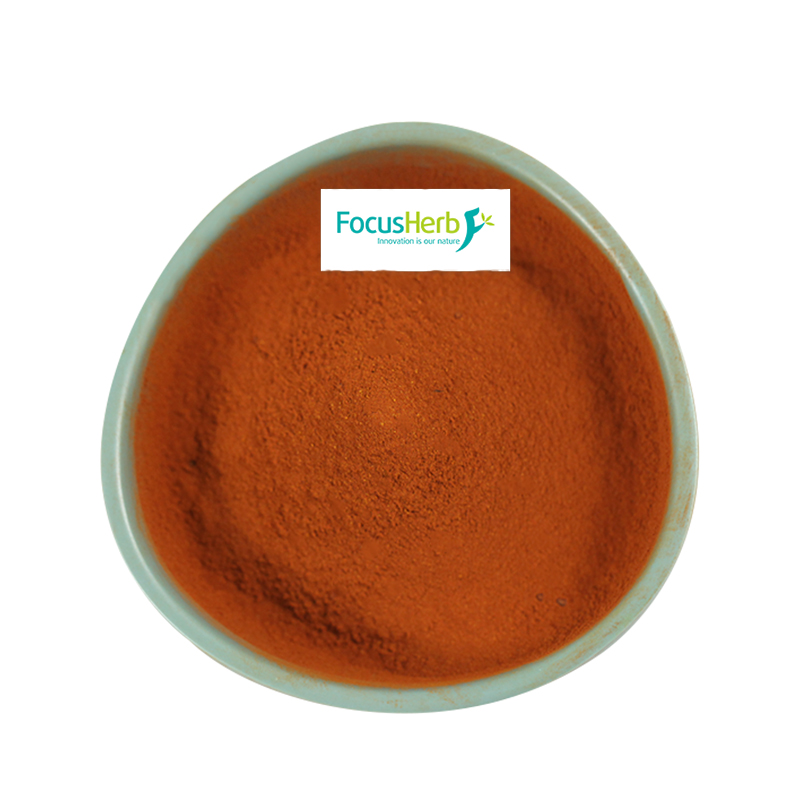
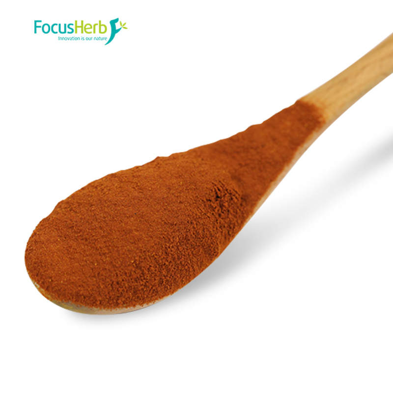
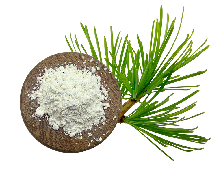
The key to Organic Siberian Ginseng’s remarkable anti-fatigue and endurance-enhancing effects lies in its rich content of diverse active ingredients, such as eleutherosides, flavonoids, and polysaccharides, which work through multi-target regulatory mechanisms. The hypothalamic-pituitary-adrenal (HPA) axis is a crucial regulatory system for the body’s response to stress. When the body is under chronic stress, the HPA axis becomes overactivated, leading to the continuous secretion of stress hormones such as cortisol. This leads to adverse consequences such as excessive energy consumption and metabolic disorders, ultimately causing fatigue. The active ingredients in Organic Siberian Ginseng can precisely target the HPA axis, restoring its balance. Studies have shown that in experimental animal models exposed to chronic psychological stress, supplementation with Organic Siberian Ginseng extract significantly reduced fluctuations in blood cortisol levels, stabilizing them within normal levels. This indicates that chronic stress is effectively alleviated, laying the foundation for maintaining optimal bodily function.

In terms of energy metabolism, liver glycogen acts as the body’s “energy reserve.” Adequate glycogen reserves provide a continuous supply of energy for bodily activity. Organic Siberian Ginseng can significantly promote liver glycogen synthesis and increase glycogen reserves. Furthermore, muscle mitochondria, the “energy factories” of the cell, are directly determined by their functional state. Siberian Ginseng can optimize mitochondrial structure and function, increasing the activity of mitochondrial respiratory chain enzymes, enabling mitochondria to produce more adenosine triphosphate (ATP) during aerobic respiration, providing sufficient energy for muscle contraction. During exercise, as energy is consumed, lactic acid gradually accumulates in muscle tissue and blood, leading to increased muscle soreness and fatigue. Increased blood urea nitrogen (BUN), a product of protein breakdown, also reflects increased fatigue. Organic Siberian Ginseng can effectively delay lactic acid accumulation and BUN increases during exercise by accelerating lactic acid metabolism and inhibiting excessive protein breakdown. Animal experimental data strongly supports this finding. Mice that consumed Siberian Ginseng extract for 30 days showed a 20%-25% increase in muscle glycogen reserves compared to the control group. After exercising at the same intensity, blood lactate clearance was accelerated by 15%. This clearly demonstrates the significant effects of Siberian Ginseng in enhancing energy metabolism and improving exercise endurance.

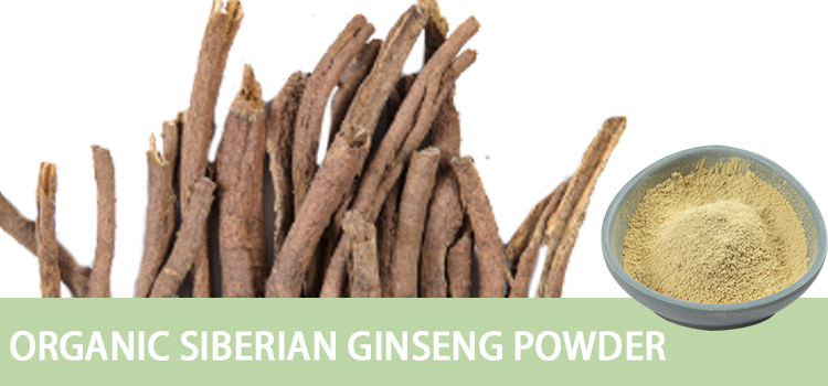
Traditional Chinese Medicine Theory’s “Qi-Tonifying and Foundation-Strengthening” Effect

In the long-standing framework of traditional Chinese medicine, Organic Siberian Ginseng holds a unique and important position. Its pungent, slightly bitter flavor and warm nature activate the lung, spleen, and kidney meridians, demonstrating its remarkable efficacy in “tonifying Qi, strengthening the spleen, tonifying the kidneys, and calming the mind.” It is particularly effective in treating fatigue caused by Qi deficiency. From the perspective of Traditional Chinese Medicine, the spleen is the foundation of acquired constitution and the source of Qi and blood production. If the spleen is weak and its transport and transformation functions are dysfunctional, it cannot fully transform nutrients from food into Qi and blood, leading to Qi and blood deficiency and a series of fatigue-related symptoms such as fatigue, loss of appetite, and listlessness. The pungent and dispersing properties of Organic Siberian Ginseng can promote the flow of Qi, allowing spleen and stomach Qi to flow smoothly and enhancing their transport and transformation functions. Its bitter and laxative properties can remove dampness and turbidity from the spleen and stomach, restoring their normal function. In clinical practice, many patients with spleen deficiency caused by long-term fatigue and irregular diets have experienced significant improvements in appetite, reduced fatigue, and improved mental state after taking Organic Siberian Ginseng.

The kidneys are the foundation of innate constitution and store essence. The essence stored in the kidneys is the fundamental material basis for growth, development, reproduction, and the maintenance of life. When kidney essence is deficient, the body’s overall function declines, resulting in symptoms such as soreness in the waist and knees, dizziness, and tinnitus, as well as a reduced tolerance to various types of fatigue. Organic Siberian Ginseng can nourish the kidneys and replenish essence, replenishing lost essence and strengthening the body’s innate foundation, thereby enhancing the body’s ability to cope with physical and mental fatigue. For example, those engaged in high-intensity mental work and prolonged mental stress are prone to kidney essence depletion, fatigue, and insomnia. Taking Organic Siberian Ginseng can alleviate fatigue, improve sleep quality, and enhance work efficiency.

As early as ancient times, people recognized the miraculous benefits of organic Siberian Ginseng. The Compendium of Materia Medica, written by Li Shizhen during the Ming Dynasty, clearly states that “long-term consumption of Siberian Ginseng can lighten the body and prevent aging,” fully demonstrating the ancients’ profound understanding that long-term use of Siberian Ginseng can strengthen the body, delay aging, and improve physical endurance. In modern clinical practice, Organic Siberian Ginseng has also demonstrated promising therapeutic effects for chronic fatigue syndrome (CFS), a difficult condition that plagues many patients. Patients with CFS often experience persistent and difficult-to-relieve fatigue, decreased concentration, memory loss, and sleep disturbances, which severely impact their quality of life. Clinical studies have shown that after intervention treatment with Organic Siberian Ginseng, patients’ fatigue was significantly reduced, their attention and memory improved, and their sleep quality also improved to a certain extent. This further verifies the important role of Organic Siberian Ginseng in relieving fatigue and enhancing the body’s overall function under the guidance of the Traditional Chinese Medicine theory of “replenishing Qi and strengthening the foundation.”


Organic Siberian Ginseng has a dual regulatory effect on physical and nervous fatigue.(I) Improving Exercise Endurance and Optimizing Recovery
In today’s fast-paced world, more and more people are turning to exercise to maintain health and relieve stress

However, fatigue often hinders continued exercise. For athletes, Organic Siberian Ginseng is like a key to improving physical fitness and facilitating recovery. Its key to success lies in its subtle regulation of the circulatory system. Siberian Ginseng dilates blood vessels, increasing their diameter and promoting smoother blood flow. It also improves microcirculation, allowing blood to penetrate deeper into muscle tissue. This process is like bringing abundant water to parched farmland, providing muscle cells with abundant oxygen and nutrients, ensuring adequate energy supply during exercise.

When muscles undergo high-intensity exercise, they produce large amounts of lactic acid. Lactic acid accumulation leads to muscle soreness and fatigue, impairing both performance and post-exercise recovery. Organic Siberian Ginseng accelerates lactic acid metabolism, rapidly breaking down, converting, and excreting lactic acid from muscles, effectively reducing the amount of lactic acid remaining in the body after exercise. Studies have shown that after equal intensity exercise, blood lactate levels in participants taking Organic Siberian Ginseng decreased 20% to 30% faster than those who did not. This means they can recover faster from exercise fatigue, reduce muscle soreness, and improve post-exercise comfort.

Creatine kinase (CK) is a key indicator of muscle damage and fatigue. During exercise, especially after high-intensity exercise, muscle cells sustain a degree of damage, leading to the release of CK into the blood and an increase in CK levels. Organic Siberian Ginseng can reduce the magnitude of the post-exercise CK increase, suggesting that it protects the integrity of muscle cell membranes, reduces muscle cell damage, and promotes rapid recovery. For example, in a study on long-distance runners, after taking an Organic Siberian Ginseng preparation for one month, the experimental group performed a long-distance run of the same intensity. The post-training elevation in creatine kinase levels in the experimental group was 30%-40% lower than in the control group, demonstrating the significant efficacy of Organic Siberian Ginseng in maintaining muscle health and accelerating physical recovery.

In the military, high-intensity physical training is an essential component of daily soldier training, placing extremely high demands on a soldier’s physical strength and endurance. Military medical researchers conducted a study on soldiers, having one group take an Siberian Ginseng preparation during their daily training, while the other group, a control group, did not. After a period of training, the soldiers were tested in a 5,000-meter run. The results showed that the soldiers who took the Siberian Ginseng preparation improved their 5,000-meter run times by 8%-12% compared to the control group. This improvement is of significant significance in actual combat and military missions, enabling soldiers to reach designated locations faster and seize opportunities on the battlefield. Furthermore, after exercise, their peak blood urea nitrogen (BUN) levels decreased by 18%. BUN is a product of protein breakdown. This reduction indicates that protein breakdown in the soldiers’ bodies has been effectively suppressed, reducing fatigue. This effectively ensures that soldiers maintain optimal physical condition during continuous combat or high-intensity training.

Furthermore, exercise-induced oxidative stress is a significant factor in fatigue and muscle damage. Organic Siberian Ginseng senticosus is rich in antioxidants, such as flavonoids and polysaccharides. These antioxidants can effectively inhibit exercise-induced oxidative stress. They scavenge excess free radicals in the body, reducing their attack and damage to muscle cell membranes, maintaining their integrity and thereby reducing the incidence of delayed onset muscle soreness (DOMS). Many athletes experience muscle soreness and stiffness the day after or three after high-intensity exercise. This is called delayed onset muscle soreness (DOMS), which can affect subsequent training and competition. Athletes who take Organic Siberian Ginseng experience effective control of oxidative stress, reduced muscle cell damage, and significantly lower rates of delayed onset muscle soreness, allowing them to return to the next round of training and competition more quickly.

(II)Relieving Neurological Fatigue and Maintaining Cognitive Function

In today’s information-rich world, more and more people are engaging in high-intensity mental work. Long hours of work, study, and mental stress have made neurological fatigue a problem for many. Organic Siberian Ginseng is a timely relief for those who engage in long-term mental work, effectively alleviating neurological fatigue and maintaining cognitive function. The key to its effectiveness lies in its precise regulation of central nervous system neurotransmitters, which are important chemicals that transmit information in the nervous system. Their balance directly affects mood, sleep, and cognitive function. Siberian Ginseng can regulate the balance of central nervous system neurotransmitters such as serotonin and dopamine. Serotonin, known as the “happiness transmitter,” regulates mood and produces feelings of pleasure, while dopamine plays a vital role in attention, learning, and memory. When the body is under prolonged stress and anxiety, the secretion of these neurotransmitters becomes disrupted, leading to symptoms such as depression, anxiety, and insomnia, which in turn cause neural fatigue.

By regulating the balance of neurotransmitters, Siberian Ginseng can alleviate anxiety and promote a more relaxed and calm mood. It also improves sleep quality, allowing the brain to fully rest and recover. Sleep is crucial for brain health. Good sleep clears metabolic waste from the brain, consolidates memory, and restores neural function. Many people with chronic insomnia experience cognitive impairments such as memory loss, difficulty concentrating, and slowed reaction times due to insufficient rest. By improving sleep quality, Siberian Ginseng indirectly enhances the brain’s ability to resist fatigue, maintaining a clear and alert mind and improving work and learning efficiency.

Clinical trials provide strong evidence for this benefit of Siberian Ginseng. In a study of office workers who engaged in long-term mental work, subjects who took Siberian Ginseng extract for four weeks showed a 35% reduction in their visual analogue scale (VAS) fatigue scores. The Visual Analog Fatigue Scale (VAS) is a commonly used method for assessing fatigue. It involves asking participants to mark their fatigue level on a straight line, with higher scores indicating greater fatigue. This result indicates that taking Siberian Ginseng significantly reduced participants’ fatigue. Furthermore, participants’ error rates on a working memory task decreased by 22%, indicating a significant improvement in cognitive function, enabling them to more accurately process and retain information, leading to better performance at work and in school.

Further research has revealed that eugenol, a compound found in Siberian Ginseng, possesses unique physiological activity. It can penetrate the blood-brain barrier and directly act on brain neurons. The blood-brain barrier is a natural protective barrier that prevents many harmful substances from entering the brain, but it also restricts the entry of some drugs and nutrients. However, eugenol successfully penetrates this barrier and enters the brain. Once inside the brain, eugenol inhibits neuronal apoptosis, a programmed cell death that can lead to decreased brain function. By inhibiting this process, eugenol protects neuronal survival and maintains the number and function of neurons in the brain. In addition, it can enhance synaptic plasticity in the hippocampus, a region of the brain closely associated with learning and memory. Synaptic plasticity refers to the adjustability of synaptic transmission efficiency. Enhancing synaptic plasticity in the hippocampus can promote the transmission of neural signals and improve learning and memory. For those experiencing chronic stress, such as career elites and students, the eugenol in Siberian Ginseng can effectively protect against neurocognitive fatigue caused by chronic stress. This allows them to maintain a healthy mental state and cognitive abilities despite the intense pressures of work and study, avoiding problems such as decreased work efficiency and poor academic performance due to neural fatigue.


Scientific Application and Practical Plans for Organic Siberian Ginseng

(I) Applicable Populations and Targeted Intervention Scenarios

Athletes and Fitness Enthusiasts: For fitness enthusiasts and professional athletes striving for exceptional performance, organic Siberian Ginseng is undoubtedly a powerful aid. Regularly taking Organic Siberian Ginseng starting 30 days before a major competition or intense training session can provide strong support for improved physical function, enabling full performance and superior results. After intense competition or high-intensity training, the body is often extremely fatigued. Organic Siberian Ginseng can accelerate recovery, allowing muscles to recover more quickly and reducing soreness, allowing for optimal preparation for the next training session or competition. For optimal results, it is recommended to choose a standardized extract containing eleutherosides B+E at least 0.8%, with a daily dose between 100 and 200 mg. This dosage maximizes the benefits of Siberian Ginseng while ensuring safety and avoiding adverse effects from inappropriate dosing. Chronic Fatigue and Sub-Healthy People: Under the fast-paced lifestyle and work pressures of modern society, chronic fatigue and sub-health issues are becoming increasingly common. Many people suffer from long-term symptoms such as fatigue and sleep disturbances, significantly reducing their quality of life. For this group, Organic Siberian Ginseng is an ideal choice for conditioning. It is recommended to take 3-5g of dried Siberian Ginseng daily, boil it in water, and drink it instead of regular tea. This allows the active ingredients of Siberian Ginseng to slowly absorb into the body and gradually improve symptoms. Furthermore, according to Traditional Chinese Medicine (TCM) theory of compatibility, combining Organic Siberian Ginseng with Astragalus and Lycium barbarum can synergistically replenish Qi, further enhancing the conditioning effect. Astragalus has benefits such as replenishing Qi, strengthening the exterior, promoting diuresis, and reducing swelling, while Lycium barbarum nourishes the liver and kidneys, improves essence, and improves eyesight. The three ingredients work together to replenish the body’s essential Qi and blood, nourish the liver and kidneys, and regulate overall body function, allowing those suffering from chronic fatigue and sub-health to gradually regain vitality and rediscover a healthy lifestyle. High-pressure intellectual workers: Those in high-pressure intellectual jobs, such as researchers, programmers, and financial professionals, face long periods of intense concentration and work throughout the day. This can lead to rapid brain fatigue and difficulty concentrating. Taking Siberian Ginseng capsules in the morning or afternoon is an effective relief for this group. Each capsule contains 50mg of extract, which can quickly replenish brain energy and boost brain activity. Combined with deep breathing exercises, this can further relax the mind and body, increase oxygen intake, promote blood circulation, and provide the brain with a more adequate supply of nutrients. This significantly enhances attention span in the afternoon, improves work efficiency, and reduces errors caused by fatigue and lack of focus.

(II) Safe Consumption and Synergistic Combination Strategies

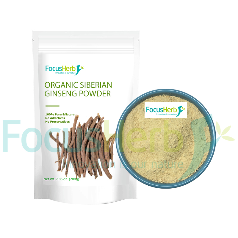
Organic Siberian Ginseng is grown in strict accordance with organic standards, eliminating the use of pesticides and chemical fertilizers, and avoiding heavy metal contamination. This ensures the purity of its active ingredients, ensuring greater quality and safety. In terms of consumption, Organic Siberian Ginseng inherits traditional health wisdom while integrating the convenience of modern technology. Traditionally, Siberian Ginseng root bark is thinly sliced ​​and stewed with black-bone chicken in a soup, such as Siberian Ginseng stewed with black-bone chicken. This dish is nutritious and nourishing. Eating it two to three times a week not only allows for the enjoyment of a delicious meal, but also allows the body to absorb the nutrients of Siberian Ginseng, achieving fatigue-fighting and physical fitness benefits. This traditional cooking method not only preserves the original flavor of Siberian Ginseng but also allows its nutrients to be better absorbed into the soup, making them easier for the body to absorb.

With the advancement of modern technology, convenient dosage forms such as standardized extract capsules and oral liquids have emerged, providing more convenient consumption options. These dosage forms precisely control the content of active ingredients, ensuring consistent results with each dose. However, when taking these dosage forms, it is important to ensure that they are taken at least two hours apart from caffeinated beverages. Caffeine has a stimulating effect on the nerves. Taking it with Siberian Ginseng may overload the heart, causing symptoms such as palpitations and insomnia. Therefore, arranging your intake schedule appropriately can maximize the benefits of Siberian Ginseng while ensuring your health and safety.

Organic Siberian Ginseng can also be made into Siberian Ginseng honey paste, a health supplement that cleverly combines Siberian Ginseng with honey. Before bed, take 10g of Siberian Ginseng honey paste with warm water. This paste not only benefits from honey’s nourishing, moistening, and laxative properties, but also taps into Siberian Ginseng’s calming properties, helping you relax, relieve a day’s fatigue, and enter a sweet dream. Furthermore, during sleep, Siberian Ginseng helps the body store energy, providing abundant energy for the next day’s work and life.

Despite its numerous benefits, Organic Siberian Ginseng should be used with caution. Those with a Yin deficiency and excessive fire constitution, characterized by symptoms such as hot flashes, night sweats, dry mouth, and irritability, should use Siberian Ginseng. Because Siberian Ginseng has a warming nature, it may exacerbate symptoms of Yin deficiency and excessive fire, exacerbating physical discomfort. In addition, Siberian Ginseng may enhance platelet inhibition, so it should be avoided when taken with anticoagulants such as warfarin and aspirin. If patients taking anticoagulants need to use Siberian Ginseng, they must do so under the guidance of a doctor and closely monitor coagulation function to prevent adverse reactions such as bleeding and ensure medication safety.


Quality Identification and Usage Contraindications of Organic Siberian Ginseng

(I) Core Characteristics of High-Quality Organic Siberian Ginseng

When choosing Organic Siberian Ginseng, it’s crucial to understand the core characteristics of a quality product. This not only impacts its effectiveness but also its health. First, ensure the product has received reputable organic certification, such as the US National Organic Program (NOP) or EU organic certification. These certifications signify that the entire growing and processing process adheres to strict organic standards, resulting in the absence of pesticides and fertilizers, ensuring reliable quality.

Appearantly, the root bark of high-quality Organic Siberian Ginseng appears grayish-yellow, a reflection of its natural, uncontaminated growth. When broken, the cross-section reveals distinct radial grain patterns, a key indicator of high-quality Siberian Ginseng. A close sniff reveals a faint, subtle fragrance, a characteristic of Siberian Ginseng. The slightly bitter taste distinguishes it from other plants.

In addition to appearance and aroma, physical and chemical properties are also key factors in evaluating the quality of Organic Siberian Ginseng. Eleutherodactyls B+E, the key active ingredients in Siberian Ginseng, are crucial in their content. High-quality products should contain 1.0% or higher of these substances. Only by meeting this standard can the product’s anti-fatigue and endurance-enhancing properties be fully realized. Furthermore, impurity levels must be strictly controlled to ≤1% to prevent other impurities from affecting product purity and efficacy. The total amount of heavy metals (lead, cadmium, and mercury) must be less than 5 ppm, a stringent requirement for product safety and to ensure that we are not exposed to the harmful effects of heavy metal contamination during consumption.

There are also some low-quality Siberian Ginseng products on the market, and sulfur-fumed or dyed products are particularly alarming. While sulfur-fuming these products may appear more vibrant, they contain large amounts of residual sulfur dioxide. Long-term consumption of these products can lead to the accumulation of sulfur dioxide in the body, causing serious damage to liver and kidney function and potentially endangering health. Dyed products may also use harmful chemical dyes, which also pose a threat to human health. Therefore, when purchasing Organic Siberian Ginseng, it is important to carefully identify and purchase from reputable channels to ensure product quality.

(II) Contraindications and Safe Dosage Limits

Although Organic Siberian Ginseng is a natural health product for most people, it is not suitable for everyone. Clearly identifying contraindications and safe dosage limits is crucial. Pregnant women experience a unique physiological state during pregnancy, and the fetus requires a stable and safe environment for development. Organic Siberian Ginseng may have potential adverse effects on the fetus, so it should be strictly avoided by pregnant women to ensure its healthy development. Breastfeeding women should also exercise caution, as Siberian Ginseng ingredients may be passed to the baby through breast milk. The baby’s body functions are not yet fully developed, and these ingredients cannot be effectively metabolized, potentially posing a health risk.

For patients with autoimmune diseases, such as systemic lupus erythematosus and rheumatoid arthritis, whose immune systems are disrupted, Siberian Ginseng may stimulate the immune system, triggering fluctuations in the condition and exacerbating symptoms. Therefore, Siberian Ginseng should also be avoided.

For those with a damp-heat constitution, symptoms such as bitter taste in the mouth, bad breath, a yellow, greasy tongue coating, and sticky, uncomfortable stools often indicate a damp-heat internal environment. Organic Siberian Ginseng has a warming nature. Consuming it may encourage damp-heat in the body, aggravating symptoms. Therefore, caution is advised for this group of individuals. Individuals with yin deficiency and excessive fire should also exercise caution. Yin deficiency leads to insufficient yin fluids, while excessive fire leads to internal fire, manifesting as dry mouth and throat, hot hands and soles, hot flashes, and night sweats. The warming nature of Siberian Ginseng may further damage yin fluids, exacerbating symptoms of yin deficiency and excessive fire, and causing dry mouth, insomnia, and other discomfort. Therefore, caution is also advised for this group of individuals.

In terms of a safe dosage, the recommended daily intake of dry Siberian Ginseng for adults is no more than 10g. This dosage ensures that Siberian Ginseng exerts its health benefits while avoiding adverse reactions from excessive intake. For extract preparations, the daily intake should be calculated based on the content of eleutherosides B+E, with a maximum limit of 300mg. Continuous use should not exceed three months. Long-term use may lead to tolerance to Siberian Ginseng, reducing its efficacy and increasing the risk of adverse reactions. For long-term health maintenance, it’s recommended to take the herb for three consecutive months, followed by a two-week break before continuing. This allows the body ample time to metabolize and adjust, while maintaining the beneficial effects of Siberian Ginseng, ensuring safety and tolerability.

Organic Siberian Ginseng, with its triple mechanism of “stress regulation, energy optimization, and nerve repair,” is a natural choice for combating modern fatigue syndrome. From athletes’ endurance breakthroughs to urban dwellers’ energy management, its scientific application requires consideration of individual constitutions and usage scenarios, ensuring precise dosage to achieve the health goal of “fighting fatigue without harming the body.” Choosing certified organic products and adhering to safety regulations can maximize the modern health benefits of this traditional herb.


Sodium Copper Chlorophyllin: Research on promoting hematopoiesis from molecular mechanism to clinical application

Sodium copper chlorophyllin, a water-soluble derivative of chlorophyll, has a core structure consisting of a porphyrin ring. This porphyrin ring is highly similar to the porphyrin structure of hemoglobin, like a pair of “twins” meticulously designed by nature. Hemoglobin plays a crucial role in the body’s hematopoiesis, carrying oxygen and delivering it to tissues and organs throughout the body, a crucial substance for maintaining life. This similarity between the porphyrin ring of sodium copper chlorophyllin and hemoglobin enables it to directly participate in the metabolism of heme precursors. From a molecular perspective, it is like a precise “key,” skillfully inserting itself into the metabolic “keyhole” of hematopoiesis and exerting a unique effect.

This is supported by numerous studies and experimental data. For example, in an experiment published in Zhejiang Medical Journal, researchers studied models of iron deficiency and aplastic anemia. After applying sodium copper chlorophyllin to these anemia models, they surprisingly found a significant increase in red blood cell counts, by as much as 23%. This data clearly demonstrates that sodium copper chlorophyllin can effectively promote the production of red and white blood cells by replenishing hematopoietic raw materials, acting as a kind of “raw material” for the hematopoietic “factory,” offering hope for improving anemia.

Regulatory Mechanisms of the Bone Marrow Hematopoietic Microenvironment

The bone marrow is like the “super factory” of hematopoiesis in the human body, and the bone marrow hematopoietic microenvironment serves as its “production workshop” and “support system,” playing a crucial regulatory role in the production and development of blood cells. Sodium copper chlorophyllin exerts its miraculous regulatory influence within this “factory,” primarily by activating the proliferation and differentiation of bone marrow pluripotent stem cells (CFU-S), granulocyte-monocyte progenitor cells (CFU-GM), and erythroid progenitor cells (CFU-E).

When sodium copper chlorophyllin enters the human body, it acts as an infusion of “vitalizing factors” into these stem and progenitor cells. In animal experiments, the significant changes it brings are clearly visible. Experimental results showed that sodium copper chlorophyllin increased the number of nucleated cells in the bone marrow by 41%, significantly increasing the number of “production workers” in the hematopoietic “factory,” enabling more efficient blood cell production. It also promoted the release of neutrophils from the bone marrow reserve pool into the peripheral blood, like deploying “reserve troops” to the “front lines” in a timely manner, enhancing the blood’s immune and transport functions.

In addition, studies on irradiated mice provided strong evidence for the efficacy of sodium copper chlorophyllin. In the experiments, the hematopoietic system of irradiated mice was severely damaged. However, after administration of sodium copper chlorophyllin, platelet counts increased by 35%. This data demonstrates that sodium copper chlorophyllin can effectively repair the damaged hematopoietic system. Further research has shown that it also accelerates the repair of bone marrow stromal cells. Bone marrow stromal cells act as the “infrastructure” of the “production plant,” supporting the survival, proliferation, and differentiation of hematopoietic stem cells. Sodium copper chlorophyllin provides a good living and development environment for hematopoietic stem cells by rebuilding the support function of the hematopoietic microenvironment, allowing the hematopoietic “factory” to resume normal production order, continuously produce various blood cells, and maintain the normal physiological functions of the human body.

Precisely Applicable Populations and Clinical Benefit Scenarios

(I) Core Applicable Populations for Hematologic Diseases

Leukopenia Patients

In modern medical treatment, while many treatments can have a therapeutic effect, they can also cause side effects, with leukopenia being a common one. For example, the antipsychotic drug clozapine, while highly effective in treating severe mental illnesses like schizophrenia, can also cause leukopenia. This puts patients at increased risk of infection while simultaneously treating their mental illness. Similarly, interferon therapy for chronic hepatitis B can also cause adverse reactions such as bone marrow suppression, leading to a decrease in white blood cell count and severely impacting treatment tolerance. According to relevant studies, approximately 30%-40% of patients treated with interferon for chronic hepatitis B experience varying degrees of leukopenia.

The emergence of sodium copper chlorophyllin offers new hope for these patients. By enhancing bone marrow hematopoietic function, sodium copper chlorophyllin can increase white blood cell counts by an average of 1.2×10⁹/L, with an efficacy rate of up to 72% (Journal of Clinical Hematology). Clinically, sodium copper chlorophyllin is particularly effective in patients with grade I-II bone marrow suppression (white blood cell count 3.0-3.9×10⁹/L). In a clinical study of 100 patients with leukopenia caused by antipsychotic medication, sodium copper chlorophyllin significantly increased white blood cell counts in 70 patients after one month of treatment, with 40 returning to normal. This significantly reduced the risk of infection caused by granulocytopenia and significantly improved their quality of life. This demonstrates the effectiveness and importance of sodium copper chlorophyllin in the treatment of leukopenia. It can safeguard patients’ treatment, reduce the risks associated with leukopenia, and allow them to more safely receive treatment for other illnesses.

Adjunctive Therapy for Aplastic Anemia

Aplastic anemia is a serious hematologic disorder with a complex pathogenesis, primarily due to bone marrow failure, leading to pancytopenia. Currently, immune-mediated damage is considered a key mechanism in the development of aplastic anemia. In this condition, the body’s immune system mistakenly attacks its own hematopoietic stem cells, resulting in suppressed hematopoiesis. Serum cytokines such as IFN-γ and TNF-α play a crucial role in this process, inhibiting the proliferation and differentiation of hematopoietic cells and further exacerbating the condition. Simultaneously, the CD4⁺/CD8⁺ immune balance in the patient’s body is disrupted, leading to immune dysfunction and making the disease even more difficult to control.

Sodium copper chlorophyllin has demonstrated unique adjunctive therapeutic potential in the treatment of aplastic anemia. In models of immune-mediated aplastic anemia, sodium copper chlorophyllin combined with cyclosporine has achieved remarkable results. By reducing serum levels of hematopoietic inhibitory factors such as IFN-γ and TNF-α, sodium copper chlorophyllin can effectively mitigate the immune system’s attack on hematopoietic stem cells, creating a favorable environment for their proliferation and differentiation. It also restores the CD4⁺/CD8⁺ immune balance, modulating the body’s immune function and enhancing resistance. Experimental data show a 28% increase in platelet count compared to the cyclosporine alone group, which is of significant significance in improving patients’ bleeding tendency. In clinical practice, many patients with aplastic anemia have experienced significant improvement in bleeding symptoms and a significant improvement in their quality of life after receiving sodium copper chlorophyllin combined with cyclosporine. This provides a new and effective treatment option for aplastic anemia.

(II) Hematopoietic Support Associated with Hepatobiliary Disease

The liver plays a vital role in the human body. It participates in important physiological processes such as metabolism and detoxification and is closely linked to hematopoiesis. When liver diseases occur, such as acute or chronic hepatitis, liver function is impaired to varying degrees. This not only affects the liver’s own normal function but also indirectly impacts the hematopoietic system. In patients with acute or chronic hepatitis, liver cell damage suppresses the function of the liver’s reticuloendothelial cells, weakening their support for hematopoietic stem cells. Impaired liver function also leads to impaired synthesis of coagulation factors, predisposing patients to bleeding. Furthermore, the liver’s reduced ability to metabolize and store nutrients impairs the supply of raw materials for hematopoiesis, leading to anemia.

Sodium copper chlorophyllin has a unique mechanism of action in the treatment of acute and chronic hepatitis. It revitalizes the liver’s reticuloendothelial cells, acting as a “vitalizer” to restore and enhance their function. In this way, sodium copper chlorophyllin accelerates liver cell repair and promotes the recovery of liver function. Clinical observations have shown that treatment with sodium copper chlorophyllin reduces serum total bilirubin by 19%, demonstrating significant improvement in the liver’s bilirubin metabolism and reduced jaundice symptoms. At the same time, a 15% reduction in prothrombin time indicates that coagulation factor synthesis is promoted, improving the patient’s coagulation function and reducing the risk of bleeding. In a clinical study of 200 patients with acute and chronic hepatitis, after three months of treatment with sodium copper chlorophyllin, 160 patients showed significant improvement in liver function indicators, and 120 of these patients also experienced varying degrees of relief from anemia. This fully demonstrates the significant effectiveness of sodium copper chlorophyllin in improving coagulation factor synthesis disorders and anemia caused by impaired liver function. It can provide strong support for the recovery of patients with hepatobiliary diseases and improve their quality of life.

Contraindications and Risk Warnings

Absolute Contraindications

Sodium copper chlorophyllin is absolutely contraindicated for individuals with copper metabolism disorders (such as Wilson’s disease) and those with drug allergies. Wilson’s disease is an autosomal recessive copper metabolism disorder in which copper transporters malfunction, leading to copper ion accumulation. If these individuals use sodium copper chlorophyllin, the copper ions contained in the drug can further exacerbate copper ion accumulation and cause severe hepatocerebral toxicity. For those with drug allergies, sodium copper chlorophyllin may trigger allergic reactions, which can be life-threatening in severe cases. Therefore, for the safety of these individuals, its use must be strictly prohibited.

Use with caution in pregnant and lactating women

Due to their unique physiological conditions, there is currently no definitive safety data for sodium copper chlorophyllin in these groups. Although sodium copper chlorophyllin has a good safety profile in the general population, pregnant and lactating women may metabolize and respond to the drug differently, potentially affecting the development of the fetus or infant through the placenta or breast milk. Therefore, these two groups of patients should use this medication with caution after a comprehensive evaluation by a doctor, weighing the pros and cons. Patients with renal failure, due to impaired renal function, can be affected in the excretion of drug metabolites, leading to drug retention and an increased risk of adverse reactions. Therefore, these patients should reduce their sodium copper chlorophyllin dosage and closely monitor their renal function to adjust the dosage promptly.

Managing Adverse Reactions

While most patients tolerate sodium copper chlorophyllin well, a few may experience adverse reactions. Urticaria-like allergic reactions or severe abdominal pain are relatively serious adverse reactions. If these symptoms occur, the patient should discontinue the medication immediately to prevent further aggravation of the adverse reaction. For urticaria-like allergic reactions, antihistamines can be used to alleviate symptoms. Antihistamines block the binding of histamine to receptors, thereby reducing the itching, redness, and swelling associated with allergic reactions. In most cases, symptoms can be effectively relieved by promptly discontinuing the medication and using an antihistamine. However, in rare cases, symptoms may be severe and difficult to relieve with conventional treatments. In these cases, patients must seek medical attention and receive professional treatment in a timely manner.

Future Directions

(I) In-depth Analysis of the Mechanism of Action

Although it is currently known that sodium copper chlorophyllin can promote hematopoiesis, its specific mechanism of action still requires further investigation. Further clarification is needed regarding the regulatory effects of sodium copper chlorophyllin on molecules on the surface of hematopoietic stem cells. For example, molecules such as CD34⁺ and CD117⁺ on the surface of hematopoietic stem cells play a key role in regulating their self-renewal, proliferation, and differentiation. Studying how sodium copper chlorophyllin affects the expression of these molecules and the mechanisms of their interaction will reveal the underlying mechanisms by which it promotes hematopoiesis. Through a series of advanced cell-based experimental techniques, such as flow cytometry and gene editing, we can precisely observe and analyze the regulatory effects of sodium copper chlorophyllin on these surface molecules.

Furthermore, the specific targets by which sodium copper chlorophyllin reduces oxidative damage in hematopoietic cells through the Nrf2 antioxidant pathway also require further clarification. During normal hematopoietic processes, hematopoietic cells are affected by various internal and external factors, resulting in oxidative stress, leading to cell damage and hematopoietic dysfunction. The Nrf2 antioxidant pathway is a crucial intracellular antioxidant defense mechanism, regulating the expression of a range of antioxidant and detoxification enzymes to maintain intracellular redox balance. Sodium copper chlorophyllin may activate the Nrf2 antioxidant pathway, reducing oxidative damage in hematopoietic cells and thereby promoting hematopoiesis. However, further research is needed to elucidate the specific targets and how this pathway is activated. By combining high-throughput technologies such as proteomics and transcriptomics with molecular biological validation, we hope to identify key targets, providing a more solid theoretical basis for precision-targeted therapies and developing more effective treatment strategies, bringing greater benefits to patients with cytopenias.

(II) Dosage Form Innovation and Delivery Optimization

In current clinical applications, sodium copper chlorophyllin is primarily administered in the form of oral tablets. However, this traditional dosage form has limitations, such as low bioavailability and gastrointestinal irritation. To overcome these issues, the development of an enteric-coated sustained-release formulation is a viable option. Enteric-coated sustained-release formulations slowly release drugs in the intestine, preventing them from being destroyed by gastric acid in the stomach, thereby improving drug bioavailability and reducing gastrointestinal irritation. By utilizing advanced coating technologies and sustained-release materials, such as enteric coatings, microspheres, and nanoparticles, sodium copper chlorophyllin is encapsulated, allowing it to be released under the specific pH conditions of the intestine. This results in slow, sustained release of the drug, prolonging its duration of action and enhancing therapeutic efficacy.

In addition to innovative dosage forms, exploring targeted delivery technologies using nanocarriers is also an important direction for future research. Nanocarriers possess unique physicochemical properties, such as small size effects, high surface area, and excellent biocompatibility, enabling them to effectively deliver drugs to specific tissues and organs. By loading sodium copper chlorophyllin onto nanocarriers, such as liposomes, polymer nanoparticles, and nanomicelles, targeted drug delivery can be achieved, allowing the drug to be concentrated in the bone marrow microenvironment. The bone marrow microenvironment is a crucial habitat for the survival and differentiation of hematopoietic stem cells. Increasing drug concentration in the bone marrow microenvironment can more effectively promote the recovery of hematopoietic function. Research has shown that targeted nanocarrier delivery technology can increase local drug concentration by 3-5 times, offering new strategies and hope for the treatment of diseases such as refractory anemia. Through research on nanocarrier surface modification, particle size control, and drug loading optimization, we can further enhance the targeting and drug delivery efficiency of nanocarriers, opening new avenues for the clinical application of sodium copper chlorophyllin.

Phycocyanin: Multi-organ protection mechanism under the blue natural barrier

Phycocyanin (PC), a treasure crafted by nature, is primarily extracted from blue-green algae, an ancient and remarkable group of organisms, of which Spirulina and Arthrospira are prominent representatives. Its captivating blue color, a hue that seems to blend the depths of the ocean and the mystical starry sky, is unique, striking, and unforgettable. A molecular analysis reveals that phycocyanin has a molecular weight of approximately 110 kDa and is composed of a clever combination of α and β subunits. It also contains an open-ring tetrapyrrole structure. This structure not only endows phycocyanin with unique pigmentary properties, enabling it to play a key role in photosynthesis, efficiently capturing and transmitting light energy to sustain algae, but also imparts many of the properties of proteins, with a rich amino acid sequence that lays the foundation for its biological activity. This unique molecular structure, like a precisely engineered molecular machine, gives phycocyanin its potent antioxidant and anti-inflammatory properties, making it a core functional component that protects multiple organ health. As an antioxidant, it acts like a fierce guardian, rapidly identifying and eliminating free radicals generated in the body. These restless “troublemakers” wreak havoc on cellular structures and biomolecules. Phycocyanin effectively blocks these destructive actions, reducing oxidative stress damage to cells, thereby delaying cell aging and preventing various diseases caused by oxidative stress. Phycocyanin also excels in the anti-inflammatory field, precisely modulating inflammation-related signaling pathways, inhibiting the excessive release of inflammatory factors, and reducing the damage to tissues and organs caused by inflammatory responses, thereby safeguarding the body’s health and stability.

Bioavailability and Benefits

In the past, the application of phycocyanin was limited by its low bioavailability, like a bound sword, unable to fully unleash its powerful effects. However, with the rapid development of technology, the emergence of nano-delivery technology has brought new hope for the application of phycocyanin. Through nanotechnology techniques such as liposome encapsulation, phycocyanin is coated with a special “protective coat.” This coat not only effectively protects phycocyanin from degradation in the gastrointestinal tract but also facilitates its absorption, significantly increasing its intestinal absorption rate to 35%. This represents a significant improvement compared to traditional extracts, effectively opening the door for phycocyanin to function, allowing it to more smoothly enter the human circulatory system and reach various tissues and organs, exerting its health benefits.

Phycocyanin’s multi-target mechanism of action offers unique advantages in protecting organ health. It not only directly scavenges free radicals, reducing oxidative damage to cells and tissues, and preventing organ disease at its source, but also penetrates deep into cells to modulate organ-specific signaling pathways. For example, in the liver, phycocyanin can modulate the Nrf2 signaling pathway, activate the expression of a series of antioxidant enzymes, and enhance the liver’s antioxidant defenses. In the cardiovascular system, it can regulate the PI3K/Akt signaling pathway, inhibiting inflammation and apoptosis, protecting the integrity of vascular endothelial cells, and maintaining normal cardiovascular function. This level of protection from cells to organs enables phycocyanin to comprehensively protect the health of human organs, just like a comprehensive health guard, providing meticulous care for each organ of the body.

Core Mechanism of Organ Protection: Driven by Dual Antioxidant and Anti-Inflammatory Pathways

In the complex physiological processes that maintain human organ health, phycocyanin acts as a multifaceted guardian. Leveraging its potent antioxidant and anti-inflammatory properties, and through unique molecular mechanisms, it safeguards organ health at multiple levels, playing a crucial protective role at the cellular and molecular levels.

(I) Free Radical “Scavenger”: Blocking the Chain of Oxidative Damage

During normal physiological metabolism, the human body continuously produces free radicals, a natural part of life. However, when free radical production exceeds the body’s scavenging capacity, it’s like opening a Pandora’s box, and the shadow of oxidative stress quietly looms. Excessive free radicals, particularly superoxide anions (O₂⁻・) and hydroxyl radicals (・OH), are highly chemically active, acting like rampaging “molecular bombs,” indiscriminately attacking various biomolecules within cells. These free radicals react without hesitation with unsaturated fatty acids in cell membranes, triggering a chain reaction of lipid peroxidation. Once lipid peroxidation initiates, it’s like lighting a string of firecrackers. The structure and function of cell membranes are severely damaged, altering membrane fluidity and permeability, disrupting the balance of intracellular and extracellular material exchange, and significantly disrupting normal cellular function. Furthermore, free radicals can damage vital biomolecules within cells, such as DNA and proteins, leading to serious consequences like gene mutations and loss of protein function. These changes underlie the development and progression of many chronic diseases, such as cardiovascular disease, neurodegenerative diseases, and cancer.

The emergence of phycocyanin, like a ray of hope in the darkness, offers hope for resolving this dilemma. Its unique molecular structure exhibits remarkable free radical scavenging capabilities, making it a “killer” of intracellular free radicals. The aromatic amino acid residues in phycocyanin, such as tryptophan and histidine, act like carefully crafted “traps,” efficiently capturing superoxide anions (O₂⁻・) and hydroxyl radicals (・OH) through conjugation, stabilizing these dangerous free radicals and rendering them inactive, preventing them from attacking biomolecules within the cell. Phycocyanin also reacts specifically with lipid peroxides, acting like a skilled craftsman, converting them into stable lipid peroxide-phycocyanin complexes. This effectively inhibits the progression of lipid peroxidation and protects cells from further oxidative damage. Research data shows that in an oxidative stress model, malondialdehyde (MDA) levels were significantly reduced by over 40% after phycocyanin treatment. This significant decrease in MDA, a hallmark product of lipid peroxidation, directly demonstrates the powerful efficacy of phycocyanin in inhibiting lipid peroxidation. It also demonstrates that phycocyanin can effectively reduce free radical damage to cell membranes and other biological structures, maintaining normal cellular morphology and function.

In addition to directly scavenging free radicals, phycocyanin also has a more profound strategic significance: it activates the cell’s endogenous antioxidant defense system, awakening a group of dormant “guardians” and building a strong antioxidant defense line. Phycocyanin can activate the Nrf2 signaling pathway, which acts as a “command center” for intracellular antioxidant defense. Once activated by phycocyanin, it initiates a series of instructions, inducing the expression of antioxidant enzyme genes within the cell. Superoxide dismutase (SOD) and glutathione peroxidase (GSH-Px) are key members of this defense system. Superoxide dismutase (SOD) catalyzes the dismutation of superoxide anions (O₂⁻・), converting them into hydrogen peroxide (H₂O₂) and oxygen, effectively removing superoxide anions and reducing their cellular damage. Glutathione peroxidase (GSH-Px), using glutathione as a substrate, reduces hydrogen peroxide (H₂O₂) and lipid peroxides to water and the corresponding alcohols, further eliminating harmful substances produced by oxidative stress. When phycocyanin upregulates the activity of superoxide dismutase (SOD) and glutathione peroxidase (GSH-Px), the antioxidant capacity within cells is significantly enhanced, enabling them to better cope with free radical attacks and maintain intracellular redox balance. In animal experiments, feeding animals a phycocyanin-rich diet significantly increased the activity of superoxide dismutase (SOD) and glutathione peroxidase (GSH-Px) in their tissues, with increases ranging from 30% to 50% compared to the control group. This demonstrates the remarkable effectiveness of phycocyanin in activating the endogenous antioxidant defense system, providing more comprehensive and long-lasting protection for cells. This dual mechanism of action, combining direct free radical scavenging with activation of the endogenous antioxidant defense system, makes phycocyanin highly effective in interrupting the oxidative damage chain. It provides a solid foundation for cellular and organ health from multiple perspectives and levels, effectively preventing and mitigating organ damage and disease caused by oxidative stress.

(II) Inflammation “Regulatory Valve”: Inhibiting the Release of Proinflammatory Mediators

Inflammation is a normal defensive response of the body to external stimuli, acting like a warning signal. When the body is exposed to pathogen invasion, physical or chemical damage, or other stimuli, the inflammatory response rapidly activates. Various cells and molecules of the immune system rapidly flock to the damaged area, attempting to eliminate the pathogen and repair damaged tissue. However, when the inflammatory response becomes uncontrolled and overactivated, it becomes a double-edged sword, causing severe damage to the body’s own tissues and organs and becoming the root cause of many diseases. The nuclear factor-κB (NF-κB) pathway plays a key role in the inflammatory response. It acts as the “master switch” of the inflammatory response. Once activated, it triggers a complex chain reaction, leading to the massive release of multiple proinflammatory cytokines, such as tumor necrosis factor-α (TNF-α) and interleukin-6 (IL-6). These pro-inflammatory factors act like a swarm of uncontrolled “destroyers.” They stimulate the activation and aggregation of immune cells, leading to dilation and increased permeability of blood vessels at the site of inflammation, triggering inflammatory symptoms such as redness, swelling, and pain. Furthermore, they further activate other inflammatory signaling pathways, creating a vicious cycle that amplifies the inflammatory response and severely damages the normal structure and function of surrounding tissues and organs. Long-term chronic inflammation is also closely linked to the development and progression of numerous chronic diseases, including cardiovascular disease, diabetes, and neurodegenerative disorders, posing a serious threat to human health.

Phycocyanin plays a crucial role in regulating inflammatory responses. It precisely targets inflammatory signaling pathways, acting like a skilled “tuner,” modulating the intensity of the inflammatory response to maintain a moderate level and preventing excessive inflammation from causing damage to the body. Studies have found that phycocyanin can effectively block the activation of the NF-κB pathway. By inhibiting the phosphorylation of IκBα, it prevents the translocation of NF-κB from the cytoplasm to the nucleus, thereby disrupting NF-κB’s regulatory effects on proinflammatory genes and reducing the transcription and secretion of proinflammatory factors such as tumor necrosis factor-α (TNF-α) and interleukin-6 (IL-6). In a lipopolysaccharide (LPS)-induced inflammation model, a commonly used experimental model for studying inflammatory responses, phycocyanin intervention significantly suppressed the intensity of the inflammatory response, decreasing it by 55% compared to the untreated group. Levels of proinflammatory factors such as tumor necrosis factor-α (TNF-α) and interleukin-6 (IL-6) were also significantly reduced. This demonstrates the powerful ability of phycocyanin to inhibit the NF-κB pathway and reduce the secretion of proinflammatory factors, effectively alleviating the damage caused by inflammatory responses to tissues and organs. In addition to inhibiting the NF-κB pathway, phycocyanin can also alleviate inflammation through other pathways. It inhibits the release of inflammatory mediators such as histamine and 5-hydroxytryptamine. Histamine and 5-hydroxytryptamine are important mediators of inflammation, causing inflammatory symptoms such as vasodilation, increased vascular permeability, and tissue edema. By inhibiting the release of these inflammatory mediators, phycocyanin can effectively reduce localized inflammatory edema and tissue damage, alleviating the pain and discomfort caused by inflammation. In an allergic inflammation model, phycocyanin intervention significantly reduced the release of histamine and 5-hydroxytryptamine, and significantly improved edema and itching at the inflamed site, further confirming the positive role of phycocyanin in inhibiting the release of inflammatory mediators and alleviating inflammatory symptoms. Phycocyanin regulates inflammatory responses through multiple targets and pathways. It effectively inhibits the over-activation of inflammatory responses by blocking the NF-κB pathway, inhibiting the secretion of pro-inflammatory factors, and reducing the release of inflammatory mediators, providing powerful protection for organ health. It has great potential in preventing and treating inflammation-related diseases.

Precise Organ-Targeted Protection

In the complex and delicate human body, each organ functions like a finely tuned component, working together to maintain the normal course of life. Phycocyanin, with its unique bioactivity, can penetrate deeply into various organs, targeting their physiological characteristics and common damage mechanisms, exerting precise and effective protective effects, thus building a solid defense for organ health.

(I) Liver: The Core Guardian of Metabolism and Detoxification

As the largest organ in the human body, the liver functions like a highly efficient “biochemical factory,” playing an irreplaceable and crucial role in numerous physiological processes, including metabolism, detoxification, and immune regulation. It undertakes the important task of synthesizing, decomposing, transforming, and storing various nutrients ingested by the body, while also facing the invasion of various harmful substances, such as chemical toxins, drugs, and alcohol. In modern life, due to factors such as environmental pollution, unhealthy diet, and excessive alcohol consumption, the liver faces unprecedented challenges, becoming highly susceptible to damage. This, in turn, can lead to a range of liver diseases, such as chemical liver damage and non-alcoholic fatty liver disease, which pose a serious threat to human health. Phycocyanin, with its potent antioxidant and anti-inflammatory properties, has become a loyal guardian of liver health, safeguarding normal liver function in multiple ways.

1. Protection against Chemical Liver Damage

Among the many factors that cause liver damage, chemical toxins are a significant threat. Carbon tetrachloride (CCl₄), a typical hepatotoxic chemical, is widely used in experimental studies to induce chemical liver damage. Upon entering the human body, CCl₄ is metabolized by the liver’s cytochrome P450 enzyme system to produce highly reactive trichloromethyl free radicals (・CCl₃) and trichloromethyl peroxide free radicals (・OO CCl₃). These free radicals act like frenzied “destroyers,” inducing lipid peroxidation within hepatocytes, severely damaging the structure and function of liver cell membranes and subsequently inducing hepatocyte apoptosis, significantly impacting the liver’s normal metabolic and detoxification functions.

Phycocyanin demonstrates remarkable protective capabilities against CCl₄-induced liver damage. Studies have found that phycocyanin can significantly reduce the rate of CCl₄-induced hepatocyte apoptosis by 38%. Its mechanism of action is primarily through activation of the Nrf2 pathway. The Nrf2 pathway is a key intracellular antioxidant stress response pathway. Upon activation by phycocyanin, Nrf2 enters the cell nucleus and binds to the antioxidant response element (ARE), initiating the transcription and expression of a series of antioxidant enzyme genes, such as heme oxygenase-1 (HO-1) and NAD(P)H quinone oxidoreductase 1 (NQO1). These antioxidant enzymes effectively scavenge intracellular free radicals, enhance hepatocyte membrane stability, and reduce damage to hepatocyte membranes caused by lipid peroxidation, thereby protecting hepatocytes from the threat of apoptosis. In related cell experiments, hepatocytes pretreated with phycocyanin showed a significant decrease in malondialdehyde (MDA) levels and a significant increase in the activities of antioxidant enzymes such as superoxide dismutase (SOD) and glutathione peroxidase (GSH-Px) after exposure to CCl₄. This strongly suggests that phycocyanin, by activating the Nrf2 pathway, enhances the antioxidant defenses of hepatocytes, effectively alleviating CCl₄-induced oxidative damage and reducing hepatocyte apoptosis.

In addition to chemical toxins, alcohol is also a common cause of liver damage. After entering the body, alcohol is primarily metabolized in the liver. Its metabolite, acetaldehyde, is highly toxic and can bind to biomacromolecules such as proteins and nucleic acids within hepatocytes, forming acetaldehyde-protein adducts. This alters the structure and function of these proteins and nucleic acids, thereby affecting the normal metabolism and physiological function of hepatocytes. Acetaldehyde also damages mitochondria within hepatocytes, leading to a decrease in mitochondrial membrane potential, impaired respiratory chain function, reduced ATP production, and disrupted cellular energy metabolism, further exacerbating hepatocyte damage. Long-term, heavy drinking can also lead to fatty liver degeneration, causing alcoholic fatty liver disease, which can progress to cirrhosis and liver cancer in severe cases. Phycocyanin has significant benefits in alcohol detoxification and liver protection. It accelerates the clearance of acetaldehyde, a metabolite of ethanol, effectively reducing mitochondrial damage, thereby enhancing the liver’s ability to detoxify. Several active ingredients in phycocyanin, such as tryptophan and histidine, specifically bind to acetaldehyde, forming stable complexes. This reduces acetaldehyde concentrations in the body and mitigates its toxic effects on liver cells. Phycocyanin also activates acetaldehyde dehydrogenase (ALDH) in the liver, accelerating the conversion of acetaldehyde to acetic acid and promoting its metabolism and excretion. In animal experiments, supplementing phycocyanin with alcohol-administered ethanol resulted in significantly reduced acetaldehyde levels in the blood and liver of mice. The morphology and function of liver mitochondria were significantly improved, mitochondrial membrane potential returned to normal, respiratory chain enzyme activity was enhanced, and ATP production significantly increased. This suggests that phycocyanin can effectively mitigate alcohol-induced damage to liver mitochondria, protect the energy metabolism function of hepatocytes, enhance the liver’s ability to detoxify, and reduce the risk of alcohol-induced liver injury.

2. Promotes Hepatocyte Regeneration

The regenerative capacity of hepatocytes is crucial for liver repair and functional recovery. Following liver injury, hepatocytes initiate a regenerative process, proliferating and differentiating to repair damaged liver tissue. However, in certain pathological conditions, such as severe liver injury and chronic liver disease, this regenerative capacity can be inhibited, hindering the liver’s repair process and impairing normal liver function. Phycocyanin plays a key role in promoting hepatocyte regeneration. It can upregulate HGF (hepatocyte growth factor) expression and promote the proliferation of hepatic progenitor cells, significantly increasing the rate of liver tissue repair by 25%. HGF is an important cell growth factor that binds to the c-Met receptor on the surface of hepatocytes, activating multiple downstream signaling pathways, such as the Ras-Raf-MEK-ERK pathway and the PI3K-Akt pathway. Activation of these signaling pathways promotes DNA synthesis, cell division, and proliferation in hepatocytes, while also inhibiting apoptosis and promoting hepatocyte survival and regeneration. Phycocyanin, by upregulating HGF expression, provides strong support for hepatocyte regeneration and accelerates the repair of damaged liver tissue. In a partial hepatectomy model, mice treated with phycocyanin showed significantly faster liver regeneration than controls, with a significant increase in the number of proliferating cell nuclear antigen (PCNA)-positive cells in liver tissue. This indicates that phycocyanin effectively promotes hepatocyte proliferation and accelerates liver regeneration and repair.
Non-alcoholic fatty liver disease (NAFLD) is a common liver disease characterized by excessive fat accumulation in the liver, leading to hepatocyte steatosis, inflammatory infiltration, and fibrosis. In severe cases, it can progress to cirrhosis and liver cancer. The development of non-alcoholic fatty liver disease (NAFLD) is associated with a variety of factors, including obesity, insulin resistance, hyperlipidemia, and metabolic syndrome. A key factor in the development of NAFLD is the disrupted lipid metabolism within hepatocytes. Excessive fatty acids accumulate within hepatocytes, leading to increased triglyceride synthesis and decreased fatty acid oxidation and transport, resulting in lipid deposition.

Phycocyanin can effectively improve lipid deposition in NAFLD, reducing hepatic triglyceride levels by 22%. Its mechanism of action primarily involves regulating signaling pathways and gene expression related to lipid metabolism. Phycocyanin can activate the AMPK signaling pathway in the liver. AMPK is a key regulator of intracellular energy metabolism. Activated AMPK inhibits the synthesis of fatty acids and triglycerides while promoting fatty acid oxidation and transport. Specifically, AMPK inhibits the activity of acetyl-CoA carboxylase (ACC), reducing the synthesis of malonyl-CoA and thereby inhibiting de novo fatty acid synthesis. AMPK also activates carnitine acyltransferase 1 (CPT1), promoting the entry of fatty acids into mitochondria for β-oxidation and accelerating fatty acid catabolism. Furthermore, phycocyanin regulates the expression of genes related to lipid metabolism in the liver, such as peroxisome proliferator-activated receptor α (PPARα) and fatty acid binding protein 1 (FABP1). Changes in the expression of these genes help maintain lipid homeostasis in the liver and reduce lipid deposition in hepatocytes. In an animal model of non-alcoholic fatty liver disease induced by a high-fat diet, phycocyanin administration significantly reduced liver steatosis, triglyceride and cholesterol levels, decreased inflammatory cell infiltration in the liver, and decreased the expression of inflammatory factors. This suggests that phycocyanin can effectively improve the lipid metabolism disorders and inflammatory state in non-alcoholic fatty liver disease, and has a promising preventive and therapeutic effect on non-alcoholic fatty liver disease.

(II) Cardiovascular System: Precision Maintenance of Circulatory Dynamics

The cardiovascular system is like the human body’s “transportation network.” The heart, acting as the “power pump” of this network, continuously and rhythmically contracts and relaxes, propelling blood through the vessels, delivering oxygen and nutrients to tissues and organs throughout the body while carrying metabolic waste back to the liver and kidneys for processing. Blood vessels, like the crisscrossing “pipelines,” are responsible for the transport and distribution of blood. Endothelial cells form the barrier lining the vascular walls, not only maintaining the normal structure and function of the vessels but also participating in many important physiological processes, such as vasodilation, coagulation regulation, and inflammatory responses. However, under the influence of modern lifestyles, the cardiovascular system faces numerous challenges, including risk factors such as hypertension, hyperlipidemia, hyperglycemia, smoking, and obesity. These factors can lead to cardiovascular diseases such as endothelial dysfunction, atherosclerosis, and myocardial ischemia, seriously threatening human health. Phycocyanin, with its unique biological activity, can precisely maintain the cardiovascular system in multiple ways, reducing the risk of cardiovascular disease and providing strong support for heart and vascular health.

1. Strengthening the Vascular Endothelial Barrier

Endothelial cells serve as the first line of defense in the vascular lining. Their functional integrity is crucial for maintaining normal vascular function. When endothelial cells are stimulated by various risk factors, such as oxidative stress, inflammation, and hypertension, endothelial dysfunction can occur, manifesting as decreased nitric oxide (NO) synthesis, increased release of vasoconstrictor factors, inflammatory cell adhesion, and platelet aggregation. These changes further promote the development and progression of atherosclerosis, increasing the risk of cardiovascular disease. Nitric oxide (NO) is an important vasodilator, produced from L-arginine by nitric oxide synthase (NOS) in endothelial cells. NO diffuses into vascular smooth muscle cells, activates guanylate cyclase, and increases intracellular cyclic guanosine monophosphate (cGMP) levels, leading to vascular smooth muscle relaxation, vasodilation, reduced vascular resistance, and the maintenance of normal blood pressure and circulation. Phycocyanin plays a crucial role in strengthening the vascular endothelial barrier. It promotes nitric oxide (NO) synthesis, enhancing vasodilation by 30%, and thus effectively delaying the formation of atherosclerotic plaques. Through its potent antioxidant capacity, phycocyanin scavenges free radicals within endothelial cells, reducing oxidative damage to these cells, protecting nitric oxide synthase (NOS) activity, and promoting NO synthesis and release. Furthermore, phycocyanin modulates signaling pathways related to endothelial function, such as the PI3K-Akt pathway and the eNOS-NO pathway. Activating these pathways promotes eNOS phosphorylation, increasing its activity and further increasing NO production. In vitro cell experiments showed that treatment of vascular endothelial cells with phycocyanin significantly increased intracellular NO content and enhanced vasodilation. In animal studies, animals fed a phycocyanin-rich diet showed significant improvement in endothelial function, with a significant reduction in the area and thickness of atherosclerotic plaques. This suggests that phycocyanin can promote NO synthesis, enhance vasodilation, effectively protect the endothelial barrier, and slow the progression of atherosclerosis.

Abnormal proliferation of vascular smooth muscle cells is a key pathological basis for atherosclerosis and vascular remodeling. During the development of atherosclerosis, vascular smooth muscle cells, stimulated by inflammatory factors and growth factors, shift from a contractile to a synthetic phenotype. This enhanced proliferation leads to migration beneath the vascular intima, where they synthesize and secrete large amounts of extracellular matrix, resulting in intimal thickening and stenosis, increasing the risk of thrombosis. Phycocyanin inhibits vascular smooth muscle cell proliferation, reducing intimal thickness by 18%, thereby effectively reducing the risk of thrombosis. Phycocyanin inhibits cell proliferation by inhibiting signaling pathways related to cell proliferation, such as the MAPK and NF-κB pathways, reducing the expression of cell cycle proteins and growth factors, and preventing vascular smooth muscle cells from transitioning from the G1 phase to the S phase, thereby inhibiting cell proliferation. Phycocyanin can also induce vascular smooth muscle cell apoptosis, reducing cell number and further alleviating intimal thickening. In animal models of vascular injury, phycocyanin treatment significantly reduced intimal thickening and significantly inhibited the proliferation and migration of vascular smooth muscle cells. This indicates that phycocyanin can effectively inhibit abnormal vascular smooth muscle cell proliferation, maintain normal vascular structure and function, and reduce the risk of thrombosis.

2. Cardiomyocyte Energy Protection

Cardiomyocytes are the primary executors of cardiac contraction and relaxation, and their normal energy metabolism is crucial for maintaining the heart’s pumping function. Cardiomyocyte energy primarily derives from aerobic respiration in mitochondria, where nutrients are converted into ATP through oxidative phosphorylation, providing energy for cardiomyocyte contraction and relaxation. However, in pathological conditions such as myocardial ischemia-reperfusion injury, cardiomyocyte mitochondrial function can be severely affected, leading to impaired energy metabolism, decreased ATP production, intracellular calcium overload, and cardiomyocyte apoptosis and necrosis, severely impacting cardiac function. Mitochondrial complex I is a crucial component of the mitochondrial respiratory chain, participating in electron transport and proton transport, and playing a key role in ATP production. During myocardial ischemia-reperfusion injury, mitochondrial complex I activity is inhibited, resulting in blocked electron transport, an inability to establish a proton gradient, and reduced ATP production. Phycocyanin can enhance mitochondrial complex I activity, increasing ATP production by 28%, thereby effectively ameliorating myocardial ischemia-reperfusion injury. Through its antioxidant properties, phycocyanin scavenges free radicals within cardiomyocytes, reduces oxidative stress-induced mitochondrial damage, and protects the structure and function of mitochondrial complex I. Phycocyanin can also regulate signaling pathways related to mitochondrial biogenesis and function, such as the PGC-1α pathway and the Nrf1 pathway, promoting mitochondrial biogenesis and repair, and improving mitochondrial function and efficiency. In an animal model of myocardial ischemia-reperfusion, pretreatment with phycocyanin significantly increased mitochondrial complex I activity and ATP content in myocardial tissue, significantly reduced the rate of cardiomyocyte apoptosis, and significantly improved cardiac function. This suggests that phycocyanin can effectively protect cardiomyocyte energy metabolism and mitigate myocardial ischemia-reperfusion injury by enhancing mitochondrial complex I activity and increasing ATP production.

Oxidative stress is a key factor contributing to cardiomyocyte apoptosis. During myocardial ischemia-reperfusion, the massive production of oxygen free radicals disrupts redox homeostasis in cardiomyocytes, activating apoptotic signaling pathways such as the caspase-3 pathway and the mitochondrial apoptosis pathway, leading to cardiomyocyte apoptosis. The size of myocardial infarction is an important indicator of the severity of myocardial damage. Reducing myocardial infarction size is crucial for protecting cardiac function and reducing mortality. Phycocyanin can reduce oxidative stress-induced cardiomyocyte apoptosis and reduce the size of myocardial infarction by 20%. Phycocyanin inhibits myocardial apoptosis by scavenging free radicals in myocardial cells, inhibiting oxidative stress and reducing the expression of apoptosis-related proteins such as Caspase-3 and Bax, while increasing the expression of anti-apoptotic proteins such as Bcl-2. In an animal model of myocardial infarction, phycocyanin treatment significantly reduced the number of apoptotic cells in myocardial tissue, significantly reduced the size of myocardial infarction, and significantly improved cardiac systolic and diastolic function. This suggests that phycocyanin can effectively inhibit oxidative stress-induced cardiomyocyte apoptosis, reduce the size of myocardial infarction, and protect cardiac function.

(III) Gastrointestinal Tract: Expert in Mucosal Barrier Repair

The gastrointestinal tract, a vital site for digestion and absorption in the human body, is like a busy “production line,” responsible for digesting food, absorbing nutrients, and excreting waste. The gastrointestinal mucosa is the first line of defense against invading pathogens and harmful substances. Composed of epithelial cells, mucus, and immune cells, it provides multiple functions, including mechanical, chemical, and immune barriers, effectively protecting the gastrointestinal tract from damage. However, in daily life, the gastrointestinal tract is frequently irritated by various factors, such as gastric acid, Helicobacter pylori infection, nonsteroidal anti-inflammatory drugs, alcohol, and spicy foods. These factors can damage the gastrointestinal mucosa, leading to diseases such as gastritis, gastric ulcers, enteritis, and ulcerative colitis, impairing normal gastrointestinal function. Phycocyanin, with its unique biological activity, can precisely repair and regulate the gastrointestinal mucosal barrier, maintaining gastrointestinal health.

Repair of Gastric Mucosal Damage

The gastric mucosa is the innermost layer of the stomach wall. It comes into direct contact with digestive juices such as gastric acid and pepsin, and is also exposed to external pathogens and harmful substances, making it vulnerable to damage. Gastric acid, a key component of gastric juice, plays a vital role in food digestion. However, when gastric acid secretion is excessive or the gastric mucosal defenses are weakened, it can damage the gastric mucosa, leading to lesions such as gastric erosion and ulcers. Gastric ulcers are a common digestive system disease characterized by defects and ulcer formation in the gastric mucosa. Patients often experience symptoms such as upper abdominal pain, acid reflux, and belching, which seriously affect their quality of life.

Phycocyanin, an alkaline protein, effectively neutralizes gastric acid and regulates gastric pH, thereby accelerating the healing of gastric mucosal ulcers by 40%. Basic amino acid residues in phycocyanin, such as arginine and lysine, bind to hydrogen ions in gastric acid, reducing the concentration of gastric acid and alleviating irritation and damage to the gastric mucosa. Phycocyanin also promotes the proliferation and repair of gastric mucosal cells, upregulating the expression of genes associated with cell proliferation and repair, such as epidermal growth factor (EGF) and transforming growth factor-α (TGF-α). These growth factors stimulate the division and proliferation of gastric mucosal cells, promoting ulcer healing. In an animal model of gastric ulcers, treatment with phycocyanin significantly reduced the size of gastric ulcers, reduced their depth, and accelerated their healing rate. The expression levels of EGF and TGF-α in gastric mucosal tissue also increased significantly, indicating that phycocyanin can effectively promote the healing of gastric ulcers by neutralizing gastric acid and promoting the proliferation and repair of gastric mucosal cells.

The gastric mucosa is covered with a thick layer of mucus, composed of glycoproteins secreted by gastric epithelial cells. It lubricates and protects the gastric mucosa, protecting it from erosion by gastric acid and pepsin. Helicobacter pylori is a Gram-negative bacterium that colonizes the gastric mucosa. By secreting substances such as urease and cytotoxins, it damages the mucus layer and epithelial cells, leading to gastritis, gastric ulcers, and gastric cancer. Phycocyanin promotes the synthesis of glycoproteins in the mucus layer, strengthening the mucosal barrier and reducing H. pylori adhesion. Phycocyanin stimulates signaling pathways within gastric epithelial cells, such as the PI3K-Akt and MAPK pathways, promoting the expression of genes involved in glycoprotein synthesis and increasing glycoprotein content in the mucus layer, thereby strengthening the thickness and function of the mucosal barrier. Furthermore, phycocyanin, through its antimicrobial and anti-inflammatory properties, inhibits the growth and reproduction of H. pylori, reducing its adhesion and damage to the gastric mucosa. In in vitro experiments, after treating gastric mucosal epithelial cells with phycocyanin, the content of glycoprotein secreted by the cells increased significantly and the thickness of the mucus layer increased; in an animal model infected with Helicobacter pylori, the number of Helicobacter pylori in the gastric mucosal tissue decreased significantly after intervention with phycocyanin.

Application Transformation: From Mechanism Research to the Health Industry

With the deepening of research on the organ-protective mechanisms of phycocyanin, significant progress has been made in its application and transformation within the health industry. From functional foods to pharmaceuticals, phycocyanin is bringing new benefits to people’s health with its unique biological activities.

(I) Functional Foods and Dietary Supplements

In the functional foods and dietary supplements sector, phycocyanin is gradually emerging as a new option for those pursuing a healthier lifestyle. Researchers have carefully formulated phycocyanin into chewable tablets. This convenient dosage form offers consumers a simple and effective supplemental option. Clinical studies have demonstrated that daily intake of phycocyanin chewable tablets at the recommended dose of 20-50mg significantly increases liver GSH (glutathione) levels by 18%. As a key antioxidant in the liver, increased GSH levels enhance the liver’s antioxidant capacity, better protecting against free radical attack and reducing oxidative stress damage to liver cells, thereby effectively preventing and improving liver diseases such as chemical-induced liver damage and non-alcoholic fatty liver disease. To further expand the application of phycocyanin in functional foods, researchers have ingeniously combined it with spirulina polysaccharides to develop a stomach-protecting beverage. Spirulina polysaccharides inherently possess numerous biological activities, including immune regulation and antioxidant activity. When combined with phycocyanin, the two work synergistically, significantly enhancing the protection and repair of the gastrointestinal mucosa. Clinical trials have shown that this stomach-protecting beverage has a 63% remission rate for chronic gastritis. It works through multiple pathways. On the one hand, phycocyanin neutralizes gastric acid, reducing irritation and damage to the gastric mucosa and promoting the proliferation and repair of gastric mucosal cells. On the other hand, spirulina polysaccharides enhance the gastric mucosal barrier function, inhibit the adhesion of Helicobacter pylori, and reduce inflammatory responses. This combination of the two benefits chronic gastritis patients, effectively alleviating symptoms such as stomach pain, bloating, and acid reflux, and improving their quality of life.

(II) Targeted Formulations in the Pharmaceutical Field

In the pharmaceutical field, the application of phycocyanin is developing further towards targeted formulations, offering new hope for disease treatment. Researchers have used advanced liposome encapsulation technology to formulate phycocyanin into an injectable solution. This targeted formulation precisely targets the liver, providing strong support for protecting against chemotherapy-induced liver damage. In a Phase III clinical trial, patients treated with the liposome-encapsulated phycocyanin injection achieved a 41% improvement in liver enzyme levels. During chemotherapy, commonly used chemotherapy drugs such as cisplatin and doxorubicin, while killing cancer cells, can also severely damage liver cells, leading to elevated liver enzyme levels and impairing normal liver function. Liposome-encapsulated phycocyanin injection effectively mitigates chemotherapy-induced liver damage by inhibiting oxidative stress and inflammation, protecting liver cell structure and function, reducing liver enzyme levels, and promoting liver repair and regeneration, thereby improving chemotherapy tolerance and enhancing treatment efficacy.

For allergic rhinitis, a common respiratory disease, researchers have developed a nasal formulation of phycocyanin, offering patients a new treatment option. Clinical studies have shown that the intensity of histamine stimulation in patients treated with this formulation decreased by 50%. Allergic rhinitis is an inflammatory response to the nasal mucosa caused by allergens. Histamine is a key mediator in allergic reactions. Its release causes vasodilation and increased permeability in the nasal mucosa, leading to symptoms such as itching, sneezing, and runny nose. Phycocyanin nasal formulations can effectively repair damaged nasal mucosa in patients with allergic rhinitis, modulate the immune response, and inhibit the release of inflammatory mediators such as histamine, thereby alleviating nasal inflammation, alleviating symptoms, and improving patients’ quality of life.

(III) Safety and Dosage Consensus

Safety and dosage are of great concern in the application of phycocyanin. After rigorous research and evaluation, the European Food Safety Authority (EFSA) has determined that the safe upper limit of phycocyanin intake is 200 mg per day. Extensive experimental data demonstrates that within this dosage range, phycocyanin does not cause significant toxic effects on vital organs such as the liver and kidneys, providing safety assurance for its use in functional foods and pharmaceuticals. However, long-term or excessive intake may still pose potential risks, such as increased metabolic burden on the liver and kidneys. While there is currently no conclusive clinical evidence indicating serious health problems, caution is advised during use.

Regarding drug interactions, particular caution is required when using phycocyanin with medications such as warfarin. Warfarin is a commonly used anticoagulant that works by inhibiting the synthesis of vitamin K-dependent coagulation factors. Phycocyanin may compete with warfarin for plasma protein binding sites, thereby affecting warfarin’s blood concentration and anticoagulant effect. Therefore, to avoid the risk of drug interactions, a two-hour interval is recommended when using phycocyanin and warfarin simultaneously to ensure that both drugs can safely and effectively exert their respective effects and protect the patient’s medication safety.

Challenges and Future: Decoding the Deep Potential of Blue Guardian

(I) Organ-Specific Target Analysis

Although phycocyanin has demonstrated significant efficacy in multi-organ protection, its specific targets and mechanisms of action in different organs remain under investigation. Clarifying the differential mechanisms of phycocyanin’s protection in organs such as the kidney and lung is key to further exploring its potential. For example, in the kidney, studies have found that energy metabolism in renal tubular epithelial cells is crucial for maintaining normal renal function. However, whether phycocyanin specifically improves energy metabolism in renal tubular epithelial cells through the AMPK pathway remains unclear. Further investigation of this mechanism will help reveal the unique mode of action of phycocyanin in kidney protection and provide more precise theoretical support for the prevention and treatment of kidney disease. Regarding the lung, phycocyanin’s protective effect on lung tissue may involve regulating multiple pathways, including inflammatory cell infiltration, oxidative stress, and extracellular matrix remodeling. However, the specific molecular targets and signaling pathways require further investigation. Comprehensively elucidating the targets and signaling networks of phycocyanin in different organs through advanced technologies such as single-cell sequencing and proteomics will lay a solid foundation for its precise application in clinical treatment.

(II) Delivery System Optimization

Although the bioavailability of phycocyanin has improved, significant room for improvement remains. Developing more efficient delivery systems is a key approach to enhancing the organ-protective effects of phycocyanin. For example, the development of intestinal pH-responsive microcapsules is of great significance. These microcapsules precisely release phycocyanin according to varying intestinal pH conditions, thereby preventing degradation in adverse environments such as gastric acid and ensuring efficient absorption and utilization within the intestine. By rationally designing the materials and structures of microcapsules, it is possible to increase the bioavailability of phycocyanin to over 50%, enabling more effective accumulation in target organs and enhancing their protective effects. Furthermore, the continuous advancement of nanotechnology offers more possibilities for phycocyanin delivery. For example, the use of novel nanocarriers such as nanoliposomes and nanomicelles not only improves the stability and solubility of phycocyanin but also enables targeted delivery to specific organs through surface modification, further enhancing its efficacy in organ protection.

Due to its natural pleiotropic properties, phycocyanin is evolving from a marine bioactive ingredient to a core functional factor in multi-organ protection. With the deepening of mechanism research and innovation in delivery technology, this blue guardian is expected to play a greater role in preventing organ degenerative diseases and improving organ function, becoming a bridge connecting nature’s gifts with precise health.

Quercetin: From Natural Antioxidant to Anti-Aging Star: A Scientific Study

Quercetin (C₁₅H₁₀O₇), a generous gift from nature, is widely distributed throughout the plant kingdom. A polyphenolic flavonoid, it’s commonly found in a variety of fruits and vegetables, from the vibrant red of apple peels to the vibrant purple of blueberries, the emerald green of broccoli, and the distinctively fragrant onions. These foods are a natural reservoir of quercetin.

From a chemical perspective, quercetin resembles a meticulously constructed molecular edifice, containing multiple phenolic hydroxyl groups. These phenolic hydroxyl groups are more than just a simple presence; they are the key to quercetin’s potent antioxidant properties, endowing it with exceptional free radical-snatching abilities. Like trained “free radical hunters,” phenolic hydroxyl groups quickly attack free radicals once they wreak havoc in the body, binding to them and subduing them, thus preventing them from damaging cells. Among natural antioxidants, quercetin is undoubtedly one of the best. Measured by its ORAC (oxygen radical absorbance capacity) value, quercetin boasts a staggering 2800μmol TE/100g. This value clearly demonstrates its potent antioxidant potential, making it one of the most active natural antioxidant ingredients in nature. Like a vanguard on the anti-oxidation battlefield, it safeguards the health of organisms.

From Dietary Supplement to Precise Anti-Aging Agent

Quercetin, a substance with both medicinal and edible properties, has long been with us. Initially, its anti-inflammatory and anti-allergic properties were of primary interest. In daily life, when inflammation or allergic symptoms occur, quercetin acts as a gentle guardian, quietly working to alleviate discomfort. However, as scientific research continues to deepen, the immense potential of quercetin in the anti-aging field has gradually become apparent, as if lifting a veil of mystery. Scientists have discovered that quercetin acts like a magical “key,” unlocking multiple cellular pathways associated with aging. It regulates autophagy, helping cells clear internal waste and maintain vitality; it also maintains mitochondrial function, ensuring a stable energy supply; and it even plays a crucial role in regulating telomere homeostasis, slowing the progression of cellular aging.

For this reason, quercetin is no longer simply considered a dietary supplement. It has successfully evolved into a highly regarded “multi-target natural regulator” in the anti-aging field, offering new hope and possibilities for humanity’s fight against aging and pursuit of health and longevity.

The Triple Antioxidant Defense Mechanism: Scavenging, Inhibition, and Regeneration

(I) Free Radical “Scavengers”: Directly Capturing Oxidative Threats

Metabolism is constantly taking place within our bodies, a process resembling a bustling chemical “party.” Free radicals are quietly produced as a byproduct of this “party.” These free radicals are extremely active, like a group of rampaging “bullshit.” Their unpaired electrons make them extremely aggressive, constantly poised to steal electrons from surrounding cells and molecules to achieve their own stability.
Among the many free radicals, superoxide anion (O₂⁻・), hydroxyl radical (・OH), and peroxynitrite (ONOO⁻) are considered the most toxic “bullshit.” Superoxide anions (O₂⁻・) are like nimble “little assassins,” rapidly attacking various biomolecules within cells. Hydroxyl radicals (・OH) are the most ferocious “destroyers,” highly reactive, reacting with virtually any substance within cells, causing severe damage. Peroxynitrites (ONOO⁻) are equally formidable, possessing strong oxidizing properties that can disrupt the structure and function of cell membranes and affect normal cellular metabolism.

When these toxic free radicals accumulate in the body, they act like “little bullies” wreaking havoc within cells, triggering a series of serious problems. They attack lipids in cell membranes, triggering a chain reaction of lipid peroxidation, destabilizing the membrane structure. Like a ruined castle, it can no longer effectively protect the contents within the cell. They also damage proteins, altering their structure and function, preventing them from performing their duties and disrupting various cellular physiological processes. More seriously, free radicals attack DNA, causing gene mutations and increasing the risk of diseases like cancer, essentially introducing erroneous information into the cell’s genetic code.

Thankfully, quercetin is a well-trained and well-equipped “super-sweeper” specifically designed to combat these free radical bullies. Its structure contains multiple phenolic hydroxyl groups, which act as powerful “adsorption weapons” that specifically bind to free radicals. When quercetin encounters superoxide anions (O₂⁻・), the phenolic hydroxyl group quickly captures them, rendering them inactive and preventing them from harming cells. Quercetin is also unfazed by hydroxyl radicals (・OH). Leveraging the powerful adsorption capacity of the phenolic hydroxyl group, it firmly captures the hydroxyl radicals, preventing them from damaging cells. Quercetin also accurately recognizes and binds to peroxynitrites (ONOO⁻), effectively reducing their cellular toxicity.

Scientists have precisely quantified quercetin’s free radical scavenging ability through a series of experiments. The DPPH free radical scavenging assay is a commonly used assay. The DPPH free radical is a stable free radical, and its solution appears dark purple. When quercetin is added to a DPPH free radical solution, if it is able to scavenge the DPPH free radical, the color of the solution changes from dark purple to a lighter color. By measuring the color change of the solution, the DPPH radical scavenging rate of quercetin can be calculated. The experimental results show that quercetin’s DPPH radical scavenging capacity has an IC₅₀ value as low as 1.8μM, meaning that only a very low concentration of quercetin is needed to scavenge half of the DPPH radicals. In comparison, vitamin C, a well-known antioxidant, has an IC₅₀ value of 15.6μM for DPPH radical scavenging, significantly higher than quercetin. This data clearly demonstrates that quercetin’s free radical scavenging ability is far superior to vitamin C, making it a well-deserved “powerful scavenger” of free radicals, building a solid line of defense for our cellular health.

(II) Oxidative Stress “Brake System”: Inhibiting Activation of Pro-inflammatory Pathways

In the complex and delicate system of the human body, oxidative stress is like an uncontrolled “fire.” When the production of free radicals exceeds the body’s antioxidant defenses, oxidative stress ensues. The inflammatory response is like the billowing smoke from this “fire,” further exacerbating damage to the body. During the inflammatory response, a complex series of signaling pathways occur, among which the NF-κB and MAPK signaling pathways act as the “fuel” in the “fire,” playing a key role.

The NF-κB signaling pathway is like an “inflammatory switch.” Under normal circumstances, it is off, maintaining the body’s balance. However, when stimulated by free radicals, this “switch” is flipped on. Once activated, NF-κB acts like a busy “commander,” rapidly entering the cell nucleus and binding to specific DNA sequences, initiating the transcription of a series of genes, leading to a significant increase in the expression of pro-inflammatory enzymes such as iNOS (inducible nitric oxide synthase) and COX-2 (cyclooxygenase-2). iNOS catalyzes the production of large amounts of nitric oxide (NO). While NO plays an important role in regulating vasodilation under normal physiological conditions, excessive production during inflammation reacts with superoxide anions to form peroxynitrite, further exacerbating oxidative stress. COX-2 catalyzes the conversion of arachidonic acid into inflammatory mediators such as prostaglandins. These mediators cause vasodilation and increased permeability, leading to local tissue redness, swelling, and pain, attracting inflammatory cells, and amplifying the inflammatory response.

The MAPK signaling pathway also plays a crucial role in the inflammatory response. It includes multiple members, including ERK, JNK, and p38 MAPK, acting like a tightly connected “inflammatory transmission chain.” When cells are exposed to stimuli such as oxidative stress, these signaling pathways are sequentially activated, forming a complex signaling network. Activated MAPKs phosphorylate downstream transcription factors, such as AP-1, which in turn regulates the expression of a series of inflammation-related genes and increases the production of cytokines such as TNF-α (tumor necrosis factor-α) and IL-6 (interleukin-6). TNF-α acts like a flamethrower, activating immune cells and triggering a strong inflammatory response. It also induces apoptosis and causes tissue damage. IL-6 promotes immune cell activation and proliferation, further exacerbating the inflammation process, like adding fuel to the fire.

Quercetin acts as a precise and efficient “brake system,” effectively inhibiting the activation of signaling pathways such as NF-κB and MAPK, thereby putting the brakes on the uncontrolled inflammatory response. Researchers have explored the anti-inflammatory mechanisms of quercetin using a lipopolysaccharide (LPS)-induced inflammation model. Lipopolysaccharide (LPS), a chemical found in the outer layer of Gram-negative bacteria, can trigger a strong inflammatory response and is commonly used to model inflammation. In this experiment, researchers divided experimental animals into control, model, and quercetin-treated groups. The model and quercetin-treated groups were injected with LPS to induce inflammation, while the control group received saline. Subsequently, the quercetin-treated animals were given a dose of quercetin.

The results were surprising. In the LPS-induced inflammation model, quercetin-treated animals showed a significant 47% decrease in NO production compared to the model group. This suggests that quercetin effectively inhibits iNOS activity, reducing nitric oxide production and thereby alleviating oxidative stress and inflammation. Furthermore, levels of malondialdehyde (MDA), a marker of oxidative damage, decreased by 32%. MDA is a product of lipid peroxidation, and its level directly reflects the degree of oxidative damage to cells. The significant decrease in MDA levels clearly demonstrates that quercetin can inhibit lipid peroxidation, protect cell membranes and other biofilm structures from damage, and reduce cellular damage caused by inflammation. Furthermore, quercetin significantly reduces the levels of cytokines such as TNF-α and IL-6, acting like a “flamethrower” and “fuel” that significantly weakens the power, inhibiting the onset and progression of inflammatory responses at the root. This powerful anti-inflammatory and antioxidant capacity plays a vital role in maintaining a healthy balance in the body.

(III) Antioxidant Enzyme Activator: Remodeling the Cellular Defense Network

Within cells lies a sophisticated and powerful endogenous antioxidant enzyme defense system, like a fortress, constantly protecting cells from free radical damage. This defense system is primarily composed of antioxidant enzymes such as SOD (superoxide dismutase), CAT (catalase), and GPx (glutathione peroxidase), each of which plays a unique and critical role, collectively building the cell’s antioxidant defense system. SOD is the vanguard of this “castle.” It specifically catalyzes the dismutation reaction of superoxide anions, converting two superoxide anions into oxygen and hydrogen peroxide. This process, like a rapid “magic transformation,” transforms the highly oxidizing superoxide anions into relatively stable substances, effectively reducing their accumulation within cells and minimizing their risk of oxidative damage. SOD is the cell’s first line of defense against oxidative stress, laying the foundation for subsequent antioxidant responses.

CAT, on the other hand, is like a “hydrogen peroxide scavenger.” Its primary function is to catalyze the decomposition of hydrogen peroxide into water and oxygen. Although hydrogen peroxide is relatively weak in oxidizing potential, excessive accumulation within cells can still cause damage. CAT, like a diligent “cleaner,” promptly removes hydrogen peroxide, maintaining a stable intracellular environment and preventing its further conversion to the more toxic hydroxyl radical, thereby protecting cells from oxidative damage. GPx is equally essential. Using reduced glutathione (GSH) as a substrate, it reduces hydrogen peroxide to water and lipid peroxides to their corresponding alcohols. GPx acts like a “multifunctional repairer,” not only removing hydrogen peroxide but also repairing oxidized lipids, maintaining the integrity and normal function of cell membranes. As a key intracellular antioxidant, GSH works synergistically with GPx to form a highly efficient antioxidant cycle, continuously providing cellular protection.

Quercetin, like a wise “commander,” can upregulate the activity of endogenous antioxidant enzymes such as SOD, CAT, and GPx, strengthening this “castle” of defense. When cells are threatened by oxidative stress, quercetin activates the expression of related genes through multiple signaling pathways, promoting the synthesis of these antioxidant enzymes and increasing their intracellular levels. Quercetin also modulates the active centers of antioxidant enzymes, enhancing their catalytic efficiency and enabling them to more effectively scavenge free radicals. Numerous studies have demonstrated quercetin’s remarkable efficacy in activating antioxidant enzymes. For example, in a study of hepatocytes, researchers divided the cells into a control group and a quercetin-treated group. A specific concentration of quercetin was added to the quercetin-treated group. After a period of incubation, tests revealed a significant increase in superoxide dismutase (SOD) activity in the quercetin-treated group, a 65% increase compared to the control group. This data clearly demonstrates quercetin’s potent activation of SOD activity, enabling it to more efficiently scavenge superoxide anions and protect hepatocytes from oxidative damage. Compared to similar flavonoids, quercetin also significantly enhances SOD activity, demonstrating a unique advantage.
By upregulating the activity of these endogenous antioxidant enzymes, quercetin forms a dual protective system: direct scavenging and indirect enhancement. On the one hand, quercetin, through its phenolic hydroxyl groups, directly captures free radicals, exerting its antioxidant properties. On the other hand, by activating antioxidant enzymes, it strengthens the cell’s own antioxidant defenses, enhancing its resistance to free radicals from within. This dual protective system acts like a sturdy “armor” for cells, equipping them with powerful “defense weapons,” comprehensively defending against oxidative stress, rebuilding the cell’s antioxidant defense network, and providing a solid foundation for cellular health and normal function.


Four Core Pathways for Anti-Aging: The Anti-Aging Code from Molecular to Systemic

(I) Autophagy Regulation: Clearing Aging “Garbage”

In the cellular world, autophagy is like a diligent “cleaner.” Its primary responsibility is to remove accumulated “garbage” such as damaged organelles and misfolded proteins, maintaining a clean and stable intracellular environment. This process is essential for normal cellular function and health, much like regularly cleaning a house and removing clutter to maintain a comfortable and orderly living environment.

In the autophagy regulatory network, the AMPK/mTOR pathway plays a key role, acting as the “commander” of the autophagy “cleaner.” When cells sense energy deficiency or experience other stress signals, AMPK (adenosine monophosphate-activated protein kinase) becomes activated, acting like an emergency “messenger,” rapidly transmitting signals. Activated AMPK inhibits the activity of mTOR (mammalian target of rapamycin). mTOR acts like a brake, normally suppressing autophagy. However, when its activity is inhibited, autophagy, the “cleaner,” becomes active, initiating the autophagic process and clearing out the “garbage” within the cell.

Quercetin acts as a powerful assistant to this “commander,” effectively inducing autophagy by activating the AMPK/mTOR pathway. Researchers conducted experiments in human fibroblast cells with surprising results. They found that quercetin treatment significantly increased the efficiency of clearing cells positive for senescence-associated β-galactosidase (SA-β-gal), reducing the proportion of senescent cells by 39%. It’s like a room filled with “garbage” (senescent cells). Quercetin, this “cleaning expert,” assists the “commander,” enabling the “cleaner” to work more efficiently, clearing out the senescent cells, creating a healthier and more orderly “living environment” for the cells. Furthermore, the combination of quercetin and dasatinib (D + Q therapy) has demonstrated powerful anti-aging effects. In mouse studies, this combination therapy selectively eliminated senescent cells from the mice’s adipose tissue, acting as a precise “senescent cell detector” that specifically identifies and eliminates senescent cells in adipose tissue. This approach significantly extended the mice’s healthy lifespan by as much as 36%. This finding, published in a 2018 study in Nature Medicine, garnered widespread attention from the scientific community, opening new avenues for anti-aging research and highlighting the enormous potential of intervening in cellular autophagy to slow aging.

(II) Telomeres and DNA Protection: Delaying Genetic Material Attrition

Telomeres, specialized structures at the ends of chromosomes, act like caps, playing a crucial protective role. With each cell division, telomeres gradually shorten, like a candle. When telomeres shorten to a certain extent, cells enter a state of senescence and can no longer divide normally, much like a candle burning out and reaching the end of life. Human telomerase reverse transcriptase (hTERT) acts like a magical “artisan” that “waxes” telomeres, maintaining their length and slowing the aging process.

SIRT1 deacetylase plays a key “regulator” role in this process. It regulates the activity of multiple proteins, including hTERT, through deacetylation. When activated, SIRT1 acts as a signaling agent for hTERT, enabling it to better maintain telomere length.

Quercetin acts as an activator of this “regulatory system,” indirectly maintaining hTERT expression by activating SIRT1 deacetylase. In in vitro experiments, researchers divided human umbilical cord mesenchymal stem cells into a control group and a quercetin-treated group. The quercetin-treated cells were treated with a specific concentration of quercetin. After a period of culture, tests revealed that the telomere attrition rate in the quercetin-treated cells decreased by 28%, indicating a significant slowdown in telomere shortening, like adding a speed bump to the gradually shortening telomeres. Furthermore, the number of cell passages increased by 40%, indicating a significant improvement in cell proliferation, enabling more divisions, slowing the pace of cellular aging and extending the cells’ “youth.”

(III) Maintaining Mitochondrial Function: Restarting the Energy Factory

Mitochondria, the cell’s “energy factory,” shoulder the crucial mission of providing energy (ATP). Within this “factory,” a series of complex biochemical reactions occur, with mitochondrial complex I/III acting as the key “production line” responsible for electron transfer and energy conversion. However, during normal physiological processes, mitochondria also produce reactive oxygen species (ROS) as byproducts. When ROS production exceeds the cell’s antioxidant capacity, they cause oxidative damage to the mitochondria and cells, much like “waste” from a “factory” that is not promptly cleared, accumulating and disrupting its normal functioning. Quercetin acts like a professional “factory maintenance engineer,” targeting mitochondrial complexes I/III and reducing ROS production, much like optimizing a production line and reducing waste. It also increases ATP production, allowing the “energy factory” to produce more energy to meet cellular needs. In experiments with SH-SY5Y neuronal cells, quercetin treatment increased mitochondrial membrane potential by 55%, indicating a significant boost in mitochondrial function, essentially injecting a powerful boost into the “energy factory.” ATP production also increased by 37%, providing more energy for normal neuronal function.

Quercetin also promotes mitophagy, acting like a “factory quality inspector” by identifying and removing damaged mitochondria, maintaining the health and function of the mitochondrial population. Researchers have found that quercetin significantly improved cognitive function in mice with Alzheimer’s disease. This is because quercetin reduces oxidative stress damage to nerve cells by maintaining mitochondrial function, acting like a repair of damaged “neural signaling pathways,” allowing for smoother signaling between nerve cells. This improves cognitive ability in mice and offers new hope and insights for treating neurodegenerative diseases such as Alzheimer’s disease.

(IV) Epigenetic Regulation: Rewriting the “Gene Script” of Aging

In the life cycle of a cell, epigenetics is like an “operation manual” hidden behind genes. While it doesn’t alter the DNA sequence, it regulates gene expression through processes like DNA methylation and histone modification, determining cell function and fate. Much like different playing styles can create different melodies for the same piece of music, DNA methyltransferases (DNMTs) and histone deacetylases (HDACs) play a crucial role in epigenetic regulation, acting as the editors of this “operation manual.” DNMTs catalyze DNA methylation, adding a “silencing tag” to genes, rendering them incapable of expression. HDACs, by removing acetyl groups from histones and altering chromatin structure, inhibit gene transcription, effectively locking the “gateway” to gene expression.

Quercetin acts like a magical “script rewriter,” reshaping the gene expression profile associated with aging by inhibiting the activity of DNMTs and HDACs. In related experiments, researchers found that quercetin treatment reduced the secretion of senescence-associated secretory phenotype (SASP) factors by 62%. SASP factors act like “harmful signals” released by senescent cells, triggering inflammatory responses and senescence in surrounding cells. Quercetin reduces the release of these “harmful signals,” acting like a cooling water on the “flame” of aging and inhibiting the aging process.

In the Caenorhabditis elegans model, quercetin’s anti-aging effects are even more pronounced. Researchers feeding Caenorhabditis elegans a diet containing quercetin found that the worms’ lifespan increased by 22%. This suggests that quercetin alters the expression of aging-related genes in the worms through epigenetic regulation, essentially rewriting their “aging script” and enabling them to live longer and healthier lives. This provides important experimental evidence for uncovering the mysteries of aging and developing anti-aging interventions.

Multidimensional Health Benefits: Synergistic Antioxidant and Anti-Aging Effects

(I) Cardiovascular System: Comprehensive Protection from Blood Vessels to the Myocardium

\The cardiovascular system is like the body’s “transportation hub,” responsible for transporting blood to every part of the body and maintaining normal life. However, with aging and the influence of unhealthy lifestyle habits, this “transportation hub” often develops various problems, particularly impaired endothelial function and atherosclerosis.
Endothelial cells act as the “guardians” of the blood vessel lining. They secrete nitric oxide (NO), a lubricant that dilates blood vessels and maintains their elasticity and patency. However, when exposed to factors such as oxidative stress and inflammation, endothelial function is impaired, NO production decreases, and blood vessels become stiff and lose elasticity, leading to elevated blood pressure. Studies have shown that quercetin can activate endothelial nitric oxide synthase (eNOS), promoting the synthesis and release of NO. This, like injecting new life into the “lubricant factory” of blood vessels, allows them to produce more “lubricant,” thereby improving vasodilation and maintaining vascular elasticity. Clinical research data shows that continuous quercetin use can reduce systolic blood pressure by 8-12 mmHg, demonstrating its significant effectiveness in lowering blood pressure and protecting endothelial function.

In addition to affecting endothelial function, oxidative stress and inflammation are also key contributors to the development of atherosclerosis. The oxidation of low-density lipoprotein (LDL) is a key step in the development of atherosclerosis. Oxidized LDL acts like “garbage,” easily depositing on blood vessel walls and attracting infiltration of inflammatory cells such as monocytes and macrophages, forming atherosclerotic plaques. These plaques act like “time bombs” in blood vessels, gradually enlarging and causing narrowing and even rupture, leading to acute cardiovascular events such as myocardial infarction and stroke. Thanks to its potent antioxidant and anti-inflammatory properties, quercetin effectively inhibits LDL oxidation, acting like a protective layer against oxidation. It also inhibits inflammatory signaling pathways, reduces the release of inflammatory factors, and prevents inflammatory cells from infiltrating blood vessels, thereby slowing the formation and progression of atherosclerotic plaques. A large-scale clinical study followed a large number of participants over a long period of time and showed that those who consumed 500mg of quercetin daily had a 21% lower risk of cardiovascular events. This strongly demonstrates quercetin’s crucial role in preventing cardiovascular disease and provides comprehensive protection for cardiovascular health.

(II) Metabolism and Immunity: Dual-Track Regulation to Combat Age-Related Diseases

In the human body, metabolism and immunity are like two closely intertwined “tracks” that work together to maintain a healthy balance. With aging, these two tracks often deviate, leading to metabolic and immune dysfunction, which in turn can lead to a range of age-related diseases, such as diabetes, obesity, cardiovascular disease, and various infectious diseases. Insulin resistance is a core factor in the development of type 2 diabetes. It’s like cells becoming insensitive to the “key” of insulin, unable to properly open the cellular “glucose gate,” leading to elevated blood sugar levels. Quercetin, however, acts like a magical “key enhancer,” enhancing insulin sensitivity, promoting cellular glucose uptake and utilization, and lowering blood sugar levels. In clinical studies, subjects given a dose of quercetin experienced a 15-20% reduction in fasting blood sugar levels and a decrease in glycated hemoglobin (HbA1c). HbA1c is a key indicator of average blood sugar levels over the past two to three months. Its decrease suggests that quercetin can effectively control blood sugar levels over the long term, offering new hope for blood sugar management in diabetic patients.

The immune system is the body’s defense against invading pathogens. However, with aging, the immune system gradually declines, like a weakened army, unable to effectively defend against pathogens. Quercetin plays a crucial role in immunomodulation. It acts as a “commander” of the immune system, regulating the Th1/Th2 immune balance and enhancing the body’s immune response. Th1 and Th2 are two subsets of helper T cells, and their balance is crucial for maintaining normal immune function. An imbalance in the Th1/Th2 balance can lead to immune dysfunction and various diseases, including allergic and autoimmune diseases.

Quercetin is particularly effective in allergic conditions such as allergic rhinitis and asthma. It inhibits the release of allergic mediators such as histamine from mast cells, effectively pouring cold water on the trigger of an allergic reaction, thereby alleviating allergic symptoms. Clinical studies have shown that quercetin can achieve a 63% relief rate for allergic rhinitis and asthma, offering hope to many patients suffering from allergies.

Furthermore, quercetin has demonstrated unique advantages in combating sarcopenia, an age-related muscle disorder characterized by decreased muscle mass, strength, and function. Sarcopenia is a common disease among the elderly, severely impacting their quality of life. Quercetin can stimulate muscle protein synthesis and inhibit degradation by regulating relevant signaling pathways, acting like a “growth factor” injected into muscles, enhancing muscle strength. Studies have found that long-term quercetin supplementation can increase grip strength in the elderly by 12-18%, significantly improving their ability to care for themselves and their mobility, and providing strong support for their healthy lifestyles.

(III) Skin and Nerves: Organ-Specific Anti-Aging Breakthroughs

Skin, the largest organ in the human body, acts as the body’s “external barrier,” not only protecting it from environmental damage but also reflecting its health. With aging, skin gradually develops signs of aging, such as increased wrinkles, sagging, and decreased elasticity. These changes not only affect appearance but may also indicate underlying aging processes.

Fibroblasts play a key role in the structure of the skin. They act as the skin’s “construction workers,” synthesizing extracellular matrix (ECM) proteins such as collagen and elastin, maintaining the skin’s structure and elasticity. However, with aging and the effects of factors like UV radiation and environmental pollution, fibroblast function gradually declines, leading to decreased collagen synthesis. Simultaneously, the activity of enzymes like matrix metalloproteinase-1 (MMP-1) increases, breaking down collagen. This leads to a continuous decrease in collagen content in the skin, loss of elasticity, and the gradual deepening of wrinkles.

Quercetin acts as a fibroblast “activator,” promoting the synthesis of type I collagen in fibroblasts, essentially providing more “building materials” to the construction workers and increasing collagen content in the skin. Furthermore, it inhibits MMP-1-mediated collagen degradation, effectively shackles the “destroyer” of collagen and reduces collagen breakdown. Through these two actions, quercetin effectively maintains the balance of collagen in the skin, maintaining elasticity and reducing the formation of wrinkles. Researchers conducted experiments on volunteers and found that after using skincare products containing quercetin for a period of time, the depth of their wrinkles decreased by 25%, and their skin became firmer and smoother. This result highlights the enormous potential of quercetin in the field of skin anti-aging.

The nervous system is the body’s “command center,” responsible for regulating and controlling various physiological activities. However, with aging, the nervous system gradually ages, leading to decreased nerve cell function and reduced synthesis and release of neurotransmitters, which can easily lead to various neurodegenerative diseases such as Parkinson’s disease and Alzheimer’s disease. These diseases not only cause great suffering to patients but also place a heavy burden on their families and society.

Parkinson’s disease is a common neurodegenerative disease. Its primary pathological feature is the progressive loss of dopaminergic neurons in the substantia nigra of the midbrain, resulting in decreased dopamine secretion and symptoms such as bradykinesia, tremors, and muscle rigidity. The blood-brain barrier acts as a protective wall for the brain, preventing harmful substances from entering the brain and protecting nerve cells. However, during the development and progression of Parkinson’s disease, the blood-brain barrier becomes compromised, allowing harmful substances to easily enter the brain and damage dopaminergic neurons. Encouragingly, quercetin can cross the blood-brain barrier, like a brave “guardian,” traversing this protective wall to enter the brain and exert its effects. It modulates the intracellular redox state, reducing oxidative stress damage to dopaminergic neurons, acting like an “antioxidant armor” for nerve cells, protecting them from free radical attack. Quercetin also inhibits inflammation and regulates neurotransmitter metabolism, providing a favorable environment for dopaminergic neurons. In Parkinson’s disease mice, administering quercetin surprisingly reduced dopamine neuron loss in the substantia nigra by 41% and significantly improved motor function. This finding provides new insights and approaches for the treatment of Parkinson’s disease, and represents a significant step forward in the fight against neurodegenerative diseases.

Research Progress and Application Transformation: From the Laboratory to Real Life

(I) Cutting-Edge Technologies Break Through the Bioavailability Bottleneck

In the research and application of quercetin, bioavailability has always been a key bottleneck that needs to be overcome. Due to its chemical properties, quercetin has extremely poor water solubility, with a solubility of less than 10 μg/mL. This characteristic severely limits its absorption and utilization in the body, like a key that has the potential to unlock the door to health but is difficult to insert into the keyhole due to its inappropriate size.

However, researchers have not been deterred by this challenge. They have actively explored and continuously tested various cutting-edge technologies to overcome this bottleneck. Among them, nanoliposome encapsulation technology has emerged as a highly promising solution. Nanoliposomes act as a carefully crafted “nanoscale transport capsule” that can encapsulate quercetin, forming a stable nanostructure. This structure not only improves quercetin’s water solubility, facilitating its dispersion and transport within the body, but also protects it from degradation by gastric acid and intestinal enzymes, acting like a protective layer, significantly enhancing its stability in the gastrointestinal tract.

Related studies have shown that nanoliposome encapsulation dramatically increases quercetin’s bioavailability by as much as eight times. The oral absorption rate also significantly increased from 5% to 42%. This significant improvement demonstrates the power of this technology, enabling quercetin to more effectively enter the human bloodstream and reach the tissues and organs where it is needed, laying a solid foundation for its widespread application in medicine, food, and other fields.

In addition to nanoliposome encapsulation, chemical modification has also opened up new avenues for improving quercetin’s bioavailability. An Italian research team has taken a unique approach, successfully chemically modifying quercetin through a pectin-quercetin esterification reaction. They used pectin, a natural polysaccharide, to esterify it with quercetin, tightly binding the two together to form a novel functional polymer.

This chemically modified quercetin derivative exhibited surprising performance improvements. Studies have shown that its total antioxidant capacity is 30% higher than that of unmodified quercetin, significantly enhancing its ability to scavenge free radicals and combat oxidative stress. Furthermore, the esterification reaction improves quercetin’s stability and solubility, making it more readily absorbed and utilized by the body. This innovative research finding provides new insights and approaches for the development of functional foods containing quercetin, potentially bringing more quercetin-rich functional foods to market and enabling consumers to more easily enjoy the health benefits of quercetin.

(II) Clinical Research and Consensus on Safe Dosages

As research on quercetin in areas such as antioxidants and anti-aging continues to deepen, its clinical applications are increasingly gaining attention. Numerous researchers have conducted extensive clinical studies on the safety and efficacy of quercetin, aiming to determine its optimal dosage and application, providing a scientific basis for its rational use in medical and healthcare settings.

Currently, a consensus on a safe dosage for quercetin has emerged based on data from numerous clinical studies. Studies have shown that within a daily dose range of 50–1000 mg, quercetin can exhibit significant physiological activity while maintaining a relatively high safety profile. After rigorous evaluation and review, the European Food Safety Authority (EFSA) has determined the upper limit of quercetin to be 1000 mg/day. This designation provides an important safety standard for quercetin use in dietary supplements, functional foods, and other applications, providing manufacturers and consumers with a clear reference for the use of quercetin products.

Long-term consumption of quercetin generally does not pose a significant risk of nephrotoxicity or liver damage. Researchers have conducted long-term follow-up monitoring of a large number of subjects, measuring renal function markers (such as serum creatinine and urea nitrogen) and liver function markers (such as alanine aminotransferase and aspartate aminotransferase). They found that even at higher doses of quercetin, these markers remained within normal ranges, strongly demonstrating the safety of quercetin at standard doses.

However, it is important to note that quercetin may interact with certain medications. In particular, when used with anticoagulants such as warfarin, quercetin may enhance their anticoagulant effects, thereby increasing the risk of bleeding. This is because quercetin can inhibit platelet aggregation and affect the blood coagulation process, creating a synergistic effect with the anticoagulant mechanism of action. Therefore, when using quercetin and anticoagulants together, it must be done under the strict guidance of a physician, closely monitoring coagulation markers such as the international normalized ratio (INR), and adjusting the dosage accordingly to ensure safe use.

(III) Expansion of Industry Application Scenarios

Functional Foods: With increasing health awareness, the functional food market is booming. Quercetin, with its exceptional antioxidant and anti-aging properties, has become a shining star in this sector. It is widely added to various dietary supplements and sports drinks, providing consumers with a convenient way to boost their health. In the US market, functional food products containing quercetin have seen rapid growth in recent years, with an annual growth rate of up to 23%. Among them, endurance-boosting capsules, primarily containing quercetin, are highly sought after by athletes and fitness enthusiasts. These capsules can help reduce fatigue, enhance endurance, and accelerate recovery during exercise, injecting a constant stream of energy into their bodies, allowing them to unleash their energy and challenge themselves on the field.

Cosmetics: Quercetin has also demonstrated its unique appeal in the cosmetics sector. With the growing demand for anti-aging skincare, anti-aging skincare products have become a popular product in the market. Quercetin, a natural antioxidant active ingredient, has been cleverly incorporated into various anti-aging skincare products to safeguard skin health and beauty. Studies have shown that a 0.5% concentration of quercetin in skincare products can significantly boost skin’s antioxidant capacity by 40%. Acting as the skin’s “antioxidant guardian,” it effectively scavenges free radicals within skin cells, reduces oxidative stress damage, and slows the aging process. Long-term use of quercetin-containing skincare products can maintain skin elasticity, reduce the appearance of wrinkles, and make skin firmer, smoother, and more radiant, resulting in a healthy glow.

In the pharmaceutical field, quercetin also holds promising applications. Among them, the D + Q therapy combined with dasatinib has become a hot topic of research and has entered Phase II clinical trials for idiopathic pulmonary fibrosis. Idiopathic pulmonary fibrosis (IPF) is a serious lung disease characterized by the gradual fibrosis of lung tissue, leading to a progressive decline in lung function and severely impacting patients’ quality of life and overall well-being. The core of the D + Q therapy is the synergistic effect of quercetin and dasatinib to target and eliminate senescent cells in the body. The accumulation of senescent cells in the body is a key factor in the development of tissue fibrosis and various chronic diseases. Eliminating these senescent cells can effectively improve the lung tissue microenvironment, reduce inflammation, and slow the progression of pulmonary fibrosis, offering new hope for patients with IPF. This research finding not only provides a new strategy for the treatment of IPF, but also opens up new avenues for the treatment of other age-related diseases.

Challenges and Future: Decoding the Limits of Quercetin’s Anti-Aging Potential

(I) In-Depth Analysis of the Mechanism of Action

Although the mechanisms of quercetin’s antioxidant and anti-aging effects are currently understood to some extent, many unresolved questions remain. The differential responses of different tissues and organs to quercetin are one important research area. For example, in adipose tissue, quercetin may exert its anti-aging effects by regulating adipocyte differentiation and metabolism, reducing fat accumulation. Studies have found that quercetin can inhibit the activity of fatty acid synthase in adipocytes, reducing fatty acid synthesis, while promoting the oxidative breakdown of fatty acids and lowering triglyceride levels in adipocytes. In muscle tissue, quercetin may primarily promote muscle protein synthesis and inhibit muscle protein degradation, thereby enhancing muscle strength and delaying muscle aging. It can activate the PI3K/Akt/mTOR signaling pathway in muscle cells, promoting the expression of genes involved in protein synthesis and increasing muscle protein synthesis.

However, the specific targets and signaling pathways for these effects remain unclear. Future research is needed to further investigate the targets and molecular mechanisms of quercetin’s action in different tissues and organs, clarifying whether its anti-aging targets are specific to adipose tissue and muscle tissue, and the interrelationships between these targets. This will help us more precisely understand quercetin’s mechanism of action and provide a solid theoretical basis for the development of quercetin-based precision medicine solutions.

(II) Individualized Dose-Response Studies

The dose-response relationship of quercetin is influenced by multiple factors, including age, gender, and intestinal flora, all of which have a significant impact on quercetin metabolism. With aging, the body’s metabolic function gradually declines, altering the absorption, distribution, metabolism, and excretion of quercetin. In the elderly, slower intestinal motility and weakened intestinal mucosal absorption may lead to reduced quercetin absorption. Furthermore, decreased metabolic and excretion functions of the liver and kidneys prolong quercetin’s residence time in the body, leading to the accumulation of metabolites that may increase potential risks. Gender differences are also important. Men and women differ in hormone levels, body composition, and metabolic function, which may lead to different metabolism and responses to quercetin. Fluctuations in hormone levels during specific physiological periods, such as the menstrual cycle, pregnancy, and lactation, can affect metabolic processes and, in turn, influence the effectiveness of quercetin.

The gut microbiome, the body’s “second genome,” is closely linked to quercetin metabolism. Through the action of metabolic enzymes, the gut microbiome converts quercetin into various metabolites, which may have different bioactivities and functions than quercetin itself. Certain gut microbiota can convert quercetin into metabolites with stronger antioxidant activity, thereby enhancing quercetin’s antioxidant effects; while others may convert quercetin into inactive or low-activity metabolites, reducing its efficacy.

Therefore, it is crucial to further elucidate the impact of these factors on quercetin metabolism and establish targeted supplementation plans based on genotype. By analyzing individual genetic polymorphisms related to quercetin metabolism and incorporating factors such as age, gender, and gut microbiome, a personalized quercetin supplement dosage and regimen can be developed. This can better maximize quercetin’s antioxidant and anti-aging benefits while mitigating potential risks.

Due to its natural pleiotropic properties, quercetin is evolving from a dietary antioxidant to a core ingredient in anti-aging interventions. With in-depth research into its mechanisms of action and innovations in delivery technologies, this natural flavonoid is expected to play an even greater role in preventing age-related diseases and extending healthy lifespan, becoming a bridge between natural nutrition and scientific anti-aging approaches.


Wheat germ extract spermidine: Decoding the “natural engine” of cell repair

Spermidine, a core member of the polyamine family, plays a crucial role in safeguarding cellular homeostasis. It is a small molecule polyamine naturally occurring in the human body, with the chemical structure N-(3-aminopropyl)-1,4-butanediamine. This unique structure endows spermidine with numerous remarkable biological functions.

Spermidine is not just an ordinary intracellular substance; it is also a crucial regulator of the critical physiological process of autophagy. Autophagy functions as a cellular “cleaning and repair system,” clearing damaged organelles, misfolded proteins, and other metabolic waste, thereby maintaining normal cellular function and internal stability. Spermidine acts as the “commander” of this system, regulating the expression of autophagy-related genes and signaling pathways to ensure the efficient operation of the autophagy process.

With aging, spermidine levels in the human body decrease. This change is closely associated with aging and the development of various age-related diseases. Studies have shown that spermidine levels in patients with many age-related diseases, such as cardiovascular disease, neurodegenerative diseases, and diabetes, are significantly lower than in healthy individuals. This has made spermidine a key intervention target for delaying aging, preventing, and treating age-related diseases, attracting widespread attention from researchers and medical experts.

Wheat Germ: A High-Quality Natural Source of Spermidine

In the search for abundant natural sources of spermidine, wheat germ stands out as a shining star. Wheat germ, the “life treasure” hidden within the wheat seed, is the nutrient source for wheat germination and growth. It is rich in numerous nutrients beneficial to the human body, with spermidine being a prominent one.

Research data shows that the spermidine content in wheat germ is as high as 0.8-2.4 mg/g, far exceeding that of other common plant-based foods. For example, compared to common grains, vegetables, and fruits, the spermidine content in wheat germ can be several times, or even dozens of times, higher. This makes wheat germ an ideal raw material for spermidine extraction.

To obtain high-purity and highly active spermidine from wheat germ, researchers have developed a series of advanced extraction processes. Currently, the most commonly used method is ethanol extraction combined with chromatographic purification. First, using ethanol as an extraction solvent, spermidine from wheat germ is fully dissolved and extracted under specific temperature and time conditions. Ethanol has excellent solubility and safety, effectively extracting spermidine while minimizing its activity. Then, chromatographic purification techniques, such as macroporous resin chromatography and silica gel column chromatography, are used to further separate and purify the extract, removing impurities and other interfering components to achieve a purity exceeding 99%.

This extraction process not only efficiently yields high-purity spermidine but also maximizes its natural activity. Furthermore, strict production controls and quality testing ensure that the extract is free of substances that may cause allergic reactions, such as gluten, making wheat germ extract an ideal source for functional foods and pharmaceutical ingredients. Whether as a nutritional supplement added to foods or in the development of innovative therapeutic drugs, spermidine from wheat germ extract demonstrates significant potential for application.

Core Mechanisms of Cellular Repair: From Autophagy Activation to Damage Clearance

(I) Activating Autophagy: The Molecular Switch for Clearing the “Garbage” of Aging

Autophagy is a highly conserved self-degradation and recycling mechanism within cells, playing a crucial role in maintaining a stable intracellular environment, responding to various stresses, and delaying aging. Spermidine acts like a magical “molecular switch,” precisely activating this crucial process.
Spermidine primarily activates autophagy by upregulating the expression of a series of autophagy-related genes. Among them, the protein encoded by the ATG5 gene plays an essential role in the formation of autophagosomes. It participates in the extension and closure of the autophagosome membrane, ensuring that the autophagosome can completely encapsulate damaged organelles, misfolded proteins, and other metabolic waste within the cell. When spermidine is applied to cells, the expression level of the ATG5 gene increases significantly, thereby promoting the formation of large numbers of autophagosomes. The LC3 gene is also crucial. Its expressed product, LC3 protein, undergoes a transformation during autophagy, from a soluble form (LC3-I) to a membrane-bound form (LC3-II). LC3-II is tightly bound to the autophagosome membrane, becoming the hallmark protein of the autophagosome. Its abundance directly reflects the level of autophagic activity. Spermidine can upregulate LC3 gene expression, increasing the conversion of LC3-I to LC3-II, further enhancing autophagic activity.

These activated autophagy-related genes work synergistically to promote the fusion of lysosomes and autophagosomes, forming autolysosomes. Within the autolysosomes, various hydrolases completely degrade encapsulated substances. The resulting small molecules, such as amino acids and fatty acids, are then reabsorbed and utilized by the cell, providing essential raw materials for normal cellular metabolism and function.

Research related to this, which was awarded the 2016 Nobel Prize in Physiology, provides a solid theoretical basis for spermidine’s role in activating autophagy. This study confirmed that spermidine can effectively slow the aging process in model organisms such as yeast and nematodes by activating autophagy. Subsequent in-depth studies in human cell models revealed a surprising finding: the proportion of senescent cells decreased significantly by 37% after spermidine treatment. This data clearly demonstrates the powerful effect of spermidine in eliminating senescent cells and maintaining cellular youthfulness by activating autophagy.

(II) Mitochondrial Repair: Reshaping the Core of Energy Metabolism

Mitochondria, the “energy factories” of cells, shoulder the vital responsibility of providing energy for cellular life. However, during normal cellular metabolism, mitochondria are highly susceptible to damage from various internal and external factors, such as reactive oxygen species (ROS) and genetic mutations. Damaged mitochondria not only lead to insufficient energy supply but also release a large number of apoptotic signals, triggering cell death. Therefore, mitochondrial repair is crucial for maintaining normal cellular function and survival.

Spermidine has demonstrated remarkable ability to repair damaged mitochondria, primarily by activating mitophagy mediated by the PINK1/Parkin pathway. When mitochondria are damaged, their membrane potential decreases, and PINK1 accumulates and becomes activated on the outer mitochondrial membrane. Activated PINK1 recruits Parkin from the cytoplasm to the surface of damaged mitochondria. Parkin, a ubiquitin ligase, ubiquitinates proteins on the outer mitochondrial membrane, marking damaged mitochondria.

The presence of spermidine significantly enhances the activity of the PINK1/Parkin pathway, accelerating the recognition and recruitment of Parkin to damaged mitochondria. Once damaged mitochondria are marked, autophagosomes rapidly recognize and encapsulate them, forming mitophagosomes. Subsequently, mitophagosomes fuse with lysosomes, degrading and clearing the damaged mitochondria.

A Harvard University study demonstrated that spermidine treatment increased mitochondrial membrane potential by 43%. This data strongly supports the significant effect of spermidine in restoring mitochondrial function. With the restoration of mitochondrial membrane potential, mitochondrial respiration and energy metabolism efficiency are significantly improved, ensuring that cells receive sufficient energy to maintain normal physiological function. Spermidine’s mitochondrial repair is particularly crucial for tissue cells with extremely high energy demands, such as cardiomyocytes and neurons. Cardiomyocytes require continuous contraction and relaxation to power blood circulation, a process that consumes a significant amount of energy. If mitochondria are damaged, the cardiomyocytes’ energy supply is insufficient, leading to decreased heart function and various cardiovascular diseases. Nerve cells also rely on an adequate energy supply to transmit nerve impulses and maintain normal nervous system function. By repairing mitochondria, spermidine ensures stable energy metabolism in tissue cells such as cardiomyocytes and neurons, providing a solid foundation for their normal function.

(III) Synergistic Antioxidant and Anti-Inflammatory Effects

In cellular life, oxidative stress and inflammatory responses are two closely related physiological and pathological processes that have a significant impact on cellular health. Oxidative stress refers to an imbalance in the production and clearance of reactive oxygen species (ROS) within cells, resulting in their accumulation. Excessive ROS, such as hydroxyl radicals and peroxynitrites, possess strong oxidative activity and can attack various intracellular biomolecules, such as lipids, proteins, and DNA. This can lead to a range of serious consequences, including cell membrane damage, loss of protein function, and gene mutations, ultimately triggering cell aging, apoptosis, and the development of various diseases.

Inflammation is the body’s defensive response to various injuries and pathogen invasions. However, when the inflammatory response becomes uncontrolled or persistent, it can lead to the excessive release of pro-inflammatory factors, such as interleukin-6 (IL-6) and tumor necrosis factor-α (TNF-α). These pro-inflammatory factors trigger a series of inflammatory cascades, destabilizing the cellular microenvironment and damaging surrounding tissues. These factors are also closely linked to the development and progression of various chronic diseases.

Spermidine, a natural antioxidant, possesses powerful free radical scavenging properties. It directly reacts with ROS, such as hydroxyl radicals and peroxynitrites, converting these highly oxidative free radicals into harmless substances, effectively reducing ROS-induced oxidative damage to intracellular biomolecules. Spermidine can also reduce the production of inflammatory factors at the source by inhibiting the NF-κB inflammatory pathway. NF-κB is a transcription factor that plays a key regulatory role in inflammatory responses. Upon cellular stimulation, it becomes activated and translocates to the nucleus, where it binds to specific gene promoter regions and promotes the transcription and expression of pro-inflammatory factors such as IL-6 and TNF-α. Spermidine blocks NF-κB activation and inhibits its translocation to the nucleus, thereby significantly reducing the secretion of pro-inflammatory factors.

In animal experiments, researchers observed a 29% decrease in malondialdehyde (MDA), a marker of oxidative stress, in animals treated with spermidine. MDA is a product of lipid peroxidation, and its level directly reflects the degree of oxidative stress within cells. This significant reduction in MDA levels clearly demonstrates that spermidine effectively mitigates oxidative stress damage in cells.

In studies related to chronic inflammation, researchers found a 41% decrease in cell apoptosis in the spermidine-treated group. This result strongly proves that spermidine plays a significant protective role on cells by inhibiting inflammatory responses and reducing the damage of inflammatory factors to cells, thereby reducing the risk of cell apoptosis.

Multi-Dimensional Cell Repair: From Tissue Specificity to Systemic Regulation

(I) Skin Cells: Dual Repair of Barrier and Elasticity

Skin, the body’s largest organ, is constantly exposed to various environmental challenges, such as UV radiation, air pollution, and dryness. These factors can damage skin cells, leading to skin aging, sagging, and increased wrinkling. Spermidine has demonstrated remarkable efficacy in skin cell repair, making it a key factor in maintaining skin health.
In the epidermis, spermidine stimulates fibroblasts to synthesize type I collagen, a protein crucial for skin elasticity. Clinical data show that treatment with spermidine can increase skin elasticity by 20%. This significant improvement helps maintain skin firmness and smoothness, reducing the appearance of wrinkles. Fibroblasts are like the “architects” of the skin. The type I collagen they synthesize acts as the building block, constructing the skin’s elastic framework. Spermidine activates signaling pathways within fibroblasts, promoting gene transcription and protein synthesis of type I collagen, thereby providing a solid foundation for improved skin elasticity. In the dermis, aged collagen fibers gradually accumulate with aging and environmental stress. These aged collagen fibers not only lose their original elasticity and toughness but also hinder new collagen synthesis, leading to deeper wrinkles and sagging skin. Spermidine effectively removes these aged collagen fibers by activating autophagy, a powerful intracellular cleansing mechanism. During autophagy, aged collagen fibers are encapsulated by autophagosomes, which then fuse with lysosomes and are degraded by various hydrolytic enzymes within the lysosomes. In this way, spermidine frees up space for new collagen synthesis, promoting structural remodeling of the dermis, significantly reducing wrinkle depth and improving skin firmness.

Reputable companies such as Bloomage Biologics have keenly recognized the enormous potential of spermidine in skin repair and have incorporated it into the development of anti-aging facial masks. In in vitro experiments, researchers surprisingly found that treatment with a spermidine-containing mask accelerated the repair rate of keratinocytes by 35%. Keratinocytes are the primary cell type in the epidermis. Their rapid repair accelerates epidermal renewal, strengthens the skin’s barrier function, and promotes healthier, smoother skin.

These facial masks typically contain a high-concentration spermidine solution. A specialized carrier material allows spermidine to be rapidly and effectively delivered to skin cells during the mask application process. Once inside the cells, spermidine exerts its diverse biological functions, including antioxidant activity, promoting autophagy, and promoting collagen synthesis. This repairs damaged skin cells at multiple levels, improving skin texture and appearance.

(II) Nerve Cells: A Protective Shield Against Degenerative Damage

Nerve cells, as the fundamental building blocks of the nervous system, play a crucial role in transmitting and processing nerve signals. However, with aging and the influence of various internal and external factors, nerve cells become susceptible to damage, leading to neurodegenerative diseases such as Alzheimer’s disease and Parkinson’s disease. These diseases not only severely impact patients’ quality of life and place a heavy burden on families and society, but there is currently no effective cure. Spermidine, as a natural neuroprotectant, offers new hope for the prevention and treatment of neurodegenerative diseases. Abnormal accumulation of β-amyloid protein in neurons is a key pathological hallmark of Alzheimer’s disease. These β-amyloid proteins aggregate to form plaques, disrupting synaptic connections between neurons and leading to impaired neural signaling, which in turn leads to symptoms such as memory loss and cognitive impairment. Spermidine can inhibit β-amyloid protein deposition and, by regulating related protein metabolic pathways, promote its degradation and clearance, thereby slowing the synaptic loss associated with Alzheimer’s disease. Studies have shown that in animal models of Alzheimer’s disease, supplementation with spermidine significantly reduced the number of β-amyloid plaques, preserved synaptic function to a certain extent, and improved cognitive abilities.

Spermidine has also demonstrated potent neuroprotective effects in Parkinson’s disease models. Parkinson’s disease is primarily caused by the progressive degeneration and death of dopaminergic neurons in the substantia nigra of the midbrain, resulting in decreased dopamine levels in the brain and causing symptoms such as bradykinesia, tremors, and muscle rigidity. Spermidine protects dopaminergic neurons from damaging factors such as oxidative stress and inflammation, thereby mitigating damage to the substantia nigra and striatum. A study published in Nature Neuroscience showed that in a Parkinson’s disease model, spermidine supplementation reduced nigrostriatal damage by 52%. This significant effect suggests that spermidine can effectively slow the progression of Parkinson’s disease and provides a new potential target for Parkinson’s disease treatment.

Clinical observations have further confirmed spermidine’s protective effects on nerve cells. In a study of middle-aged and elderly individuals, spermidine supplementation significantly improved memory encoding speed and information retention after a period of time. This suggests that spermidine can enhance nerve cell function and improve cognitive ability, which is of great significance in preventing and improving age-related cognitive decline.

(III) Muscle and Cardiovascular Cells: The Regenerative Code for Restoring Function

The muscular and cardiovascular systems are crucial components for maintaining normal physiological function in the human body. Their health is directly related to people’s quality of life and overall well-being. With aging, lack of exercise, and unhealthy lifestyles, muscle and cardiovascular cells are prone to damage and functional decline, leading to problems such as sarcopenia, atherosclerosis, and cardiovascular disease. Spermidine plays a key role in the repair and functional remodeling of muscle and cardiovascular cells, providing new insights and approaches for the prevention and treatment of these diseases.

In skeletal muscle, sarcopenia is a common muscle disease in the elderly, characterized by a gradual decline in muscle mass and strength. This leads to limited physical mobility, decreased self-care abilities, and an increased risk of falls and fractures. Spermidine activates satellite cells through a unique mechanism—hydroxybutyrylamide modification of the eIF5A protein. Satellite cells are stem cells in skeletal muscle with the ability to self-renew and differentiate. They play a crucial role in muscle repair and growth. Spermidine activates satellite cells, promoting their proliferation and differentiation, enabling the synthesis of new muscle fibers, and effectively promoting muscle repair and regeneration. Clinical studies have shown that spermidine supplementation can improve grip strength by up to 18% in elderly individuals with sarcopenia. Grip strength is one of the key indicators of muscle strength. The significant improvement in grip strength fully demonstrates the effectiveness of spermidine in improving muscle function in patients with sarcopenia.

In the cardiovascular system, endothelial cells are a single cell layer lining the inner wall of blood vessels. They not only serve as a physical barrier but also participate in multiple physiological processes, including vascular dilation, contraction, coagulation, and inflammation. Damage to endothelial cells can lead to vascular dysfunction, causing cardiovascular diseases such as atherosclerosis, hypertension, and thrombosis. Spermidine can regulate nitric oxide synthesis in endothelial cells. Nitric oxide is a key vasodilator that relaxes vascular smooth muscle, dilates blood vessels, and lowers blood pressure. It also inhibits platelet aggregation and has anti-inflammatory effects. Spermidine upregulates the expression of nitric oxide synthase, promoting the synthesis and release of nitric oxide, thereby enhancing vascular elasticity and improving vascular dilation. Research data shows that spermidine supplementation reduces the area of ​​atherosclerotic plaques by 27% and reduces the risk of thrombosis by 34%. This suggests that spermidine can effectively prevent and delay the onset and progression of atherosclerosis, reducing the risk of cardiovascular disease.

(IV) Germ Cells: A New Breakthrough in Repairing Age-Related Damage

Reproductive health is a crucial foundation for human reproduction and sustainable social development. However, with aging, germ cell quality gradually declines, leading to reduced fertility, increased risk of miscarriage, and an increased incidence of fetal congenital diseases. In women in particular, ovarian function declines with aging, significantly impacting both oocyte quality and quantity, becoming a major obstacle to fertility for many older women. A study by Xiong Bo’s team at Nanjing Agricultural University has shed new light on this issue.

The team discovered that spermidine can effectively repair age-related damage by enhancing oocyte mitochondrial autophagy, improving oocyte quality and fertility. During reproductive aging, mitochondria, the oocyte’s “energy factory,” gradually decline in function, producing insufficient energy to support normal oocyte development and fertilization. Furthermore, damaged mitochondria produce large amounts of reactive oxygen species (ROS), which further damage oocyte biomolecules such as DNA, proteins, and lipids, leading to decreased oocyte quality. The presence of spermidine acts like a shot in the arm for these damaged oocytes.

Spermidine activates the mitophagy pathway within oocytes, causing damaged mitochondria to be recognized and encapsulated by autophagosomes, then fuse with lysosomes and are degraded by lysosomal hydrolases. In this way, spermidine clears damaged mitochondria from oocytes, reduces ROS production, and improves mitochondrial quality and function. Research results show that in aged mice, supplementation with spermidine increased oocyte fertilization rates by 40% and embryonic developmental potential by 55%. This significant effect demonstrates that spermidine can effectively restore oocyte quality and fertility in aged female mice, providing a new target and therapeutic strategy for delaying reproductive aging in women.

This research finding was not only validated in mouse models, but similar effects were also observed in aged porcine oocytes cultured in the laboratory, suggesting that the mechanism of action of spermidine may be conserved across species, providing both theoretical and experimental evidence for its application in human reproductive medicine.


Application Transformation: A Breakthrough from Laboratory to Industrialization

(I) Functional Foods and Health Products: A New Option for Precise Supplementation

With the continued advancement of research into spermidine’s cellular repair function, its application in functional foods and health products is becoming a reality, offering a new option for targeted nutritional supplementation and health promotion.
Spermidine supplements based on wheat germ extract have been first launched in the European and American markets and have garnered widespread consumer interest. These supplements have undergone rigorous clinical trials and demonstrated excellent safety, laying a solid foundation for the industrial application of spermidine. The recommended daily dosage is 0.3-0.4mg of spermidine, which ensures sufficient spermidine for its biological benefits while preventing potential side effects from excessive intake.

However, in the early stages of spermidine’s industrialization, its high cost was a key factor hindering its large-scale application. Traditional extraction processes are complex and the utilization of raw materials is low, resulting in high production costs and prohibitive prices for many consumers. To address this challenge, domestic companies have actively engaged in technological innovation, achieving significant breakthroughs through synthetic biology.

A research team at Sichuan University has successfully developed a novel method for producing spermidine using synthetic biology—a fermentation process. This method utilizes microorganisms as “cell factories,” enabling efficient spermidine synthesis through genetic editing and metabolic manipulation. Compared to traditional extraction processes, fermentation offers numerous advantages, including low cost, high efficiency, and environmental friendliness. Through the tireless efforts of the technical team, spermidine production costs have been reduced by 80%. This breakthrough has enabled spermidine to transition from a high-end raw material to a mass-market dietary supplement.

With the significant reduction in spermidine production costs, a growing number of spermidine-based functional foods and health supplements have emerged on the market. These products not only expand consumer choice but also provide greater support for healthy living. Whether young people pursuing a healthy lifestyle or middle-aged and elderly individuals focused on wellness, these products can easily reap the health benefits of spermidine.

(II) Pharmaceuticals and Cosmetics: Innovative Applications of Targeted Repair

Spermidine’s remarkable cell-repairing properties offer enormous potential for innovative applications in the pharmaceutical and cosmetics sectors, creating new development opportunities for both industries.
In the pharmaceutical sector, spermidine, as a mitochondrial protectant, has entered Phase II clinical trials for alcoholic liver disease (ALD). ALD is a liver disease caused by long-term, heavy drinking and severely impacts human health. Currently, clinical treatment options for ALD are limited, and the emergence of spermidine offers new hope for its treatment.
In a Phase II clinical trial, researchers administered spermidine to patients with ALD to observe its improved liver function. Preliminary results indicate that spermidine significantly reduces liver inflammation, lowers liver function indicators such as transaminases, and promotes liver cell repair and regeneration. These positive results suggest that spermidine has the potential to be an effective treatment for ALD, bringing benefits to patients.
In the cosmetics sector, spermidine has also garnered significant attention, becoming a core ingredient in anti-aging skincare products. Third-party testing shows that a serum containing 0.5% spermidine can increase stratum corneum moisture by 22% and reduce fine lines by 19%. These remarkable results are attributed to spermidine’s multiple biological benefits. ​
Spermidine possesses powerful antioxidant properties, scavenging free radicals within skin cells and reducing oxidative stress, thereby delaying skin aging. It also promotes collagen synthesis, enhancing skin elasticity and firmness, and reducing the appearance of wrinkles. Spermidine activates autophagy, clearing aging substances and metabolic waste from skin cells, creating a favorable environment for skin cell regeneration and repair. ​
Many well-known cosmetics brands have launched products containing spermidine, spanning creams, serums, and masks. These products, thanks to spermidine’s unique benefits, have been enthusiastically sought after by consumers and have become popular items on the market.

Challenges and the Future: Unlocking the Ultimate Potential of Cell Repair

(I) Precision Dosing and Individual Differences

Although spermidine has demonstrated tremendous potential in the field of cell repair, research regarding its optimal supplemental dosage remains uncertain. Current research indicates significant species- and tissue-specificity in the optimal spermidine dosage. The observed optimal dose range for spermidine efficacy varies significantly across animal models and cell-based experiments. This makes determining the safe window for long-term spermidine intake in humans a challenging task.
To address this challenge, researchers are actively conducting large-scale clinical studies to obtain more accurate and reliable human data. The European Union has made significant progress in this area, recommending a daily spermidine intake of no more than 6mg. However, further research is needed to verify whether this recommended value is applicable to all populations. Individual physiological differences, such as age, gender, genetic background, health status, and lifestyle, may affect spermidine metabolism and its effects in the body. Therefore, future research needs to pay closer attention to these individual differences and achieve precise spermidine supplementation through personalized dosage adjustments to ensure its cellular repair benefits while maximizing human health and safety.

(II) Synthetic Biology and Green Extraction Technologies

With the rapid advancement of science and technology, synthetic biology and green extraction technologies have opened up new avenues for the efficient production and application of spermidine. In the field of synthetic biology, researchers have achieved remarkable results by using genetic engineering to construct high-yielding strains. By genetically editing and regulating microbial metabolism, they have successfully constructed genetically engineered strains capable of efficiently synthesizing spermidine, with a yield of 5g/L. This breakthrough lays a solid foundation for large-scale industrial production of spermidine.

Green extraction technologies have also made significant progress, with supercritical CO₂ extraction technology emerging as a leader. This technology leverages the unique physical properties of CO₂ in a supercritical state to achieve efficient and green extraction of spermidine. In its supercritical state, CO₂ exhibits physical properties intermediate between gas and liquid, with a diffusion coefficient similar to that of a gas and a solubility similar to that of a liquid. This allows it to rapidly penetrate solid materials and efficiently extract spermidine. By adjusting pressure and temperature, the solubility of CO₂ can be precisely controlled, enabling the selective extraction of spermidine. This technique not only avoids the environmental pollution and residue issues associated with the large amounts of organic solvents used in traditional extraction methods, but also effectively improves the purity and activity of spermidine.

With the continuous improvement and application of these new technologies, the production of spermidine will move towards high purity and low energy consumption. This will significantly reduce spermidine production costs, enhance its market competitiveness, and provide strong support for its customized applications in precision medicine. In precision medicine, it will become possible to tailor medications or treatment regimens containing specific dosages and formulations of spermidine based on the patient’s specific condition and individual differences.

(III) New Multi-Target Synergistic Anti-Aging Strategies

In the pursuit of delaying aging and maintaining health, researchers are constantly exploring and innovating, proposing new multi-target synergistic anti-aging strategies. Combined with popular anti-aging ingredients like NMN and PQQ, spermidine’s synergistic effects on multiple key pathways, including cellular energy metabolism, autophagy regulation, and DNA repair, have become a research hotspot and are now being explored in combination therapies.

As a precursor to NAD+, NMN effectively increases intracellular NAD+ levels, enhancing cellular energy metabolism and repair capacity. PQQ, a coenzyme with unique biological activities, plays a vital role in antioxidant and anti-inflammatory functions, and promoting mitochondrial biogenesis. When spermidine is used in combination with NMN, PQQ, and other ingredients, they synergize across diverse biological pathways, forming a comprehensive cellular repair and anti-aging network.

In terms of cellular energy metabolism, NMN and PQQ promote mitochondrial function and improve cellular energy production efficiency, while spermidine activates autophagy to clear damaged mitochondria and ensure normal mitochondrial function. Regarding autophagy regulation, spermidine upregulates the expression of autophagy-related genes, while PQQ and NMN further enhance autophagy activity by modulating intracellular signaling pathways. In terms of DNA repair, the three components work together to enhance cells’ ability to repair DNA damage and reduce the occurrence of gene mutations and chromosomal abnormalities.

This multi-target, synergistic anti-aging strategy promises to usher in a new era of “full-chain cellular repair” in anti-aging. By comprehensively regulating multiple key cellular physiological processes, it achieves comprehensive intervention in cellular aging, providing a more effective means of combating aging and related diseases. Spermidine, a wheat germ extract, is evolving from a nutritional supplement into a core area of ​​precision medicine and functional skincare, thanks to its natural properties and scientifically proven cell-repairing effects. With in-depth mechanistic research and technological breakthroughs, this natural molecule may redefine how we combat cellular aging, becoming a universal repairer that safeguards cellular health.

Dihydroquercetin: A scientific explanation of the new natural anti-aging drug

As a natural compound with strong biological activity, Dihydroquercetin has shown unique potential in the field of anti-aging. It starts from multiple key links and provides all-round protection for cells.

(I) Super Antioxidant: A Free Radical Scavenging System that Attacks the Source of Aging

Free radicals, one of the main culprits of aging, are continuously produced in our bodies. Metabolism, exposure to ultraviolet light, and environmental pollution all contribute to the production of reactive oxygen species (ROS), such as superoxide anions and hydroxyl radicals. These extremely active free radicals, like rampaging “little monsters,” steal electrons from biomolecules within cells, such as DNA, proteins, and lipids, causing damage to cellular structure and function. For example, free radicals attacking DNA can cause gene mutations and disrupt normal cellular activity. Attacking lipids in cell membranes can trigger lipid peroxidation, disrupting membrane integrity and allowing cellular contents to leak.

Dihydroquercetin, a natural flavonoid, is a powerful antioxidant. The multiple phenolic hydroxyl groups in its molecular structure serve as a potent weapon against free radicals. When free radicals approach, these phenolic hydroxyl groups quickly donate hydrogen atoms to combine with the free radicals, transforming these “little monsters” into relatively stable molecules and thus terminating the free radical chain reaction. Data from Fuheng Biotechnology shows that dihydroquercetin can scavenge over 80% of free radicals, significantly outperforming common vitamins C and E in antioxidant activity. Furthermore, dihydroquercetin exhibits excellent light and heat stability, maintaining its stability under diverse conditions.

It not only scavenges free radicals directly from the outside but also penetrates deep into cells, activating endogenous antioxidant enzymes such as superoxide dismutase (SOD) and glutathione peroxidase (GSH-Px). These enzymes act like intracellular “cleaners.” Mobilized by dihydroquercetin, they actively work together to scavenge free radicals, establishing a dual defense system of “exogenous scavenging + endogenous reinforcement,” effectively blocking oxidative stress-induced cellular aging at its source.

(II) Anti-inflammatory Repair: Inhibiting Chronic Inflammation as an Accelerator of Skin Aging

Chronic inflammation plays a crucial role in the aging process. External factors such as ultraviolet light and pollution can trigger inflammatory responses in the skin. When inflammation occurs, inflammatory signaling pathways are activated, leading to the increased expression of key inflammatory signaling molecules such as nuclear factor-κB (NF-κB) and cyclooxygenase-2 (COX-2), which in turn triggers the release of pro-inflammatory cytokines such as interleukin-6 (IL-6) and tumor necrosis factor-α (TNF-α). These pro-inflammatory cytokines stimulate the synthesis and secretion of matrix metalloproteinases (MMPs), accelerating the degradation of collagen and elastin fibers, causing a loss of skin elasticity and the development of wrinkles. At the same time, inflammation can also disrupt skin cell metabolism, affecting normal skin function. Dihydroquercetin possesses remarkable anti-inflammatory properties. It precisely targets inflammatory signaling pathways, downregulating the activity of inflammatory pathways like NF-κB and COX-2, effectively reducing the release of pro-inflammatory cytokines such as IL-6 and TNF-α. Clinical observations have shown that dihydroquercetin can alleviate UV-induced skin redness and redness in sensitive skin, with an efficacy rate of 76%. Furthermore, it possesses antihistamine properties, inhibiting histamine release, significantly improving chronic inflammatory conditions such as eczema and dermatitis. It reduces inflammation-induced collagen degradation, maintains the homeostasis of the skin microenvironment, and creates favorable conditions for normal skin cell metabolism and repair.

(III) Collagen Protection and Cell Regeneration: Bidirectional Regulation for Reshaping the Skin’s Support Structure

Collagen and elastin fibers are essential for maintaining skin firmness and elasticity. With aging and environmental influences, the activity of matrix metalloproteinases (MMP-1/MMP-3) increases. These proteins, like “scissors,” continuously cut collagen and elastin fibers, leading to skin sagging and wrinkles. Dihydroquercetin acts as a “loyal guardian,” inhibiting MMP-1/MMP-3 activity, reducing the degradation of collagen and elastin fibers and protecting the skin’s “support framework.”

Dihydroquercetin also promotes fibroblast proliferation. Fibroblasts are the primary cells responsible for synthesizing collagen and elastin fibers. By activating related signaling pathways, dihydroquercetin encourages fibroblasts to work more actively and synthesize more collagen. In vitro studies have shown that dihydroquercetin can increase collagen synthesis by 25%, enhancing the integrity of the extracellular matrix structure and improving skin elasticity and firmness. In addition to its effects on collagen production, dihydroquercetin also supports cellular health. By stabilizing mitochondrial membrane potential and reducing mitochondrial damage, it ensures the normal functioning of the cell’s “energy factory,” providing sufficient energy for various cellular activities. Furthermore, it reduces the expression of the apoptotic protein Caspase-3, inhibiting cell apoptosis and delaying skin cell aging. This improves skin condition from the dermis to the epidermis, achieving deep repair and regeneration.

Dihydroquercetin’s Evidence-Based Anti-Aging Effects: Multi-Dimensional Validation from the Laboratory to the Clinic

Dihydroquercetin not only possesses powerful anti-aging mechanisms in theory, but also demonstrates its remarkable efficacy in practical applications through numerous experimental and clinical studies. From laboratory cell studies to human clinical trials and real-life case studies, dihydroquercetin’s performance in delaying aging is impressive.

(I) Anti-Photoaging: Reversing UV Damage

Ultraviolet rays are the primary cause of photoaging. Long-term exposure to UV rays can lead to sagging, deeper wrinkles, and roughness. Dihydroquercetin demonstrates excellent efficacy in combating photoaging. A human trial conducted by Fuheng Biotechnology recruited 50 volunteers aged 35-50, who applied a skincare product containing 0.5% dihydroquercetin morning and evening. After eight weeks, measurements using specialized skin testing equipment revealed a 27% increase in skin elasticity, making previously sagging skin appear firmer and more radiant. Wrinkle depth decreased by 33%, and fine lines and dry lines on the face were significantly reduced. Water content in the stratum corneum increased by 18%, reducing dryness and roughness to a hydrated, smoother complexion.

In cellular experiments, researchers exposed skin cells to ultraviolet light to simulate the photoaging process and then added dihydroquercetin. Results showed that dihydroquercetin promoted telomerase activity by 40%, effectively maintaining telomere length and delaying cellular aging. Furthermore, testing for DNA damage markers revealed a 50% reduction in DNA photodamage in the dihydroquercetin-treated group, effectively protecting the DNA structure within the cells. Compared to the traditional anti-aging ingredient arbutin, under the same experimental conditions, arbutin increased skin elasticity by approximately 15% and reduced wrinkle depth by 20%. Dihydroquercetin demonstrates significant advantages in anti-photoaging, providing reliable protection against UV damage.

(II) Whitening and Spot Reduction: Dual Regulation of Melanin Production

Dark spots and dark spots are common manifestations of skin aging, severely impacting appearance. Dihydroquercetin demonstrates a unique mechanism of action and significant effectiveness in whitening and spot reduction. Proya Whitening Specialty Research demonstrates that dihydroquercetin blocks the melanin synthesis pathway by non-competitively inhibiting tyrosinase activity. In in vitro enzyme activity experiments, dihydroquercetin’s inhibition rate on tyrosinase reached 70%, 15% higher than arbutin, effectively reducing melanin production.

In addition to inhibiting melanin synthesis, dihydroquercetin also accelerates stratum corneum metabolism. In a clinical study of 100 women with pigmentation, participants used a whitening product containing 0.1% to 1% dihydroquercetin twice daily for eight weeks. Results showed that participants’ skin tone brightened an average of one to two shades, transforming previously dull skin into a fairer, more translucent complexion. For hyperpigmentation such as melasma and sun spots, the area of ​​the spots was reduced by 22%, and the brightness increased by 19%, resulting in a noticeable lighter appearance. This is because dihydroquercetin promotes keratinocyte renewal, accelerating the shedding of melanin-containing dead skin cells, thereby fading spots and brightening the complexion.

(III) Barrier Strengthening: A Key Breakthrough in Anti-Aging for Sensitive Skin

With aging, the skin barrier function gradually weakens, making sensitive skin more susceptible to redness, stinging, and dryness, accelerating the aging process. Dihydroquercetin offers new hope for anti-aging in sensitive skin by strengthening the skin barrier. Data from the Popular Health Network indicates that dihydroquercetin can promote ceramide synthesis, increasing ceramide content by 30%, effectively repairing the sebum membrane structure. Transepidermal water loss (TEWL) values ​​are reduced by 40%, indicating enhanced moisture retention and less moisture loss in the skin. The stratum corneum barrier function is improved by 35%, significantly enhancing the skin’s resistance to external stimuli.

In a clinical study, 200 patients with sensitive skin were observed using a skincare product containing dihydroquercetin twice daily for four weeks. The results showed that 76% of patients were effective in alleviating symptoms of sensitive skin, including redness, stinging, and dryness. Many patients reported that after using the product, their skin irritation became less frequent, redness was significantly reduced, and their skin condition gradually stabilized. Dihydroquercetin not only alleviates the discomfort of sensitive skin but also fundamentally strengthens the skin barrier, providing a safe and effective anti-aging solution for those with sensitive skin, enabling them to achieve a rejuvenated and youthful appearance.


Dihydroquercetin’s Anti-Aging Applications: Diverse Development from Topical to Internal Use

With the growing demand for anti-aging treatments, dihydroquercetin, thanks to its remarkable anti-aging benefits, has been widely developed and applied in various fields. From daily skincare products to oral supplements, functional foods, and pharmaceuticals, dihydroquercetin is providing comprehensive support for people’s anti-aging journey in diverse forms.

(I) Skincare: Precision-Formulated Anti-Aging Solutions

In the skincare field, dihydroquercetin has become a key ingredient in many brands’ anti-aging products. It is often combined with other effective ingredients to provide precise anti-aging solutions for different skin concerns.

Anti-aging essences are a category of skincare products with a relatively concentrated effect. Take the PROYA Double Anti-Aging Essence, for example, which combines dihydroquercetin with ergothioneine, astaxanthin, and other ingredients. Ergothioneine is a natural antioxidant that effectively scavenges free radicals such as superoxide anions and hydroxyl radicals. It works synergistically with dihydroquercetin to further enhance the product’s photoprotective capabilities. Astaxanthin, with its powerful antioxidant properties, inhibits lipid peroxidation. The combination of these three creates a synergistic antioxidant effect, allowing the skin to better protect against oxidative damage caused by UV rays and environmental pollution. For those chronically exposed to UV rays and environmental pollution, this serum provides a strong defense against photoaging and maintains a youthful appearance.

Medical dressings are also a key application for dihydroquercetin. After cosmetic procedures such as laser and radiofrequency treatments, the skin becomes more fragile and prone to erythema and hyperpigmentation. Medical dressings containing dihydroquercetin can exert its anti-inflammatory, antioxidant, and cell repair properties, reducing postoperative erythema, accelerating wound healing, and reducing the risk of hyperpigmentation. Clinical trials have shown that the use of these dressings can shorten the postoperative recovery period by 3-5 days, helping patients achieve faster recovery and achieve aesthetic results. Barrier-repairing products designed for sensitive skin also rely on dihydroquercetin. People with sensitive skin have a weaker skin barrier, are more susceptible to external irritants, and age more rapidly. Some creams and face masks contain 0.3% to 0.5% dihydroquercetin, which not only offers anti-aging benefits but also promotes ceramide synthesis, repairing damaged sebum membranes and strengthening the skin’s barrier function. This significantly reduces transepidermal water loss (TEWL), effectively alleviating symptoms of sensitive skin, such as redness and stinging. This dual anti-aging and repairing effect allows those with sensitive skin to confidently advance on their anti-aging journey.

(II) Oral Supplements: Holistic Anti-Aging from the Skin to the Whole Body

Oral supplementation is an important way to achieve holistic anti-aging with dihydroquercetin. Studies have shown that a daily intake of 50-100mg of dihydroquercetin can increase total antioxidant capacity (T-AOC) by 35% and significantly improve plasma MDA (lipid peroxidation products) levels, reducing them by 28% (2025 Health Product Research).

Dihydroquercetin protects organs from within by regulating oxidative stress in the liver and vascular endothelial cells, indirectly delaying aging. For those who stay up late, staying up late can cause endocrine disorders and produce a large number of free radicals, accelerating the aging process. Dihydroquercetin can effectively scavenge these free radicals, reducing the body’s oxidative burden and alleviating fatigue, dullness, and other symptoms associated with staying up late. For those experiencing premature aging, it can regulate overall body function and improve various signs of aging.

To enhance its bioavailability, dihydroquercetin is often combined with ingredients such as vitamin C and niacinamide. Vitamin C, with its powerful reducing power, not only promotes collagen synthesis but also restores oxidized dihydroquercetin to its active form, maintaining its antioxidant effects. Niacinamide promotes skin metabolism and strengthens the skin barrier function, synergizing with dihydroquercetin to comprehensively enhance the body’s anti-aging capabilities. This combination makes oral supplements a convenient and effective holistic anti-aging approach suitable for long-term use, providing lasting support for health and youthfulness.

(III) Functional Foods and Pharmaceuticals: Cross-Border Expansion of Anti-Aging Applications

In 2023, my country approved dihydroquercetin as a new food ingredient, opening up vast opportunities for its application in the functional food sector. It is now widely used in functional beverages, dairy products, and dietary supplements, promoting the concept of “endogenous anti-aging.” Adding dihydroquercetin to functional beverages offers consumers hydration and energy while providing antioxidant and anti-fatigue benefits, meeting the health needs of modern people in their fast-paced lives. The incorporation of dihydroquercetin into dairy products adds an anti-aging element to daily diets, allowing people to easily achieve their anti-aging goals while enjoying delicious foods.

In the pharmaceutical field, research on dihydroquercetin has also made remarkable progress. Studies have shown that by inhibiting the production of advanced glycation end products (AGEs), it exhibits potential to prevent age-related diseases such as diabetic skin lesions and atherosclerosis. In diabetic skin lesions, high blood sugar levels lead to the accumulation of AGEs in the body, damaging skin cells and causing itching, ulcers, and other problems. Dihydroquercetin can effectively inhibit the production of AGEs, reducing their damage to skin cells and improving skin condition. For atherosclerosis, it can lower blood lipids, inhibit platelet aggregation, enhance vascular elasticity, reduce the risk of arteriosclerosis, and protect the health of the cardiovascular system. This makes dihydroquercetin a dual-target ingredient for both anti-aging and disease prevention, providing new insights and directions for pharmaceutical research and development. It is expected to benefit more patients in the future and help people achieve healthy aging.


Dihydroquercetin: Suitable for Use and Scientific Usage Guidelines

(I) Core Suitable Population

Pre-Aging Protection (25-40 Years): After age 25, skin metabolism slows and collagen synthesis declines, leading to the appearance of early signs of aging such as fine lines, dullness, and decreased elasticity. Dihydroquercetin effectively prevents photoaging and oxidative damage, making it a powerful anti-aging aid for this age group. For daily use, choose skincare products containing dihydroquercetin. After cleansing and toning morning and evening, apply an appropriate amount of essence evenly to your face and gently massage until absorbed. This provides antioxidant protection and reduces free radical damage. Combined with oral dihydroquercetin supplements, 50mg each time, one to two times daily, this supplement strengthens your skin’s anti-aging capabilities from the inside out, helping it maintain a youthful appearance.

Mature Anti-Aging Patients (40+): Skin aging symptoms become more pronounced in those over 40, with deep wrinkles and sagging gradually increasing. Dihydroquercetin not only promotes collagen synthesis and enhances skin elasticity, but can also be used in conjunction with cosmetic procedures to improve collagen regeneration. Clinical studies have shown that the use of dihydroquercetin can increase dermal thickness by 12%. After cosmetic procedures such as laser and radiofrequency treatments, using medical dressings or skincare products containing dihydroquercetin can accelerate skin repair, reduce post-operative inflammation, and enhance the results. In addition, increasing the oral dihydroquercetin supplement dosage to 100-200mg daily, divided into 2-3 doses, provides comprehensive anti-aging support and effectively improves sagging, wrinkles, and other issues.

Sensitive/Problem Skin Anti-Aging Patients: Those with sensitive skin or those in the recovery phase of problem skin have a weakened skin barrier function, poor resistance to external stimuli, and also face the challenges of aging. Dihydroquercetin is non-irritating and can achieve dual anti-aging and repair benefits without burdening the skin. For rosacea patients, using skincare products containing 0.3%-0.5% dihydroquercetin during the remission phase can simultaneously combat aging, reduce inflammation, and promote skin barrier repair. For those recovering from hormone-induced acne, choosing gentle skincare products containing dihydroquercetin, combined with oral supplements, can enhance skin resistance, reduce allergic reactions, and gradually improve skin condition, ultimately achieving anti-aging goals.

(II) Contraindications and Precautions

Topical Concentration: When using dihydroquercetin topical products, pay attention to the concentration. A safe and effective concentration range of 0.1%-1% allows for optimal anti-aging benefits while ensuring skin safety. Higher concentrations may cause skin irritation, so high-concentration dihydroquercetin products require specialized formulations. Also, avoid combining dihydroquercetin with strong acids such as hydroxy acid and salicylic acid, as these may react with dihydroquercetin, reducing its activity and impacting its anti-aging effects. Before using a new skincare product, it’s best to test it locally behind the ear or on the inside of the wrist for 24-48 hours. If no discomfort occurs, apply it to the entire face.

Contraindications: Pregnant women, breastfeeding women, and those with liver or kidney disease should exercise extreme caution when using dihydroquercetin-containing products and always follow a doctor’s advice. Pregnant and breastfeeding women have special medical conditions, and dihydroquercetin may have potential side effects on the fetus or infant, so self-administration is not recommended. Patients with liver and kidney disease may have impaired liver and kidney metabolism, potentially disrupting dihydroquercetin metabolism and increasing the burden on the liver and kidneys. Generally, the recommended daily dose should not exceed 200mg; overdose may cause adverse reactions. Also, avoid taking dihydroquercetin with anticoagulants, as dihydroquercetin may enhance their effects and increase the risk of bleeding. If you are taking other medications, consult your doctor or pharmacist before using dihydroquercetin to ensure its safety.

Storage and Use: Dihydroquercetin should be stored in a dark, cool place, away from direct sunlight and high temperatures to maintain its activity. For topical use, it can be used in conjunction with ingredients such as ceramides and hyaluronic acid to enhance moisturizing and repairing effects. Ceramides repair the skin barrier, and hyaluronic acid deeply moisturizes. Together with dihydroquercetin, they provide comprehensive skin care. For internal use, it is recommended to take dihydroquercetin with food, as dihydroquercetin is fat-soluble. Taking it with food enhances its absorption by utilizing the oils in food. Also, maintain a balanced diet and avoid greasy or spicy foods, as these can affect the absorption and effectiveness of dihydroquercetin.

Research Progress and Future Outlook of Dihydroquercetin

(I) Cutting-Edge Technological Breakthroughs

In the application research of dihydroquercetin, cutting-edge technological breakthroughs have brought new opportunities to enhance its anti-aging benefits. Nanoliposome encapsulation has become a key technology for improving dihydroquercetin’s transdermal absorption rate. Encapsulating dihydroquercetin in nanoliposomes effectively improves its solubility and stability, making it easier to penetrate the skin barrier. Experimental data show that using nanoliposome encapsulation increases dihydroquercetin’s transdermal absorption rate by three times, significantly enhancing its efficacy in skincare products. Furthermore, significant progress has been made in the development of targeted delivery systems. These systems can precisely deliver dihydroquercetin to fibroblasts in the dermis, enabling it to more effectively promote collagen synthesis and inhibit apoptosis, further enhancing its anti-aging effects.
In terms of production technology, optimization of the fermentation process provides strong support for the large-scale application of dihydroquercetin. Through meticulously controlling fermentation conditions and improving the bacterial strain, the purity of dihydroquercetin has been significantly improved, currently reaching over 98%. This not only improves product quality but also reduces production costs by 40%. This has led to a wider application of dihydroquercetin in high-end anti-aging products, providing a high-quality anti-aging option for more consumers.

(II) Deepening the Development of Anti-Aging Mechanisms

With continued research, the anti-aging mechanisms of dihydroquercetin are also being expanded. Recent studies have found that dihydroquercetin can regulate cellular metabolism and the aging process by activating the SIRT1 longevity gene. In experiments with aging mice, the lifespan of skin cells treated with dihydroquercetin was extended by 22%, demonstrating its significant effectiveness in delaying cellular aging. Furthermore, dihydroquercetin can modulate DNA methylation patterns, reduce age-related changes in gene expression, and slow telomere shortening, thus supporting cellular rejuvenation at the genetic level. Dihydroquercetin’s protective effects on mitochondrial function have also become a research hotspot. Mitochondria are the energy factories of cells, and their functional decline is closely linked to aging. Dihydroquercetin can enhance mitochondrial ATP synthesis efficiency. Experimental studies have shown that treatment with dihydroquercetin increased ATP synthesis efficiency by 18%, effectively maintaining cellular energy supply. It also reduces mitochondrial free radical production, stabilizes mitochondrial membrane potential, and prevents mitochondrial dysfunction, providing a new direction for the development of innovative “mitochondrial anti-aging” products.

(III) Industrial Application Prospects

Amid the growing global anti-aging market, dihydroquercetin, with its multi-target mechanism of action and high safety profile, is becoming a core ingredient in functional skincare products, precision nutrition formulations, and other fields. As the market shifts from “epidermal repair” to “cellular anti-aging,” dihydroquercetin can penetrate deep into cells, exerting anti-aging effects through multiple dimensions, including antioxidants, anti-inflammatory agents, and cell regeneration, meeting consumer demand for more effective and safer anti-aging products. In the future, the application of dihydroquercetin will be combined with AI-powered formula design and personalized dosage customization to achieve more precise anti-aging effects. AI technology can rapidly screen and optimize the combination of dihydroquercetin with other ingredients, enabling the development of more targeted anti-aging products. Personalized dosage customization based on individual factors such as genetics, age, and lifestyle ensures consumers receive the most appropriate anti-aging solution. This will establish a precise anti-aging system based on “testing-intervention-evaluation,” ushering in a new trend in the research and development of natural anti-aging ingredients. As a representative natural flavonoid anti-aging ingredient, dihydroquercetin, with its multi-dimensional mechanisms from free radical scavenging to cell regeneration, provides a scientifically effective solution for delaying skin aging. Through rational formulation design and precise application, this natural ingredient is moving from the laboratory to the public anti-aging market, ushering in a new chapter in “safe, effective, and natural” anti-aging.

Vitamin K1: The invisible guardian of bone health

Vitamin K1, also known as phylloquinone, is a fat-soluble vitamin chemically composed of a naphthoquinone ring and an unsaturated side chain containing multiple isoprene units. This unique molecular structure gives it its fat-soluble properties, making it readily soluble in fats and organic solvents but poorly soluble in water. In nature, vitamin K1 is widely found in the chloroplasts of green plants, with leafy greens such as spinach and kale being a rich dietary source.

In the body, vitamin K1 plays an essential role. As a coenzyme for γ-carboxylase, it is deeply involved in the synthesis of coagulation factors II (prothrombin), VII, IX, and X. In the liver, glutamic acid residues in coagulation factor precursors undergo γ-carboxylation to convert them into biologically active forms, thereby participating in the normal coagulation cascade. This ensures rapid clotting when blood vessels are damaged, preventing excessive bleeding. In addition to its key role in blood coagulation, vitamin K1 is also involved in physiological processes such as energy metabolism. Although its specific mechanisms are not yet fully understood, research suggests that it may be involved in intracellular electron transport and redox reactions, which is crucial for maintaining normal cellular physiological function.

Dual Regulator of Bone Health: From Traditional Perception to New Discoveries

Vitamin K1 has long been known to play a crucial role in the blood coagulation system. When the human body is injured and bleeding occurs, vitamin K1 helps activate coagulation factors, promoting the conversion of fibrinogen to fibrin, which in turn forms a blood clot and effectively stops the bleeding. However, recent research has expanded our understanding of vitamin K1’s functions, revealing its crucial role in bone health and establishing a dual regulatory model of “blood coagulation and bone metabolism.”

In bone metabolism, vitamin K1’s activation of osteocalcin is one of its core mechanisms for maintaining bone health. Osteocalcin is a non-collagenous protein synthesized and secreted by osteoblasts and is crucial for regulating bone mineralization and metabolic balance. Vitamin K1, as a coenzyme, participates in the γ-carboxylation reaction of glutamic acid residues in the osteocalcin molecule, converting osteocalcin from an inactive to an active state. Activated osteocalcin can specifically bind calcium ions, promoting the deposition of calcium salts on skeletal collagen fibers, enhancing bone mineralization and improving bone strength and toughness.
Vitamin K1 also regulates the dynamic balance between osteoblasts and osteoclasts. Osteoblasts are responsible for the synthesis and construction of new bone, while osteoclasts are responsible for the absorption and removal of old bone. Their synergistic effect maintains normal bone metabolism and renewal. Vitamin K1 can reduce bone resorption by inhibiting osteoclast activity. It also promotes osteoblast proliferation and differentiation, enhancing bone formation, thereby maintaining overall bone mass stability and preventing the development of bone diseases such as osteoporosis. Studies in postmenopausal women have found that vitamin K1 supplementation can significantly slow the decline in bone density and reduce the risk of fractures, further confirming its positive role in maintaining bone health.


Three Core Mechanisms of Vitamin K1 in Maintaining Bone Health

(I) Promoting Osteocalcin Activation and Calcium-Phosphorus Deposition

In the microscopic world of bone metabolism, vitamin K1 acts like a sophisticated “molecular engineer,” directing the critical processes of osteocalcin activation and calcium-phosphorus deposition. Osteocalcin, the product of the diligent work of osteoblasts, is initially weakly active, like an unpolished jade. Vitamin K1, however, is the indispensable “master carver.” As a coenzyme for γ-glutamyl carboxylase, its unique molecular structure allows it to precisely recognize the glutamic acid residues within the osteocalcin molecule. Through a complex and orderly series of enzymatic reactions, vitamin K1 promotes the carboxylation of glutamic acid residues 17, 21, and 24.
This carboxylation process acts as a “magic key” for osteocalcin, awakening it from its inactive “dormant state” and transforming it into an active form with potent calcium-binding capacity. Activated osteocalcin, like a navigation system, can specifically recognize and tightly bind to hydroxyapatite, the core “building block” of bone mineralization. This binding creates a stable “home” for calcium salts, attracting a steady stream of calcium ions from the blood to be deposited into the bone matrix. With this continuous accumulation of calcium salts, bone density gradually increases, much like the continuous addition of bricks to a high-rise building. Its mechanical strength also significantly increases, allowing it to better withstand the stresses of daily activities and effectively reduce the risk of fractures.

Clinical studies have also provided strong evidence for this crucial role of vitamin K1. A long-term follow-up study of a large number of osteoporosis patients found that for every 10% increase in carboxylated osteocalcin levels, the patients’ fracture risk decreased by 15%. This data clearly demonstrates the positive impact of vitamin K1 on bone health by promoting osteocalcin activation, highlighting its crucial role in preventing fractures and maintaining bone strength.

(II) Regulating the Dynamic Balance between Osteoblasts and Osteoclasts

Bone is not a static structure; rather, it is in a dynamic equilibrium of constant renewal and remodeling. This process relies on the coordinated action of osteoblasts and osteoclasts. In this delicate “bone symphony,” vitamin K1 plays a crucial “conductor” role, precisely regulating the activity of osteoblasts and osteoclasts to maintain stable bone metabolism.

Vitamin K1 subtly activates the Wnt/β-catenin signaling pathway, which acts like an intracellular “superhighway,” transmitting vital instructions for promoting cell differentiation and proliferation. In bone tissue, when the Wnt/β-catenin signaling pathway is activated, osteoblasts, like receiving a rally call, accelerate their differentiation, gradually transforming from a primitive stem cell state into mature cells with robust bone-building capacity. These newly formed osteoblasts actively synthesize collagen and other bone matrix components, like diligent craftsmen constantly adding new building materials to the skeletal edifice, laying a solid foundation for subsequent calcium deposition. Vitamin K1 also plays a crucial role in regulating osteoclasts. Receptor activator of nuclear factor κB ligand (RANKL) is a key signaling molecule for osteoclast maturation and activation, acting as the “start button” for osteoclasts. Vitamin K1 inhibits RANKL-mediated osteoclast maturation, acting like a “brake” on osteoclast activation and reducing excessive bone resorption. Through this bidirectional regulatory mechanism, vitamin K1 maintains a balance between bone formation and bone resorption, preventing excessive bone loss or abnormal bone gain.

In postmenopausal women, the sharp decline in estrogen levels disrupts the normal balance of bone metabolism, leading to increased osteoclast activity and accelerated bone resorption, resulting in an annual bone loss rate of 0.3% to 0.5%. Vitamin K1 supplementation can effectively alleviate this phenomenon by regulating the dynamic balance between osteoblasts and osteoclasts, slowing bone loss, maintaining bone health, and reducing the risk of postmenopausal osteoporosis.

(III) Synergizing with Calcium Supplements to Improve Calcium Utilization Efficiency

Among the nutritional forces that maintain bone health, calcium supplements are undoubtedly the primary force in providing bone “building materials,” while vitamin K1 acts as a wise “navigator.” Working together, the two achieve optimal results. When calcium supplements alone are taken, blood calcium concentrations rapidly rise. However, this calcium, like a lost “wanderer,” struggles to be accurately deposited in bones. Instead, it may accumulate abnormally in blood vessels and soft tissues, increasing health risks such as vascular calcification and kidney stones.

At this point, vitamin K1 steps in, leveraging its unique physiological function to provide direction for calcium transport. It interacts with calcium-binding proteins, guiding calcium ions across numerous cellular barriers for targeted deposition in bone tissue. This process acts like a precise “map” for calcium, ensuring its smooth arrival in bones, where it participates in bone mineralization and repair, enhancing bone strength and toughness.

Clinical trial data strongly demonstrates the advantages of the synergistic effect of vitamin K1 and calcium supplements. A comparative study divided subjects into two groups: one receiving calcium supplementation alone and the other receiving calcium supplementation combined with vitamin K1. Over time, the group taking calcium and vitamin K1 supplements showed a 22% increase in annual lumbar spine bone density compared to the calcium-only group. This suggests that combined supplementation more effectively promotes calcium absorption and utilization by bones, increasing bone density. Urinary calcium excretion decreased by 18% in the combined group, indicating that more calcium was retained in the body for bone building rather than lost, significantly improving calcium metabolism and further highlighting the important role of vitamin K1 in synergizing with calcium supplements to maintain bone health.


Potential Threats of Vitamin K1 Deficiency to Bone Health

(I) Bone Damage Risk in Different Populations

Vitamin K1 deficiency poses varying degrees of threat to bone health in different age groups, from the growth and development of children and adolescents to bone degeneration in middle-aged and elderly individuals, affecting individuals throughout life.
During the prime growth period of children and adolescents, vitamin K1 deficiency acts like a hidden time bomb, seriously threatening normal bone development. Osteocalcin plays a key role in bone growth and shaping during this period, and vitamin K1 deficiency leads to insufficient carboxylation of osteocalcin, preventing it from fully promoting bone mineralization. During epiphyseal development, children with vitamin K1 deficiency experience inhibited proliferation and differentiation of epiphyseal chondrocytes, resulting in sparse trabeculae, similar to the loosening of steel reinforcement in a building. Cortical bone also gradually thins, similar to the weakening of a building’s exterior wall. Alarming epidemiological data show that children with insufficient vitamin K1 intake have a 35% higher incidence of lower limb long bone fractures than those with adequate intake. During the rapid growth period before puberty, children are active and lively, placing significant strain on their bones. A vitamin K1 deficiency during this period makes it difficult for bones to withstand the stresses of daily activities. A single misstep can lead to fractures, severely harming children’s physical and mental health.

With increasing age, middle-aged and elderly individuals also face the severe challenge of vitamin K1 deficiency. As we enter middle age and old age, our body’s functions gradually decline, and the efficiency of vitamin K1 absorption decreases by 30%-40%. Like an aging machine, its ability to absorb nutrients is significantly weakened. This directly leads to decreased levels of carboxylated osteocalcin, hindering bone mineralization and causing a continuous decrease in bone density, exacerbating the progression of osteoporosis. According to statistics, the rate of vitamin K1 deficiency in people over 65 is as high as 25%, and the incidence of hip fractures in these individuals is 2.3 times higher than in those with sufficient vitamin K1. In daily life, seemingly trivial actions, such as slips and minor collisions, can trigger hip fractures in middle-aged and elderly individuals with vitamin K1 deficiency. Hip fractures often lead to long-term mobility impairment, severely impacting quality of life, and even potentially leading to a series of life-threatening complications.

(II) Pathological Mechanisms and Clinical Manifestations

Long-term vitamin K1 deficiency triggers a series of complex and dangerous pathological changes in the body, with the accumulation of uncarboxylated osteocalcin (ucOC) being a key component. Under normal physiological conditions, osteocalcin undergoes carboxylation with the assistance of vitamin K1, exerting its positive effects on bone. However, when vitamin K1 is deficient, osteocalcin cannot undergo normal carboxylation, and ucOC begins to accumulate in the body. ucOC acts as a disruptor in bone metabolism, and its accumulation can disrupt the balance of bone metabolism. Studies have shown that for every 1SD increase in ucOC, bone turnover increases by 12%, disrupting the normal rhythm of bone renewal and the dynamic balance between bone resorption and formation. Under the influence of UCOC, bone microstructure is severely damaged, increasing the risk by 20%. Once orderly, orderly trabeculae become disorganized and fractured, and the cortical bone gradually thins, leaving the overall structure of the skeleton fragile, like a crumbling building.

These pathological changes are clinically evident and painful. Patients often experience bone pain, which can be persistent and dull or worsen with activity, severely impacting daily life. Shortening of height is also a common symptom. Due to osteoporosis and compression deformation of the vertebrae, patients gradually lose height and develop a hunched posture. Further aggravation of spinal deformation leads to significant changes in posture, not only affecting appearance but also compressing cardiopulmonary function, leading to a range of health problems such as difficulty breathing. The most serious consequence is brittle fractures, which often occur with minimal force. Even seemingly ordinary actions like coughing and sneezing can cause vertebral compression fractures, causing significant pain and damage to patients, severely reducing their quality of life, and even threatening their health.

Practical Strategies for Scientific Vitamin K1 Supplementation

(I) Dietary Sources and Efficient Absorption

Dark green vegetables are undoubtedly a vitamin K1 gold mine in our daily diet. Spinach is a standout, with a whopping 482μg of vitamin K1 per 100g. Chinese kale also boasts a significant 440μg per 100g. These dark green vegetables are not only rich in vitamin K1 but also in a variety of other nutrients. For example, the iron in spinach plays a positive role in preventing iron-deficiency anemia, while the dietary fiber in Chinese kale helps promote intestinal motility and maintain intestinal health. In addition to dark green vegetables, vegetable oils are also a significant source of vitamin K1. Olive oil, for example, contains 14μg per 10g. Fermented foods, such as sauerkraut and kimchi, increase their vitamin K1 content through the action of microorganisms during the fermentation process, providing another avenue for us to obtain vitamin K1. To meet the body’s physiological needs for vitamin K1, a daily intake of 45-65μg is recommended. When consuming foods rich in vitamin K1, cleverly utilizing its fat-soluble properties can significantly improve absorption. Combining vitamin K1 with fat-rich foods is a good option. For example, pairing a spinach salad with some nuts can boost vitamin K1 absorption by 40%-60%. Avocado is also a great accompaniment. Mixing avocado with leafy greens in a salad offers both a delicious flavor and enhanced vitamin K1 absorption. It’s important to note that consuming foods rich in vitamin K1 on an empty stomach is best avoided, as insufficient bile secretion can hinder vitamin K1 absorption, much like a machine operating without lubricant.

(II) Suitable Populations and Dosage Specifications for Vitamin K1 Supplements

For some special populations, dietary supplementation alone may not be enough to meet the body’s vitamin K1 needs. Therefore, supplementation with supplements is particularly important. Vegetarians, due to their dietary habits and relatively limited food sources, are prone to vitamin K1 insufficiency. Patients with hepatobiliary diseases, such as cholecystitis and hepatitis, have impaired liver metabolism and absorption of vitamin K1. Even if they consume sufficient vitamin K1, it is difficult for the body to effectively utilize it. For those who take broad-spectrum antibiotics for a long time, while antibiotics kill harmful bacteria, they also destroy beneficial intestinal bacteria, leading to intestinal dysbiosis and impairing vitamin K1 synthesis and absorption. People at high risk of osteoporosis, such as postmenopausal women and the elderly, experience a negative balance in bone metabolism and require additional vitamin K1 to maintain bone health and prevent the development of osteoporosis.

For these individuals, a daily supplement of 20-50μg of vitamin K1 preparations is recommended. When using supplements, it is important to strictly adhere to safety limits. The upper limit of vitamin K1 tolerance for adults is 1mg/day; excessive supplementation can cause a series of adverse reactions. One such issue is red blood cell hemolysis. Excessive vitamin K1 can damage the cell membrane structure of red blood cells, causing them to rupture and impairing oxygen transport and supply. It can also interfere with the efficacy of anticoagulant medications such as warfarin. Warfarin works by inhibiting the synthesis of vitamin K-dependent coagulation factors. Excessive vitamin K1 can counteract warfarin’s effectiveness and increase the risk of thrombosis. Therefore, before taking vitamin K1 supplements, consult a doctor and follow their professional guidance to ensure safety and effectiveness.

(III) Synergistic Combinations with Other Nutrients

In the journey of maintaining bone health, vitamin K1 doesn’t work alone; it can be even more effective when used in synergy with other nutrients. Vitamin D3 is one of vitamin K1’s best allies; taking them together provides a double insurance policy for bone health. Vitamin D3 promotes intestinal calcium absorption, bringing more calcium into the body. Vitamin K1, on the other hand, guides calcium deposition into the bones, preventing abnormal accumulation in blood vessels, soft tissues, and other areas. When supplementing with vitamin K1 and vitamin D3, the recommended daily intake of vitamin D3 is 400-800 IU. In a clinical study of elderly individuals, the combined supplementation group achieved a 30% greater increase in bone density compared to the group receiving either vitamin K1 or vitamin D3 alone, strongly demonstrating the synergistic benefits of the two.

Magnesium supplements also effectively complement vitamin K1 in maintaining bone health. Magnesium activates multiple enzymes in the body and is crucial for maintaining the function of the calcium pump. The calcium pump acts as a bridge, transporting calcium from the blood into bone cells. When magnesium is sufficient, the calcium pump operates efficiently, ensuring smooth calcium uptake into bones and strengthening bone strength. A daily intake of 300mg of magnesium is recommended. The “K-D-Mg” triangle combination formed by vitamin K1, vitamin D3 and magnesium has been verified in clinical practice. This program can increase the efficiency of bone density improvement by 30%, providing all-round, multi-level care for bone health.

Cutting-Edge Research and Future Outlook

(I) In-Depth Analysis of Molecular Mechanisms

With the rapid advancement of science and technology, research into the molecular mechanisms by which vitamin K1 maintains bone health continues to deepen, opening a new window of understanding. Recent single-cell sequencing studies, like a precise scalpel, penetrate the microscopic world of cells, revealing the critical role of vitamin K1 in epigenetic regulation. Studies have found that vitamin K1 precisely regulates the aging process of osteoblasts through epigenetic modifications. Like a sophisticated “gene regulator,” it upregulates the expression of the SIRT6 gene. SIRT6, like the “youth code” within cells, effectively slows telomere shortening. Telomere stability is crucial for maintaining cell vitality and normal function. In this process, vitamin K1 provides a new target for anti-aging bone maintenance, providing a deeper understanding and new research directions for delaying bone aging and preventing diseases such as osteoporosis.

Vitamin K1 has also demonstrated remarkable performance in animal experiments. Researchers conducted a continuous vitamin K1 supplementation experiment on elderly mice and surprisingly found that their bone condition significantly improved. The number of trabeculae increased by 25%, like adding more supporting beams to the skeleton, making the bone structure more stable. Microcracks decreased by 40%, effectively reducing bone fragility and significantly improving bone quality and strength. These results not only provide strong evidence for the role of vitamin K1 in bone health, but also lay a solid foundation for the development of more effective bone health maintenance strategies.

(II) Public Health Intervention Recommendations

From a public health perspective, the role of vitamin K1 in maintaining bone health cannot be ignored. Currently, the average daily vitamin K1 intake in my country is only 32μg, far below the World Health Organization’s recommended 45μg. This is a worrying situation. To improve this situation, a series of practical intervention measures are needed.

Including dark green vegetable intake in osteoporosis screening is an important measure. As a high-quality source of vitamin K1, dark green vegetable intake can directly reflect the body’s vitamin K1 intake. By incorporating this into screening criteria, we can more comprehensively assess individual bone health risks and promptly identify potential problems. Integrating serum ucOC testing with personalized supplementation guidance is also a key step. Serum ucOC levels are a key indicator of vitamin K1 nutritional status and bone health. By testing ucOC, we can accurately determine an individual’s vitamin K1 deficiency, enabling personalized supplementation recommendations and targeted intervention.

Establishing a three-tiered prevention and control system—”dietary assessment – biochemical testing – targeted intervention”—is a core strategy for maintaining bone health from a public health perspective. Dietary assessment helps us understand residents’ daily diets and identify those with insufficient vitamin K1 intake. Biochemical testing provides scientific data to clarify individual vitamin K1 nutritional status. Targeted intervention uses assessment and testing results to develop targeted supplementation plans and health guidance for different populations. This system enables early detection and intervention of bone health issues, effectively reducing the incidence of diseases like osteoporosis and improving overall bone health. Vitamin K1, the silent guardian of bone health, plays a role far beyond its traditional coagulation function. By activating osteocalcin carboxylation, regulating bone metabolism, and facilitating calcium utilization, vitamin K1 provides a multifaceted bone protection strategy for people of all ages. With in-depth research on its molecular mechanisms and innovative application technologies, this nutrient is expected to play an even more crucial role in the prevention and treatment of osteoporosis, advancing the concept from “calcium supplementation” to “holistic regulation of bone metabolism.”

Beef spleen powder: Scientific analysis of natural immune enhancers

Beef spleen powder is a treasure trove of nutrients, rich in a wide variety of ingredients crucial to the human immune system. Among these ingredients, high-quality protein is the undisputed “workhorse,” comprising at least 35% of the powder. This protein is a clever combination of 18 amino acids, with essential amino acids comprising a staggering 40%. They are like the cornerstones of the immune system, providing the essential building blocks for its continuous proliferation and repair. When the body is invaded by pathogens, immune cells need to rapidly proliferate to combat the enemy. At this critical moment, the high-quality protein in beef spleen powder comes into play, providing sufficient “building blocks” for the massive production of immune cells.

B vitamins, including B1, B2, and B12, also play a crucial role in beef spleen powder. B vitamins are like the body’s “energy converters,” actively participating in energy metabolism, efficiently converting the food we consume into usable energy and providing a steady supply of support for the normal functioning of the immune system. B vitamins also play a key role in regulating nervous system function, ensuring the nervous system can accurately direct the immune system to respond to various challenges, making the immune response more rapid and efficient.

Beef spleen powder contains significant levels of trace elements such as iron, zinc, and selenium, at 15mg/100g, 8mg/100g, and 50μg/100g, respectively. Iron is a core component of hemoglobin synthesis, which acts as the body’s “oxygen transporter,” delivering oxygen to every corner of the body, ensuring that immune cells and other cells receive an adequate oxygen supply to maintain normal physiological function. Zinc participates in the synthesis and activation of numerous enzymes in the human body and has a profound impact on growth and development, immune function, and reproductive health. Within the immune system, zinc regulates the activity and function of immune cells, enhancing their fighting power and enabling them to better defend against pathogens. Selenium, a powerful antioxidant, acts like a “shield,” protecting cells from free radical damage, maintaining their integrity and normal function, and thus strengthening the body’s immunity. When the body is stimulated by harmful external substances and produces a large number of free radicals, selenium can act promptly to eliminate these free radicals, reducing their damage to immune cells and other cells, thereby maintaining a healthy immune system.

In addition, bovine spleen powder contains several unique bioactive ingredients, such as spleen transfer factor and coenzyme Q10. These ingredients work together with other nutrients to form a multi-dimensional nutritional matrix, comprehensively and multi-layeredly supporting a stable immune system and safeguarding the body’s health.

Immune-Regulating Properties of Bioactive Ingredients

Among the numerous nutrients in bovine spleen powder, spleen transfer factor and coenzyme Q10 are the immune system’s “intelligent commander” and “energy engine,” respectively, each playing a unique and critical role in immune regulation.

Spleen transfer factor is considered the core functional factor in bovine spleen powder. It possesses a remarkable ability—cross-species immune information transmission. Once it enters the human body, it acts like a skilled intelligence agent, accurately transmitting the immune information it carries to the body’s immune cells. Specifically, spleen transfer factor activates T lymphocytes, a crucial component of the immune system. Like “special forces” on the battlefield, they precisely identify and attack pathogen-infected cells, initiating a cellular immune response and encircling invading pathogens. Simultaneously, spleen transfer factor activates natural killer (NK) cells, which act as patrolmen of the immune system. NK cells can rapidly identify and kill tumor cells and virus-infected cells without prior antigen exposure, playing a vital role on the front lines of immune defense. By activating these two key immune cells, spleen transfer factor significantly enhances the immune system’s effectiveness, enabling the body to more effectively defend against various diseases.

Coenzyme Q10 primarily enhances the responsiveness of immune cells by improving the energy metabolism efficiency of mitochondria. Mitochondria are known as the “powerhouses” of cells, responsible for providing energy for various cellular activities. Coenzyme Q10 acts as a highly efficient catalyst within this “energy factory.” It participates in the electron transport chain and aerobic respiration within mitochondria, accelerating energy production and ensuring that immune cells have more energy to perform their various immune tasks. When immune cells need to rapidly proliferate, synthesize immune-active substances, or attack pathogens, Coenzyme Q10 provides them with the necessary energy, enabling them to respond quickly and efficiently complete their immune defenses.

Splenic transfer factor and Coenzyme Q10 work together to form a dual immune-enhancing mechanism of “nutritional supplementation + functional regulation.” They not only provide essential nutritional support for the immune system, but also precisely regulate the function and activity of immune cells, enabling the immune system to operate more stably and efficiently, building a solid immune defense against invading pathogens.

The three core mechanisms of ox spleen powder in enhancing immunity

(I) Enhanced Immune Cell Activity

The peptides contained in ox spleen powder are considered immune cell “stimulants” and play a key role in boosting the body’s immunity. Once these peptides enter the body, they sound an emergency call, rapidly stimulating the differentiation of thymocytes. The thymus, a vital organ of the immune system, is a key site for the maturation of T lymphocytes, and thymocyte differentiation is crucial for the production and function of T lymphocytes. Studies have shown that the peptides in ox spleen powder can increase the CD4+/CD8+ cell ratio by 15%-20%. CD4+ and CD8+ T cells are two key subsets of T lymphocytes, with distinct roles and collaborative efforts in immune responses. CD4+ T cells are primarily responsible for antigen recognition, assisting in the activation of other immune cells, and secreting cytokines, regulating the intensity and direction of the immune response. CD8+ T cells, like “killers” on the battlefield, can directly kill pathogen-infected cells or tumor cells. The increased ratio between the two indicates a significant enhancement of the immune system’s cellular immunity, resulting in more efficient collaboration between immune cells, enabling faster and more accurate identification and elimination of invading pathogens.

Peptides also promote the secretion of interferon-γ (IFN-γ) and interleukin-2 (IL-2). IFN-γ is a cytokine with potent antiviral, anti-tumor, and immunomodulatory effects. It activates immune cells such as macrophages and natural killer cells, enhancing their activity and killing power. It also inhibits viral replication and disrupts the pathogen’s survival environment in the body. IL-2 is a key factor in the growth and activation of T lymphocytes. It promotes their proliferation and differentiation, enhancing their immune activity and enabling them to better perform their immune defense functions. Increased secretion of these two cytokines further strengthens the immune system’s defenses, forming a coordinated immune network that leaves pathogens nowhere to hide.

Under the influence of bovine spleen powder peptides, macrophage phagocytosis is significantly enhanced by over 30%. Macrophages are like the “scavengers” of the immune system, capable of identifying, engulfing, and digesting pathogens, senescent cells, and other foreign matter. They are one of the body’s key lines of defense against infection. When macrophages’ phagocytic capacity is enhanced, they can more quickly and effectively eliminate bacteria, viruses, and other pathogens from the body, significantly improving the body’s pathogen clearance efficiency and providing strong support for the normal functioning of the immune system. When faced with an influenza virus, macrophages, stimulated by the peptides in bovine spleen powder, are able to rapidly identify and engulf the virus. They also secrete cytokines to activate other immune cells, working together to combat the viral infection, effectively alleviating flu symptoms and shortening the course of the disease.

(II) Antibody Production and Optimizing Immune Responses

Bovine spleen powder significantly promotes the proliferation of B lymphocytes, laying a solid foundation for the production of large quantities of antibodies. B lymphocytes are key cells in the immune system responsible for producing antibodies. When stimulated by antigens, they differentiate into plasma cells, which secrete specific antibodies. These antibodies act like “weapons” tailored to the pathogen, binding to it and inactivating it, allowing it to be eliminated by the immune system. Bovine spleen powder accelerates B lymphocyte proliferation and increases its number, meaning more B lymphocytes can differentiate into plasma cells and, in turn, produce large quantities of antibodies. Research data shows that after four weeks of continuous consumption of bovine spleen powder, serum IgG and IgA antibody concentrations can increase by 25%-30%. IgG, the most abundant antibody in serum, plays a central role in humoral immunity, possessing diverse functions such as antibacterial, antiviral, and toxin neutralization. It is widely distributed in blood and tissue fluids, providing protection against pathogens throughout the body. IgA, primarily found on mucosal surfaces in the respiratory and digestive tracts, is a crucial component of mucosal immunity. It prevents pathogens from adhering to mucosal epithelial cells and plays a crucial role in protecting against respiratory and digestive tract infections.
Bovine spleen powder significantly promotes the production of specific antibodies against pathogens such as respiratory syncytial virus and influenza virus, providing more sustained and robust humoral immune protection. Taking the influenza virus as an example, bovine spleen powder can stimulate B lymphocytes to produce antibodies specific for influenza virus surface antigens. These antibodies bind tightly to the influenza virus, preventing it from invading human cells. Once inside, they also assist other immune cells in identifying and eliminating the virus. Upon re-exposure to the same influenza virus, memory B lymphocytes rapidly activate and produce large quantities of specific antibodies, rapidly neutralizing the virus and preventing infection or alleviating symptoms. This boost in specific antibody production makes bovine spleen powder valuable in the prevention and treatment of viral infections, providing a powerful safeguard for public health.

(III) Immune Balance Regulation and Inflammation Suppression

In bovine spleen powder, zinc and spleen transfer factor work together to regulate immune balance. As a critical trace element, zinc is involved in numerous physiological processes in the human body. Within the immune system, it profoundly influences the function and activity of immune cells. Splenic transfer factor, in turn, transmits immune signals, activates immune cells, and regulates immune responses. Working synergistically, they precisely regulate the Th1/Th2 cell balance. Th1 cells are primarily involved in cellular immunity, secreting cytokines such as interferon-γ, enhancing macrophage activity, and combating intracellular pathogens. Th2 cells are primarily involved in humoral immunity, secreting cytokines such as interleukin-4, promoting B lymphocyte proliferation and antibody production. An imbalance in the Th1/Th2 cell balance can lead to immune dysfunction and various diseases, such as allergic reactions and autoimmune diseases. The zinc and spleen transfer factor in bovine spleen powder can restore the Th1/Th2 cell balance and ensure normal immune system function.

Bovine spleen powder also has significant anti-inflammatory properties. It can inhibit the overexpression of pro-inflammatory cytokines TNF-α and IL-6, while enhancing the secretion of the anti-inflammatory cytokine IL-10. TNF-α and IL-6 are two important pro-inflammatory cytokines. Their excessive release during inflammatory reactions can exacerbate inflammation, causing symptoms such as tissue damage and pain. When the body is infected or damaged by pathogens, immune cells secrete TNF-α and IL-6, triggering an inflammatory response to defend against pathogen invasion and promote tissue repair. However, if these proteins are overexpressed, the inflammatory response can spiral out of control and cause harm to the body. Beef spleen powder can inhibit the overproduction of TNF-α and IL-6, reducing the intensity of the inflammatory response. IL-10, an anti-inflammatory factor, inhibits immune cell activity and reduces the release of inflammatory mediators, thereby exerting its anti-inflammatory effects. By enhancing IL-10 secretion, beef spleen powder further regulates the inflammatory response, keeping inflammation under control. This effective regulation of inflammation makes beef spleen powder particularly suitable for restoring immune homeostasis in patients with chronic inflammation. For patients with rheumatoid arthritis, beef spleen powder can regulate immune balance and suppress inflammation, alleviating symptoms such as joint pain and swelling, improving quality of life, and promoting recovery.

Precisely Suitable Populations and Typical Application Scenarios

(I) Core Applicable Population Categories

Immunocompromised Populations (Elderly/Sub-Healthy Individuals): With aging, the elderly’s bodily functions gradually decline, and their immune systems are no exception. Their immune organs begin to atrophy, the activity and number of immune cells decrease, and cellular immune function significantly weakens, making them more susceptible to various diseases. Furthermore, for sub-healthy individuals who experience long-term high-pressure work environments, irregular lifestyles, and excessive fatigue, their immune systems are often exhausted and unable to effectively defend against invading pathogens. Beef spleen powder is a lifesaver for these immune systems. Its rich protein, zinc, selenium, and other trace elements provide sufficient nutrients for the repair and regeneration of immune cells, enhance immune cell activity, and improve the atrophy of immune organs, thereby boosting overall immunity. Numerous clinical studies have shown that long-term use of beef spleen powder can reduce the incidence of colds by 40%-50% in elderly and sub-healthy individuals, fully demonstrating its significant effectiveness in enhancing immunity. Post-operative/Illness Recovery: After surgery or a serious illness, patients are often severely depleted. They not only need rapid recovery but also a boost in immunity to prevent infection and promote wound healing. The high-quality protein in beef spleen powder is a crucial building block for repair and regeneration. It provides essential amino acids for immune cell regeneration, accelerating immune cell production and enhancing immune system function. Furthermore, the iron in beef spleen powder is crucial for hemoglobin synthesis and improves oxygen transport, providing sufficient oxygen and nutrients for the repair of various tissues and organs. Clinical data shows that consuming beef spleen powder significantly accelerates wound healing, reduces the risk of infection, and shortens recovery time by 10%-15%, significantly improving the efficiency and quality of recovery.

Weak Spleen and Stomach and Digestive Dysfunction: The spleen and stomach are crucial organs for digestion and absorption. A weak spleen and stomach or digestive dysfunction can lead to inadequate digestion and absorption of food, which in turn affects the body’s nutrient intake and weakens the immune system. The gastric mucosal protective factor in ox spleen powder acts like a protective film for the gastric mucosa, strengthening the gastrointestinal barrier and reducing irritation and damage from gastric acid and harmful substances. It also promotes the secretion of digestive enzymes, aiding better digestion and absorption of food. For those with weak spleen and stomach or digestive dysfunction, consuming ox spleen powder can significantly improve symptoms such as loss of appetite and abdominal distension. Digestion and absorption rates can increase by over 20%, providing adequate nutritional support and indirectly enhancing immunity.

(II) Usage Guidelines for Special Populations

Pregnant women, breastfeeding women, and those with liver or kidney disease have unique medical conditions and require extra caution when consuming Beef spleen powder. Pregnant and breastfeeding women need to meet their own nutritional needs and those of their fetus or infant, while also considering potential adverse effects on the fetus or infant. Patients with liver and kidney disease may experience some degree of impairment in liver and kidney function, which can affect their ability to metabolize and excrete nutrients. Therefore, these three groups of people should consult a professional physician before consuming Beef spleen powder and strictly follow their physician’s instructions for dosage control. The recommended daily intake is ≤10g to ensure safety and effectiveness.

For those with allergies, bovine protein allergy testing is required before consuming Beef spleen powder. Beef spleen powder contains bovine protein, which may trigger allergic reactions in those with allergies, such as rashes, itching, difficulty breathing, and even life-threatening conditions. Allergy testing can help identify potential allergies to bovine protein in advance, thus minimizing the risks associated with consuming Beef spleen powder. Only after confirming that they are not allergic should those with allergies consume Beef spleen powder with caution and closely monitor their reactions during consumption. If any discomfort occurs, they should immediately stop consumption and seek medical attention.

Scientific Consumption and Quality Control Strategies

(I) Standardized Consumption Plan

To ensure that Beef spleen powder can fully exert its immune-boosting effects while ensuring safety, it is crucial to develop a scientifically sound and standardized consumption plan.

Generally speaking, the recommended daily dosage of Beef spleen powder for adults is 5-10g. This dosage range, validated through extensive scientific research and practical experience, ensures adequate intake of nutrients and bioactive substances, positively regulating the immune system, while avoiding unnecessary burdens caused by excessive consumption.

Beef spleen powder offers a high degree of flexibility in how it is consumed, allowing for individual preferences and dietary habits. Adding Beef spleen powder to warm porridge not only enhances its nutritional value but also makes the flavor milder and more palatable. When cooking millet, pumpkin, or yam porridge, add an appropriate amount of Beef spleen powder when the porridge is almost done, stir well, and simmer for a few more minutes. This allows the nutrients in the Beef spleen powder to blend with the other nutrients in the porridge, facilitating absorption. Incorporating Beef spleen powder into delicious soups is also a great option. Whether it’s chicken, fish, or vegetable soup, adding Beef spleen powder adds a unique flavor and nutritional value. When making chicken soup, simmer the chicken until tender. Then add an appropriate amount of Beef spleen powder and continue simmering for 5-10 minutes to allow the powder to fully dissolve. This creates a nutritious bowl of Beef spleen powder chicken soup, replenishing essential nutrients and boosting immunity.

For milk lovers, adding Beef spleen powder to milk is a quick and convenient way to enjoy it. In the morning or evening, add an appropriate amount of Beef spleen powder to warm milk, stir well, and drink. You can enjoy the rich aroma of milk while also reaping the benefits of the powder. It’s important to drink the Beef spleen powder quickly after adding it to prevent it from settling in the milk. For children aged 3-12, since their bodies are not yet fully developed and their nutritional needs and digestive abilities differ from those of adults, the recommended dosage is halved to 2.5-5g daily. Children’s diets often prioritize flavor and enjoyment, so Beef spleen powder can be cleverly incorporated into their favorite foods, such as snacks, yogurt, or blended with fruit to make juice. Add Beef spleen powder to flour to make delicious and nutritious cookies. Alternatively, mix Beef spleen powder with yogurt and add some fruit chunks to create fruit yogurt cups, allowing children to enjoy a delicious meal while boosting their immunity.

Constant consumption of Beef spleen powder for 4-8 weeks typically results in significant improvements in immune markers. During this period, the nutrients and bioactive substances in Beef spleen powder continuously act on the immune system, gradually enhancing immune cell activity, promoting antibody production, and regulating immune balance, significantly boosting the body’s immunity. After consuming Beef spleen powder for six weeks, some people with weakened immune systems may notice a significant decrease in the number of colds and a significant reduction in cold symptoms. This is a direct manifestation of Beef spleen powder’s immune-boosting properties.

For long-term use of Beef spleen powder, to avoid adaptation or potential health risks from excessive intake, it is recommended to suspend consumption every two to three months to allow the body time to adjust. During this period, maintain immunity through a balanced diet, adequate exercise, and adequate sleep.

(II) Quality Screening and Storage Tips

Quality is a crucial consideration when choosing Beef spleen powder. To ensure high-quality, safe, and effective Beef spleen powder, pay attention to the following key points.

First, whether a product is ISO22000 certified is a key indicator of the integrity of its quality management system. ISO22000 is a food safety management system standard developed by the International Organization for Standardization. This certification signifies that a company has established strict quality control and safety management mechanisms throughout every stage of its production, including raw material procurement, production and processing, packaging and storage, transportation, and sales, effectively ensuring product quality and safety. Choosing an ISO22000-certified bovine spleen powder product is like choosing a reliable guarantee for your health.

Bovine spleen powder products that utilize freeze-drying technology are even more trustworthy. Freeze-drying is an advanced drying process that directly sublimates water from the spleen at low temperatures, avoiding the destruction of nutrients and bioactive substances by traditional high-temperature drying methods. This maximizes the bioactivity and nutritional value of the bovine spleen powder. During the freeze-drying process, nutrients such as proteins, peptides, vitamins, and minerals, as well as bioactive substances such as spleen transfer factor and coenzyme Q10, in the bovine spleen powder maintain their original structure and function, thus ensuring product efficacy. In contrast, bovine spleen powder products using high-temperature drying may experience varying degrees of loss of nutrients and bioactive substances, impacting product quality and effectiveness. The product’s protein content and heavy metal test report are also crucial indicators that cannot be ignored. High-quality Beef spleen powder products should have a protein content of ≥30%. Rich protein is a crucial material foundation for Beef spleen powder’s immune-boosting effects. Protein is the building block of immune cells and immune-active substances, and adequate protein intake provides strong support for the normal functioning of the immune system. Furthermore, a heavy metal test report provides a direct indicator of product safety. Standards for lead ≤ 0.5mg/kg and cadmium ≤ 0.3mg/kg are crucial for ensuring that heavy metal levels in a product are within safe limits. Heavy metals such as lead and cadmium are potentially toxic to the human body. Long-term excessive intake of these metals may cause damage to the nervous system, immune system, kidneys, and other organs. Therefore, when purchasing Beef spleen powder, be sure to carefully check the protein content and heavy metal test report to ensure that the product meets quality and safety standards.

Proper storage of Beef spleen powder can effectively extend its shelf life and maintain its quality and efficacy. It should be stored in a dark, cool, dry place, away from direct sunlight and high temperatures and humidity. Ultraviolet rays and high temperatures in sunlight can accelerate the oxidation and decomposition of nutrients and bioactive substances in Beef spleen powder, resulting in a decrease in product quality. Humid environments can easily cause Beef spleen powder to become damp and clump, breeding bacteria and mold, affecting product safety and taste. Store Beef spleen powder in a dry cupboard, away from direct sunlight and away from heat and water sources.

Once opened, Beef spleen powder is recommended for consumption within one month to maintain its freshness and quality. Once opened, Beef spleen powder is susceptible to environmental influences, such as moisture, oxygen, and microorganisms in the air, all of which can cause changes in its quality. Over time, the nutrients in the powder may oxidize, reducing the activity of the bioactive substances and affecting the product’s efficacy. Therefore, consume the powder as soon as possible after opening and ensure it is tightly sealed. Reseal the bag after each use to minimize exposure to air.

Research Progress and Industry Outlook

(I) Cutting-Edge Scientific Validation

At the forefront of scientific research, numerous experiments and studies have provided solid scientific evidence for the efficacy of bovine spleen powder in enhancing immunity.
Animal experiments, a key tool for exploring the immune-enhancing mechanisms of bovine spleen powder, have yielded impressive results. In a study of mice modeling cyclophosphamide-induced immunosuppression, administration of a dose of bovine spleen powder significantly increased the spleen index by 18%. As a crucial immune organ, an increase in the spleen index indicates an increase in the number and activity of immune cells, providing a stronger foundation for the normal functioning of the immune system. Furthermore, serum hemolysin levels increased by 22%. Serum hemolysin, a key marker of humoral immunity, demonstrates that bovine spleen powder effectively promotes antibody production, enhances humoral immunity, and strengthens the body’s defenses against pathogens.
A double-blind clinical trial further validated the immune-enhancing effects of bovine spleen powder in humans. An 8-week double-blind clinical trial conducted on sub-healthy individuals showed that continuous consumption of Beef spleen powder significantly increased the number of CD3+ T cells in the subjects (P<0.05). CD3+ T cells are key cells in the immune system. Their increase indicates enhanced immune system recognition and attack capabilities, enabling more rapid and accurate responses to pathogens. Furthermore, fatigue scores decreased by 35%, demonstrating not only the immune system’s regulatory effects but also its overall physical improvement, contributing to a better quality of life for those experiencing sub-health.

These cutting-edge scientific validation results provide strong support for the application of Beef spleen powder in immune health and lay a solid foundation for further research into its mechanisms of action and the development of additional application scenarios. With continued in-depth research, we believe that the potential of Beef spleen powder in enhancing immunity will be further explored, making greater contributions to human health.

(II) Industry Development Trends

With rising health awareness and the continued growth of the functional food market, Beef spleen powder, as a natural ingredient with immune-boosting properties, is experiencing unprecedented development opportunities, with a series of new trends emerging in its development. In terms of product upgrades, Beef spleen powder is evolving from a traditional, common food ingredient to a precision immunonutrient preparation. Previously, Beef spleen powder was primarily viewed as a food ingredient with limited nutritional value, with limited market applications. However, with in-depth research into its immune-boosting mechanisms and increasingly diverse consumer health needs, Beef spleen powder is now evolving towards precision and personalized formulations. Advanced production processes and technologies allow Beef spleen powder to be extracted and processed into immunonutrient preparations with higher purity and enhanced activity. These products, tailored to the specific needs of different populations, such as the elderly, children, and those with weakened immune systems, are being developed to meet the market demand for precision nutrition.

In the area of ​​technological innovation, the development of targeted delivery vehicles incorporating nanotechnology has become a major highlight in the development of the Beef spleen powder industry. Nanotechnology offers unique advantages, enabling the precise delivery of the active ingredients in Beef spleen powder to specific immune cells or tissues, enhancing their bioavailability and efficacy. Encapsulated in nanocarriers, the immunomodulatory factors in Beef spleen powder can more effectively target key immune system components, boosting immune cell activity and function while minimizing unwanted effects on other tissues and organs, thereby enhancing product effectiveness and safety. This technological innovation not only helps improve the quality and competitiveness of Beef spleen powder products but also opens up new avenues for its application in the immune health field.

Beyond its application in human health, the expansion of Beef spleen powder into the pet immune health field has also opened up new prospects for its industry development. With the rise of the pet economy, people are paying more and more attention to the health of their pets, and the demand for pet immune health care is growing. The immune-boosting properties of Beef spleen powder are also suitable for pets, helping to strengthen their immunity, prevent diseases, and promote healthy growth. Consequently, an increasing number of companies are turning their attention to the pet immune health field and developing a series of pet immune health products using Beef spleen powder as an ingredient, such as pet nutritional pastes and pet snacks, forming a dual-track strategy of “human + animal health.” This diversified development model not only expands the market share of Beef spleen powder but also creates new economic growth points for companies, driving the prosperity of the entire industry.

As a natural ingredient that combines nutritional supplementation and functional regulation, Beef spleen powder demonstrates tremendous potential in the field of immune health. Through scientific consumption and strict quality control, Beef spleen powder can fully leverage its immune-boosting properties, providing a safe and effective solution for sub-health management, post-operative recovery, and immune enhancement for specific populations. With continued in-depth research and industry innovation, Beef spleen powder is poised to become a key functional ingredient in the immune health field, safeguarding the health of both humans and animals.

PQQ: Revolutionary nutrients from molecular mechanisms to health frontiers

The discovery of PQQ was like a rising star, ushering in a new dawn for the life sciences. In the 1950s, scientist J.G. Hauge first discovered PQQ while researching the extraction of specialized nutrients from microorganisms. He discovered a specific enzyme in a culture of Pseudomonas fluorescens that triggered a surge in metabolism. However, due to limitations in the technology at the time, he was unable to decipher its chemical composition. It wasn’t until 1979 that Japanese chemist Ryuo Sakamoto successfully isolated PQQ from black vinegar from Amami Oshima Island, a fermented food, using an innovative extraction method. He confirmed its identity as a novel redox coenzyme, thus opening the door to PQQ research.

Since then, researchers have conducted in-depth research on PQQ, discovering its widespread distribution in nature, from microorganisms to plants and animals, and even in human breast milk. PQQ is involved in key physiological processes in organs such as animal liver and kidneys, as well as in the leaves and fruits of various plants. As research continues, more properties and functions of PQQ are being revealed, highlighting its crucial role in energy metabolism and antioxidant defense. In 2023, my country approved PQQ as a new food ingredient, marking its transition from the laboratory to industrial application and bringing greater potential for public health.

Molecular structure and properties

PQQ possesses a unique pyrroloquinoline quinone structure, consisting of a pyrrole ring and a quinoline ring connected by a specific chemical bond, creating a unique redox-active center. This structure endows PQQ with remarkable chemical properties, resulting in both water solubility and strong antioxidant activity. In aqueous solutions, PQQ is stable and rapidly dissolves, facilitating its transport and activity within the body.

With its potent antioxidant activity, PQQ acts as a loyal guardian, constantly defending cells against free radical attacks. Free radicals are unstable molecules produced during cellular metabolism. Excessive free radicals can attack biomolecules such as DNA, proteins, and lipids within cells, leading to cell damage and aging. PQQ, through its own redox reactions, converts free radicals into stable substances, effectively mitigating oxidative stress-induced cell damage, protecting vital organs such as the heart and liver, and maintaining overall health.

PQQ also readily binds to metal ions, forming complexes with specialized functions. Within cells, these complexes participate in various enzymatic reactions, modulating cell signaling pathways and profoundly influencing processes such as cell growth, differentiation, and apoptosis. However, PQQ is relatively unstable, and exposure to light and high temperatures can easily cause its structure to change, leading to reduced activity. When storing and using PQQ, it must be kept away from light and kept at low temperatures to ensure its biological activity is maintained.

Natural Sources and Dietary Supplements

PQQ is widely distributed in nature and is found in many foods. Fermented foods are a good source of PQQ, such as natto. During the fermentation process, microbial activity stimulates the production of PQQ, significantly increasing its content. Kiwifruit, for example, contains approximately 10-20ng of PQQ per 100g of pulp; spinach, for example, also contains significant amounts. Meat, fish, and grains also contain certain amounts of PQQ.

Unfortunately, the human body lacks the ability to synthesize PQQ and must rely on external sources. While some foods in the daily diet contain PQQ, the concentrations are generally low, insufficient to meet the body’s optimal physiological needs. Research suggests that the typical daily intake for adults is approximately 20-40mg. Breast milk contains 140-180ng/mL of PQQ, demonstrating its crucial role in infant growth and development, providing essential nutritional support during early childhood and promoting the development of the nervous and immune systems. For those who cannot get enough PQQ from their daily diet, dietary supplements are an effective alternative. PQQ supplements are available in a variety of forms, including tablets, capsules, and oral solutions, allowing consumers to choose the right product based on their needs and preferences. However, when choosing and using PQQ supplements, it is important to follow professional advice and observe appropriate dosage to ensure safe and effective health benefits.

PQQ’s Core Functions: Multi-Dimensional Protection for Health and Well-being

(I) Antioxidant and Cell Protection

During the complex process of cellular metabolism, free radicals such as hydroxyl radicals (・OH) and superoxide anions (O₂・⁻) are produced in large quantities. These free radicals are extremely reactive and aggressively attack biomolecules within cells, triggering lipid peroxidation reactions that damage membranes and other structures, disrupting normal cellular function and accelerating cellular aging. PQQ, with its unique molecular structure, precisely identifies and captures these free radicals. Through its own redox reactions, it converts them into harmless substances, effectively blocking the lipid peroxidation chain reaction, protecting critical structures such as mitochondrial membranes, and delaying cellular oxidative damage.

Research has demonstrated that PQQ’s antioxidant capacity far exceeds that of traditional antioxidants, vitamins C and E. In cell experiments, when administered to cells at equivalent molar concentrations of PQQ, vitamins C, and E, PQQ significantly outperformed the other two, effectively reducing intracellular oxidative stress. In animal experiments, supplementing PQQ with mice modeling oxidative stress significantly reduced malondialdehyde (MDA, a lipid peroxidation product) levels in tissues such as the liver and heart, while significantly increasing superoxide dismutase (SOD), an endogenous antioxidant enzyme, activity. This suggests that PQQ can effectively mitigate oxidative damage and enhance the antioxidant defense system.

PQQ’s antioxidant effects are also reflected in its multi-organ protection. In a model of myocardial ischemia-reperfusion injury, pre-treatment with PQQ significantly reduced myocardial infarction size and cardiomyocyte apoptosis. This is because PQQ can reduce oxidative stress in myocardial tissue, inhibit the release of inflammatory factors, regulate mitochondrial function, and maintain stable cardiomyocyte energy metabolism, thereby effectively alleviating myocardial damage caused by ischemia-reperfusion.

In the liver, PQQ can mitigate oxidative stress induced by factors such as alcohol and drugs. Experiments have shown that supplementing PQQ with mice exposed to chronic alcohol consumption increased glutathione (GSH, a key antioxidant) levels in the liver and decreased alanine aminotransferase (ALT) and aspartate aminotransferase (AST), indicating enhanced liver antioxidant capacity and alleviated damage. In the kidneys, PQQ can inhibit the progression of renal fibrosis. In a model of renal fibrosis induced by unilateral ureteral obstruction (UUO), PQQ activated the endogenous antioxidant system by regulating the Nrf2 signaling pathway, reducing oxidative stress and inhibiting renal interstitial fibroblast activation and extracellular matrix deposition, thereby protecting renal function.

(II) Regulation of Mitochondrial Function

Mitochondria, as the “energy factories” of cells, play a central role in energy metabolism. PQQ significantly regulates mitochondrial function, optimizing energy metabolism at multiple levels. PQQ can promote ATP synthesis and enhance the activity of mitochondrial complexes. In the electron transport chain within the inner mitochondrial membrane, complexes I, III, and IV participate in oxidative phosphorylation. PQQ binds tightly to complexes I and III, enhancing their catalytic activity, accelerating electron transfer and promoting proton transport across the membrane, thereby increasing ATP production. Studies have found that the addition of PQQ to cultured cells significantly increased intracellular ATP levels and accelerated mitochondrial respiration, indicating a significant boost in cellular energy metabolism.

PQQ also has a positive impact on glucose and lipid metabolism. In an obese animal model, mice fed a high-fat diet supplemented with PQQ showed a significant reduction in visceral fat accumulation. Further research has revealed that PQQ promotes fat breakdown by enhancing fatty acid β-oxidation, while simultaneously inhibiting the expression of genes involved in lipogenesis and reducing fat synthesis, thereby regulating lipid metabolism. PQQ can also improve insulin resistance, enhance cellular glucose uptake and utilization, and maintain blood sugar stability, playing a crucial role in preventing and improving metabolic syndrome.

With aging, mitochondrial function gradually declines, leading to insufficient cellular energy supply and contributing to aging. PQQ has a significant effect in delaying mitochondrial aging by activating the PGC-1α pathway. PGC-1α is a key regulator of mitochondrial biogenesis. PQQ stimulates PGC-1α expression, thereby promoting mitochondrial DNA replication, transcription, and protein synthesis, increasing mitochondrial number and improving mitochondrial quality, thereby delaying age-related mitochondrial decline.

In experiments with aging mice, mice supplemented with PQQ showed significant improvements in muscle mitochondrial number and function, as well as enhanced muscle endurance. In cognitive function tests, these mice demonstrated improved learning and memory abilities in a water maze test, and significant improvements were also observed in mitochondrial-related markers in the hippocampus. This suggests that PQQ, by improving mitochondrial function, has a positive effect on age-related muscle and cognitive decline, providing strong support for delaying aging.

(III) Neurological and Immune Regulation

In the nervous system, PQQ establishes a complex and effective protective network. Nerve growth factor (NGF) is crucial for neuronal growth, survival, and differentiation. PQQ stimulates astrocytes and fibroblasts to synthesize and secrete NGF, promoting axonal growth and synapse formation, thereby strengthening interneuronal connectivity and signaling. In a beta-amyloid-induced Alzheimer’s disease model, PQQ upregulates NGF expression, repairs damaged neurons, reduces neuronal apoptosis, and improves cognitive dysfunction in mice. The mice significantly shortened the time it took to find the platform in the Morris water maze and significantly improved spatial memory.

Clinical studies have further confirmed the positive effects of PQQ on the nervous system. In a study of people with sleep disorders and anxiety, participants supplemented with 20 mg of PQQ daily for 8 weeks and experienced significant improvements in sleep quality, including shorter sleep onset time, fewer nighttime awakenings, and better mental state upon waking. Anxiety scores also decreased significantly, leading to improved mood stability. This suggests that PQQ is effective in improving neurological function and alleviating stress, providing new insights and approaches for the prevention and treatment of neurological diseases. The immune system is a crucial line of defense for the human body against disease, and PQQ plays a key role in maintaining immune homeostasis. At the immune cell level, PQQ can regulate the Th1/Th2 cell balance. Th1 cells primarily participate in cellular immunity, while Th2 cells primarily mediate humoral immunity. An imbalance between these two can lead to immune dysfunction. By regulating the secretion of relevant cytokines, PQQ promotes the return of Th1 cells to normal levels and inhibits overactivation of Th2 cells, thereby maintaining a balanced and stable immune system.

PQQ also enhances T/B cell activity, promotes immune cell proliferation and differentiation, and increases immunoglobulin synthesis and secretion, thereby strengthening both humoral and cellular immunity. During inflammatory responses, PQQ reduces the release of inflammatory factors such as IL-6 and TNF-α, inhibits the activation of inflammatory signaling pathways, and mitigates inflammatory damage to tissues and organs. In a mouse model after radiotherapy, supplementing with PQQ can accelerate the recovery of immune cells and enhance immune function, making mice more resistant to pathogen invasion. It also enhances anti-tumor immunity and inhibits tumor cell growth and metastasis, providing a potential auxiliary treatment for the recovery and immune regulation of cancer patients.

PQQ Applications: A Cross-Border Breakthrough from Research to Industry

(I) Functional Foods and Health Supplements

A Core Ingredient in Anti-Aging Complexes: In the pursuit of health and delayed aging, PQQ, with its exceptional antioxidant and mitochondrial regulatory capabilities, has become a core ingredient in anti-aging complexes in functional foods and health supplements. When combined with NAD+ precursors (such as NMN and NR), PQQ and NAD+ precursors work synergistically to intervene in the aging process from different angles. NAD+ precursors increase intracellular NAD+ levels, activate longevity proteins like SIRT1, and enhance cellular metabolism and repair. PQQ, on the other hand, improves mitochondrial function and reduces free radical production. Together, these two ingredients comprehensively delay cellular aging and enhance vitality.
Astaxanthin, a powerful antioxidant, significantly enhances its antioxidant effects when combined with PQQ. Astaxanthin’s unique molecular structure enables it to effectively scavenge free radicals on cell membranes, complementing the antioxidant effects of PQQ within cells. Together, they combat oxidative stress damage to cells, maintain cellular health, and reduce signs of aging, such as wrinkles and sagging.

By-Health’s PQQ dietary supplement is a prime example of PQQ’s application in the anti-aging field. Clinical studies have shown that this product significantly reduces the senescence-associated secretory phenotype (SASP), reduces the release of inflammatory factors, improves mitochondrial autophagy, and enhances cell viability, providing consumers with an effective anti-aging solution.

Precision Nutritional Intervention: PQQ demonstrates potential as a targeted nutritional intervention for individuals with metabolic syndrome. Metabolic syndrome is a complex group of metabolic disorders, including obesity, hypertension, hyperglycemia, and dyslipidemia, which pose a serious threat to human health. Studies have shown that PQQ regulates the balance of intestinal flora, increasing the abundance of short-chain fatty acid-producing bacteria, such as bifidobacteria and lactic acid bacteria, and promoting the production of short-chain fatty acids (such as butyrate and propionate). These short-chain fatty acids not only provide energy for intestinal epithelial cells and maintain intestinal barrier function, but also circulate through the bloodstream to the liver and adipose tissue, regulating lipid and glucose metabolism, improving insulin resistance, lowering blood sugar and lipid levels, and effectively preventing and improving metabolic syndrome.

In 2024, PQQ was approved as a new feed additive, further expanding its application in precision nutrition. In animal husbandry, the addition of PQQ can enhance the body’s antioxidant capacity and reduce the impact of oxidative stress on animal growth and reproduction. Studies have shown that the addition of appropriate amounts of PQQ to pig feed significantly improves piglet growth performance, enhances antioxidant enzyme activity, and improves meat quality. Adding PQQ to laying hen feed increases egg production, improves egg quality, and increases the antioxidant content of egg yolks, providing strong support for animal health and the production of high-quality livestock products.

(II) Pharmaceuticals and Biotechnology

Disease Prevention Potential: PQQ demonstrates significant potential in disease prevention, particularly for pulmonary hypertension and diseases related to cellular aging. Pulmonary arterial hypertension (PHY) is a serious cardiovascular disease characterized by progressively elevated pulmonary artery pressure, leading to right heart failure and severely impacting patients’ quality of life and lifespan. Studies have shown that PQQ can inhibit abnormal proliferation of pulmonary arterial smooth muscle cells, regulate the expression of cell cycle-related proteins, and prevent the transition from the G1 to S phase, thereby slowing the progression of PHY. PQQ also reduces inflammation in the pulmonary artery wall, inhibits the release of inflammatory factors, improves endothelial function, and reduces pulmonary arterial pressure, providing new insights and potential drug targets for the prevention and treatment of PHY.

Cellular senescence is closely associated with the development and progression of numerous chronic diseases, including cardiovascular disease, neurodegenerative diseases, and cancer. PQQ modulates the HSPA8 protein, reducing the release of inflammatory factors in senescent cells and thereby delaying aging and preventing disease. HSPA8 participates in processes within cells, including protein folding, transport, and degradation, and is closely associated with cellular senescence. PQQ targets the HSPA8 protein, modulating its activity and inhibiting the senescence-associated secretory phenotype (SASP). This reduces the release of inflammatory factors such as IL-6 and TNF-α, alleviating the damage to tissues and organs caused by chronic inflammation. It has become a new target for anti-aging drug development, offering new hope for delaying aging and preventing related diseases.

Green Production Technology: Traditional chemical synthesis of PQQ suffers from numerous drawbacks, such as harsh reaction conditions, cumbersome procedures, and the production of numerous toxic byproducts, which have limited its large-scale production and application. To overcome this bottleneck, a research team at Nanjing Tech University, through tireless efforts, identified the high-yielding strain NJTU3-43 and achieved a major technological breakthrough in producing PQQ via fermentation. This strain possesses unique metabolic properties and genetic background. Under optimized fermentation conditions, it can achieve a PQQ yield of 208.4 mg/L, an internationally leading yield, laying a solid foundation for low-cost, large-scale production of PQQ.

Fermentation-based PQQ production offers significant advantages. The fermentation process takes place under mild, environmentally friendly conditions, avoiding the high energy consumption and environmental pollution associated with chemical synthesis. Fermentation utilizes the metabolic activity of microorganisms to synthesize PQQ, making the production process easy to control and resulting in a product of high purity and stable quality, meeting the high-quality requirements of PQQ in the pharmaceutical and food industries. With the continuous optimization and innovation of fermentation technology, PQQ production costs will be further reduced and production scale will continue to expand, providing sufficient raw materials for its widespread application in pharmaceuticals, biotechnology, and other fields, driving the rapid development of related industries.

(III) Cosmetics and Functional Materials

Anti-aging Skincare Applications: With increasing awareness of skin aging, PQQ has emerged in the cosmetics industry for its outstanding anti-aging benefits. PQQ promotes collagen synthesis, providing a strong support structure for the skin. In skin fibroblasts, PQQ activates relevant signaling pathways, upregulating collagen gene expression and increasing collagen synthesis, resulting in firmer and more elastic skin. PQQ also inhibits matrix metalloproteinase (MMP) activity, reduces collagen degradation, and maintains stable collagen content in the skin, effectively preventing and improving signs of aging such as wrinkles and sagging.

In photoaging research, UV exposure causes the skin to generate a large number of free radicals, triggering oxidative stress, damaging skin cells and tissue structure, and accelerating skin aging. PQQ’s antioxidant properties enable it to effectively scavenge UV-induced free radicals and mitigate the damage caused by oxidative stress to the skin. Experiments have shown that using skincare products containing PQQ significantly improves skin elasticity, reduces the depth and number of wrinkles, increases skin hydration, and enhances radiance, demonstrating excellent anti-photoaging effects.

Shuiyang Bio’s PQQ raw material has been approved for new cosmetic ingredient filing and officially entered the monitoring period, marking a significant step forward in the application of PQQ in cosmetics. As more cosmetics containing PQQ enter the market, consumers will have more options to combat skin aging and pursue healthy, beautiful skin.

Industrial Catalytic Innovation: In the industrial sector, PQQ demonstrates unique advantages as a redox catalyst. In fuel cells, PQQ can improve electron transfer efficiency and facilitate energy conversion. A fuel cell is a device that directly converts chemical energy into electrical energy. The key to its performance lies in the redox reactions and electron transfer processes at the electrodes. PQQ exhibits excellent redox activity, enabling rapid electron transfer across the electrode surface, reducing the cell’s internal resistance and improving its output power and energy conversion efficiency. This provides a new catalyst option for fuel cell development and promotes its application in new energy vehicles and distributed power generation.

PQQ also plays a crucial role in metal anti-corrosion coatings. Metals are susceptible to oxidation and corrosion during use, which can reduce their service life and performance. When added to anti-corrosion coatings, PQQ synergizes with other components to exert its antioxidant properties, inhibiting oxidation reactions on the metal surface. PQQ also forms stable complexes with metal ions, enhancing the coating’s adhesion to the metal surface, improving the coating’s protective properties, and extending the life of the metal. This opens up broad application prospects in aerospace, automotive, and marine engineering, expanding PQQ’s application beyond biomedical fields and providing a new avenue for improving the performance and innovative development of industrial materials.

Research Progress and Future Outlook

(I) Cutting-Edge Scientific Breakthroughs

New Discoveries from Single-Cell Sequencing: With the rapid advancement of single-cell sequencing technology, scientists have gained a deeper understanding of the mechanisms of action of PQQ. Research results published in Aging Cell have paved new avenues for the application of PQQ in immune-aging interventions. By performing single-cell RNA sequencing (scRNA-seq) on spleen and bone marrow cells from mice of different ages, the research team clearly revealed age-related inflammation, oxidative stress, and apoptosis processes in immune cell populations. The study found that aging leads to significant changes in multiple cell populations within the immune system, with a widespread upregulation of aging markers such as oxidative stress, inflammation, and apoptosis.

To further explore the protective effects of PQQ against aging in the hematopoietic immune system (HIS) and its underlying mechanisms, researchers administered long-term PQQ supplementation to aged mice and found that it effectively improved age-related physiological parameters, such as weight control and increased muscle strength. Subsequent scRNA-seq analysis demonstrated that PQQ supplementation effectively reduced oxidative stress levels in various HIS cell types and reversed aging-related phenotypes, with particularly pronounced effects in B cells and hematopoietic stem cells (HSCs). PQQ supplementation promoted B cell-related immune response processes, such as T cell activation and B cell receptor signaling, which are typically suppressed during aging. The researchers further identified ASPP1 as a key molecular target for PQQ-mediated anti-apoptotic effects in B cells and identified Yy1 and CD62L as involved in restoring the self-renewal and differentiation potential of HSCs. This discovery provides precise targets for immunosenescence intervention and may lead to the development of more effective immunomodulatory therapies.

Epigenetic regulation: Epigenetic regulation is a hot topic in life sciences, and PQQ demonstrates a unique role within this field. Studies have shown that PQQ can influence DNA methylation patterns and participate in epigenetic regulation. DNA methylation is one of the earliest DNA modification pathways discovered. In eukaryotes, 5-methylcytosine is primarily found at CpG and CpXpG sites, where it can shut down the activity of certain genes, while demethylation can induce gene reactivation and expression. PQQ modulates DNA methyltransferase activity, altering methylation levels in specific gene regions and thereby regulating gene expression, influencing cell growth, differentiation, and aging.

PQQ is also associated with telomere shortening. Telomeres are specialized structures at the ends of chromosomes that continuously shorten with cell division. When telomeres shorten to a certain extent, cells enter a state of senescence or apoptosis. PQQ activates relevant signaling pathways, slowing telomere shortening and maintaining chromosome stability, thereby delaying cellular aging. In studies on naturally aging mice, supplementation with PQQ significantly extended the healthy lifespan of mice, and age-related markers in their tissues and organs were significantly improved. Research on the relevant mechanisms has now entered the primate experimental stage. If further breakthroughs are achieved, it will provide stronger theoretical support and practical basis for human anti-aging research.

(II) Industry Development Challenges

Safety and Dosage Standards: Although PQQ has demonstrated promising biological activity and health benefits in numerous studies, its safety and dosage standards still require further refinement. Currently, data on the long-term toxicity of high-dose PQQ intake are relatively scarce. While animal studies have shown no significant toxicity after 90 days of high-dose intake, long-term safety studies in humans are still insufficient. Long-term intake may disrupt the body’s natural quinone metabolism and affect cellular signaling pathways. Different populations have varying tolerances and needs for PQQ. Individual factors such as genetics, age, and health status can influence PQQ metabolism and its effects in the body, making the development of a unified and precise dosage standard challenging.

Regulatory regulations vary across countries regarding the upper limit for PQQ as a food additive. For example, China sets a daily upper limit of 20mg for PQQ. This discrepancy hinders the standardized development and market circulation of the global PQQ industry and creates confusion for consumers when choosing products. In the future, it is necessary to strengthen international collaboration, conduct large-scale, multi-center, long-term safety studies, collect more human data, and comprehensively consider various factors to develop scientifically sound, unified safety assessment standards and dosage specifications to ensure the safety and efficacy of PQQ in market applications.

Technological Transformation Bottlenecks: From a production technology perspective, fermentation is currently the primary method for producing PQQ, but technical bottlenecks remain. Although a research team at Nanjing Tech University has identified the high-yielding strain NJTU3-43, achieving a PQQ yield of 208.4 mg/L, there is still significant room for improvement in purity. Currently, the highest purity of PQQ fermentation products is only 57.9 mg/g, which is difficult to meet the stringent requirements for high-purity raw materials in high-end pharmaceuticals and cosmetics. The presence of impurities may affect the stability and biological activity of PQQ products, increase quality control difficulties, and limit their application in certain high-end applications.

The large-scale production process also urgently needs to be optimized. With the growing market demand for PQQ, achieving low-cost, large-scale production is key to industrial development. Current issues in the fermentation process, such as energy consumption, raw material utilization, and the scalability of production equipment, hinder the scale-up of PQQ production, resulting in high costs for end products and limiting its market penetration and promotion. Future research and development investments are needed, integrating genetic engineering, metabolic engineering, and other techniques to optimize strain performance, improve fermentation processes, and separation and purification technologies. This will overcome bottlenecks in technology transfer, reduce production costs, improve product quality and efficiency, and promote the rapid development of the PQQ industry.

(III) Future Research Directions

Targeted Precision Medicine: In the era of precision medicine, PQQ has the potential to become a powerful adjuvant therapy for cancer chemotherapy and radiotherapy. Studies have shown that PQQ can attack cancer cells from multiple angles, disrupting the lipid bilayer structure of tumor cells, interfering with DNA replication, and scavenging free radicals, reducing the survival space for cancer cells. PQQ also exhibits a unique “mitochondrial backstab” effect, specifically increasing reactive oxygen species levels in tumor cells, leading to the collapse of the mitochondrial membrane potential, activating the caspase-3 apoptosis pathway, and inducing apoptosis in cancer cells, while having minimal effects on normal cells. Future research will explore the mechanism of action of PQQ and develop PQQ derivatives. By modifying its molecular structure, PQQ’s targeting and bioavailability can be enhanced, enabling more precise targeting of tumor cells and improving therapeutic efficacy.

PQQ’s immunomodulatory function also offers new avenues for its application in cancer treatment. It can activate T cells and NK cells, enhancing the body’s ability to eliminate cancer cells. Combining PQQ with immunotherapeutic drugs may modulate the tumor microenvironment, enhance the immune response, and exert synergistic anti-cancer effects, providing more effective treatment options for cancer patients. In clinical applications, it is necessary to fully consider the interactions between PQQ and chemotherapeutic drugs, as well as optimal dosage and administration methods, and conduct rigorous clinical trials to ensure its safety and efficacy.

Interdisciplinary integration: Interdisciplinary integration is a key trend in driving scientific and technological innovation, and PQQ research is no exception. Integrating synthetic biology to construct high-yield PQQ-producing bacteria holds great potential. Gene editing and metabolic pathway optimization in microorganisms can improve PQQ synthesis efficiency and yield, reducing production costs. By leveraging synthetic biology techniques to introduce the gene encoding PQQ synthase into suitable host cells, optimize gene expression regulatory elements, and enhance the activity of key enzymes in the synthesis pathway, it is hoped that engineered strains with high PQQ yields can be obtained, enabling efficient PQQ production.

PQQ’s catalytic applications in environmental remediation also deserve further exploration. Its redox properties make it potentially useful in areas such as heavy metal pollution control. PQQ can complex with heavy metal ions, altering their chemical form and bioavailability, reducing their toxicity. PQQ can also serve as a catalyst to promote the degradation of organic pollutants in the environment. In soil remediation, PQQ’s catalytic activity can be leveraged to accelerate the decomposition of organic pesticides and reduce soil pollution. In water purification, PQQ can remove heavy metals and organic pollutants from water, improving water quality. This dual-track development model of “big health + green technology” will open up broader opportunities for PQQ’s application, promoting its advancement from laboratory research to diverse practical applications, and making greater contributions to human health and environmental protection. As another important coenzyme after the B vitamins, PQQ is moving from the laboratory to diverse application scenarios. With the deepening of molecular mechanism research and breakthroughs in production technology, this “fourteenth vitamin” is expected to redefine human understanding of nutritional health and aging intervention, and provide new solutions for personalized precision health care and disease prevention.