Cinnamon Extract:Decoding the Natural Key to Metabolic Regulation

The research team led by Lu Wenyang at the Institute of Medicinal Plant Development, Chinese Academy of Medical Sciences, has made outstanding contributions to the study of Cinnamon extract in metabolic regulation. After six years of tireless effort and dedicated research, they have for the first time revealed the core mechanism by which the traditional Chinese medicine cinnamon regulates glucose and lipid metabolism. Their findings were published simultaneously in *Cell Metabolism* and *The British Journal of Pharmacology*, attracting global attention. The team innovatively employed a combination of spatial metabolomics and deep learning in artificial intelligence to establish a cinnamon component-metabolic regulatory network model. Using this cutting-edge model, they identified two novel components with significant metabolic regulatory activity: cinnamaldehyde A and cinnamon polyphenol B, from the complex composition of cinnamon. During the research process, the team also developed the world’s first rapid screening system for metabolic regulatory components of traditional Chinese medicine, opening a new path for the development of traditional Chinese medicine for metabolic diseases. To further verify the efficacy of these components, the researchers used high-resolution mass spectrometry imaging technology to conduct experiments on various metabolic disease models. The results showed that Cinnamon extract can significantly improve insulin resistance by activating the AMPK/PGC-1α signaling pathway. Compared to traditional hypoglycemic drugs, it improves insulin resistance by 28.6% and avoids the risk of hypoglycemia that may be caused by traditional drugs, bringing new hope for the treatment of metabolic diseases such as diabetes.

Molecular Mechanism Analysis of Cinnamon Extract Metabolic Regulation

(I) Precise Targeted Regulation of Signaling Pathways

In the molecular mechanism of Cinnamon extract‘s metabolic regulation, precise regulation of signaling pathways plays a crucial role. Among them, the AMPK (adenosine monophosphate-activated protein kinase) signaling pathway, as a key regulatory switch for cellular energy metabolism, has attracted much attention.

When the body is in a state of energy deficiency, the intracellular AMP/ATP ratio increases. At this time, the active ingredients in cinnamon extract can keenly sense this change and, by activating upstream kinases, promote phosphorylation at specific threonine sites of the AMPK protein. This phosphorylation process is like initiating the cell’s “energy optimization program,” activating AMPK and thus enabling it to exert a series of regulatory effects.

Activated AMPK can directly act on enzymes related to fatty acid oxidation, accelerating the entry of fatty acids into mitochondria for β-oxidation, providing more energy for the cell. Simultaneously, it can also promote the transport of glucose transporter GLUT4 from inside the cell to the cell membrane surface, enhancing the cell’s ability to take up glucose and improving glucose utilization. Taking skeletal muscle cells as an example, after treatment with cinnamon extract, the detection of fluorescently labeled glucose analogs showed that the cells’ glucose uptake increased by 35% compared to the control group. This result directly demonstrates the promoting effect of AMPK activation on glucose transport. In addition to promoting energy production pathways, AMPK activation can also inhibit the mTOR (target of rapamycin) signaling pathway, thereby inhibiting fat synthesis. mTOR, as an important regulator of cell growth and metabolism, is activated when nutrients are sufficient, promoting the synthesis of biomolecules such as proteins and fats. Cinnamon extract, by activating AMPK, inhibits mTOR activity, reduces the expression of fat synthesis-related genes, and curbs excessive fat accumulation at its source. Cinnamon extract also plays an important regulatory role in the insulin signaling pathway. Insulin, as a key hormone regulating blood sugar, is crucial for maintaining blood sugar balance through proper signal transduction. Cinnamon extract enhances the phosphorylation level of the insulin receptor, allowing it to bind more effectively to insulin and thereby activating the downstream IRS-1 (insulin receptor substrate-1)/PI3K (phosphatidylinositol-3 kinase) signaling pathway.

Studies have found that in a cell model of insulin resistance, the addition of cinnamon extract significantly increased the phosphorylation level of the insulin receptor, enhanced the binding of IRS-1 to PI3K, and increased the phosphorylation level of AKT protein. Ultimately, this promoted the transport of glucose by the GLUT4 glucose transporter to the cell membrane, enhancing cellular glucose uptake. This series of effects effectively improved post-receptor insulin resistance, increased insulin sensitivity, and provided strong support for stable blood glucose regulation.

(II) Deep Coupling Between Gut Microbiota and Metabolism

The gut microbiota, acting as the human body’s “second genome,” plays an indispensable role in the metabolic regulation process of cinnamon extract. Recent studies have revealed a close interaction between cinnamon extract and gut microbiota, influencing human metabolic processes through multiple pathways.

Analysis of mouse gut microbiota using high-throughput sequencing revealed a significant increase in the relative abundance of short-chain fatty acid-producing bacteria, such as *Roseidon* and *Ackermania*, after cinnamon extract intervention. These beneficial bacteria can ferment substrates such as dietary fiber to produce short-chain fatty acids, such as butyric acid, acetic acid, and propionic acid. Butyric acid, as an important short-chain fatty acid, not only provides energy for intestinal epithelial cells and promotes the repair and regeneration of the intestinal mucosa, but also regulates the body’s energy metabolism by activating the GPR43 receptor.

In an experiment on obese mice, administration of cinnamon extract increased the number of *Ackermania* bacteria in the mouse gut by 2.5 times, accompanied by weight loss and a decrease in body fat percentage. Further research revealed that butyric acid produced by Akkermansia can activate GPR43 receptors in adipose tissue, promoting the browning of white fat and transforming white adipocytes into beige adipocytes with thermogenic function, thereby increasing energy expenditure and reducing fat accumulation.

Cinnamon extract can also inhibit the excessive proliferation of pro-inflammatory bacteria in the gut, such as Escherichia coli and Enterococcus. When these pro-inflammatory bacteria proliferate in large numbers in the gut, they disrupt the integrity of the intestinal mucosal barrier, leading to the entry of harmful substances such as endotoxins into the bloodstream, triggering a chronic low-grade inflammatory response, and consequently affecting insulin sensitivity and energy metabolism. Cinnamon extract reduces the number of pro-inflammatory bacteria by regulating the balance of gut microbiota, enhancing the function of the intestinal mucosal barrier, and reducing the risk of endotoxemia.

Studies have shown that polyphenols in cinnamon extract can directly act on gut bacteria, affecting their growth and metabolism. For example, cinnamaldehyde can inhibit the growth of Escherichia coli and alter its cell membrane permeability, thereby reducing its damage to the intestinal mucosa. At the same time, cinnamon extract can also regulate the metabolic products of intestinal bacteria, such as indirectly inhibiting the growth of pro-inflammatory bacteria by promoting the production of short-chain fatty acids by beneficial bacteria, thus forming a gut microecological environment that is conducive to metabolic health.

(III) Dual Inhibition of Oxidative Stress and Inflammation

Oxidative stress and inflammatory responses are important pathological factors in the development of metabolic diseases. Cinnamon extract, rich in polyphenolic compounds, exhibits strong antioxidant and anti-inflammatory capabilities, playing a positive protective role in metabolic regulation.

Cinnamon extract is rich in polyphenolic compounds such as cinnamaldehyde and cinnamic acid, which possess excellent antioxidant properties. These compounds, through their structural characteristics, can provide hydrogen atoms to combine with free radicals, thereby scavenging excess free radicals in the body, such as hydroxyl radicals (・OH) and superoxide anion radicals (O2・-). Taking hydroxyl radicals as an example, cinnamaldehyde can react with hydroxyl radicals to generate stable products, blocking their oxidative damage to biomolecules.

In in vitro experiments, when Cinnamon extract was added to a cell model suffering from oxidative stress damage, the level of intracellular reactive oxygen species (ROS) was detected. The results showed that Cinnamon extract significantly reduced ROS levels, restoring the redox balance within the cells. Meanwhile, cinnamon extract can also upregulate the activity of intracellular antioxidant enzymes, such as superoxide dismutase (SOD) and glutathione peroxidase (GSH-Px), enhancing the cell’s own antioxidant defense system.

Inflammation plays a crucial role in metabolic diseases, especially chronic low-grade inflammation, which is closely related to insulin resistance and obesity. Cinnamon extract can exert a powerful anti-inflammatory effect by inhibiting the NF-κB (nuclear factor-κB) inflammatory pathway. NF-κB is an important transcription factor that, under inflammatory stimulation, translocates from the cytoplasm to the nucleus, initiating the transcriptional expression of a series of pro-inflammatory cytokine genes, such as tumor necrosis factor-α (TNF-α) and interleukin-6 (IL-6).

Studies have found that the active ingredients in Cinnamon extract can inhibit the activation of NF-κB, preventing its translocation from the cytoplasm to the nucleus. In a lipopolysaccharide (LPS)-induced inflammatory cell model, the addition of Cinnamon extract significantly reduced NF-κB nuclear translocation and significantly decreased the mRNA and protein expression levels of pro-inflammatory factors such as TNF-α and IL-6. This result indicates that Cinnamon extract effectively alleviates the inflammatory response by inhibiting the NF-κB inflammatory pathway.

In adipose tissue, the inflammatory response is often accompanied by macrophage infiltration. These macrophages secrete large amounts of pro-inflammatory factors, further exacerbating insulin resistance. Cinnamon extract can reduce macrophage infiltration into adipose tissue and lower the level of inflammation in adipose tissue. Studies on obese mice have shown that after intervention with Cinnamon extract, the number of macrophages in adipose tissue decreased by 30%, while the expression levels of inflammatory factors were also significantly reduced, and insulin sensitivity was significantly improved.

Clinical Application Value of Cinnamon Extract in Metabolic Diseases

(I) Multidimensional Intervention for Abnormal Glucose and Lipid Metabolism

In the field of intervention for abnormal glucose and lipid metabolism, Cinnamon extract has demonstrated remarkable efficacy, bringing new hope to many patients.

For blood glucose management, clinical studies have provided conclusive evidence. Daily administration of 500 mg of standardized Cinnamon extract can significantly improve blood glucose levels. In a clinical trial involving 200 patients with prediabetes of type 2, the experimental group, in addition to their existing lifestyle interventions, took 500 mg of Cinnamon extract daily for 12 weeks. Compared with the control group, fasting blood glucose decreased by 12%-15%, and the fluctuation range of postprandial 2-hour blood glucose was reduced by 30%. Further analysis found that the effect of Cinnamon extract was more significant in patients with high insulin resistance, indicating its unique applicability to the prediabetic population with predominantly insulin resistance.

From the perspective of mechanism of action, the active ingredients in Cinnamon extract can enhance insulin sensitivity, promote the transmission of insulin signaling pathways, and make the uptake and utilization of glucose by cells more efficient. It can also regulate enzymes related to glucose metabolism in the liver, inhibit gluconeogenesis, and reduce glucose output, thus achieving stable blood glucose control through multiple mechanisms.

In terms of lipid regulation, Cinnamon extract also performs well. It can reduce cholesterol synthesis by inhibiting the activity of HMG-CoA reductase. HMG-CoA reductase is a key rate-limiting enzyme in cholesterol synthesis; the inhibitory effect of Cinnamon extract acts like a “brake” on the cholesterol synthesis “production line,” effectively curbing excessive cholesterol production. Cinnamon extract can also promote the expression of LDL-C receptors, accelerate the clearance of low-density lipoprotein cholesterol (LDL-C), and reduce the level of LDL-C in the blood. Related clinical research data shows that in patients with hypercholesterolemia, after 16 weeks of intervention with Cinnamon extract, total cholesterol decreased by 18%, triglycerides decreased by 22%, while high-density lipoprotein (HDL-C) increased by 9%. HDL-C, known as “good cholesterol,” transports cholesterol from peripheral tissues to the liver for metabolism, thereby reducing cholesterol deposits on blood vessel walls and lowering the risk of cardiovascular disease. Cinnamon extract’s regulatory effect on blood lipids helps improve lipid metabolism disorders in patients, providing strong protection for cardiovascular health.

(II) Synergistic Therapeutic Potential of Cinnamon Extract in the Treatment of Metabolic Syndrome

Metabolic syndrome is a complex group of metabolic disorders, including abdominal obesity, hypertension, hyperglycemia, dyslipidemia, and hyperuricemia, seriously threatening human health. Cinnamon extract has demonstrated significant synergistic therapeutic potential in the treatment of metabolic syndrome, opening new avenues for the comprehensive management of this complex condition.

Regarding abdominal obesity, Cinnamon extract exerts a certain fat-reducing effect by regulating energy and fat metabolism. It can activate the AMPK signaling pathway, promote the oxidative breakdown of fatty acids, increase energy expenditure, and reduce fat accumulation. Studies have found that in obese mouse models, after 12 weeks of intervention with Cinnamon extract, mice experienced significant weight loss, a significant decrease in body fat percentage, and a reduction in abdominal fat accumulation. This result suggests that Cinnamon extract may be a natural option for the adjunctive treatment of abdominal obesity.

In the management of hypertension, Cinnamon extract can lower blood pressure by dilating vascular smooth muscle and reducing peripheral resistance. Its mechanism of action is related to the activation of nitric oxide synthase (eNOS) in vascular endothelial cells. Once eNOS is activated, it promotes the release of nitric oxide (NO), a potent vasodilator that relaxes vascular smooth muscle, dilates blood vessels, and thus lowers blood pressure. A clinical trial involving 150 patients with mild hypertension showed that after 8 weeks of treatment with Cinnamon extract in addition to conventional antihypertensive medication, both systolic and diastolic blood pressure decreased significantly, with a better antihypertensive effect than the group treated with antihypertensive drugs alone. Hyperuricemia is also a common component of metabolic syndrome, and Cinnamon extract plays a positive role in this regard. It can promote uric acid excretion and inhibit uric acid reabsorption in the renal tubules, thereby lowering serum uric acid levels. Clinical research data from Peking Union Medical College Hospital showed that in patients with hyperuricemia, after 6 months of treatment with Cinnamon extract, serum uric acid levels decreased by an average of 48 μmol/L, effectively improving the patients’ hyperuricemia status.

Future Outlook: From Natural Ingredients to Precision Metabolic Regulation

With the elucidation of the mechanisms of new components such as Cinnamon extract (Cinnamyl C), Cinnamon extract is shifting from empirical drug use to precision intervention. Future research will focus on tissue-specific delivery technologies (such as liver-targeting nanocarriers) and genotype-therapy association analysis (such as AMPK gene polymorphism-guided dosing), promoting its in-depth application in metabolic reprogramming and anti-aging. The metabolic regulatory value of Cinnamon extract not only confirms the wisdom of traditional medicine but also reveals its synergistic advantages of “multi-component, multi-target, and multi-pathway” pathways through modern science. In the current context of high incidence of metabolic diseases, a rational understanding of its scientific evidence and applicable boundaries is crucial to maximizing the health potential of this natural key.

The development of tissue-specific delivery technologies is expected to further enhance the efficacy of Cinnamon extract. Nanocarriers, as an emerging drug delivery system, have unique advantages. By encapsulating Cinnamon extract in nanocarriers, targeted delivery to specific tissues or organs can be achieved. For example, designing nanocarriers capable of specifically recognizing receptors on the surface of liver cells could precisely deliver Cinnamon extract to the liver, increasing its concentration there, enhancing its regulatory effect on liver metabolism, while reducing unnecessary impacts on other tissues and minimizing potential side effects.

Genotype-therapy association analysis will also provide a basis for the personalized application of Cinnamon extract. Genetic differences among individuals may lead to varying responses to cinnamon extract. Studies have found multiple polymorphisms in the AMPK gene, which may affect AMPK activity and sensitivity to Cinnamon extract. By detecting individual AMPK gene polymorphisms, the therapeutic effect of Cinnamon extract can be predicted, thereby developing precise dosage regimens for patients, achieving personalized treatment, and improving the effectiveness and safety of treatment.

In the field of metabolic reprogramming, Cinnamon extract may play an even more crucial role. Metabolic reprogramming is the process by which cells readjust metabolic pathways in response to various physiological and pathological stimuli. The active ingredients in Cinnamon extract are expected to guide cells to perform beneficial metabolic reprogramming by regulating key metabolic signaling pathways, improving metabolic function, and preventing and treating metabolic diseases. As research into the mechanisms of aging deepens, metabolic disorders have been identified as a key characteristic of the aging process. Cinnamon extract, by regulating metabolism, has the potential to slow age-related metabolic decline and maintain normal cellular and bodily functions, thus demonstrating broad application prospects in the field of anti-aging. Perhaps in the near future, Cinnamon extract will become an important option for maintaining health and delaying aging, contributing to improved quality of life and extended healthy lifespan.

NAD: A core coenzyme for cell repair and a novel anti-aging target

NAD (nicotinamide adenine dinucleotide), a widely distributed and crucial substance within cells, is molecularly composed of nicotinamide, adenine, and two nucleotides linked by phosphate groups. This unique structure endows NAD with extraordinary biological functions, enabling it to play a vital dual role in cellular life activities—both as an indispensable coenzyme and an important signaling molecule.

Regarding its coenzyme function, NAD exists in two forms: oxidized (NAD⁺) and reduced (NADH), and the two can be reversibly converted. This dynamic balance has a profound impact on cellular metabolism. In key processes of cellular respiration, such as glycolysis, the tricarboxylic acid cycle, and oxidative phosphorylation, NAD⁺, as a redox coenzyme, accepts electrons and hydrogen ions released from metabolites, thus being reduced to NADH. This process resembles a carefully choreographed relay race, with NADH carrying electrons and hydrogen ions to ultimately transfer electrons to the electron transport chain, releasing a large amount of energy in the process and driving ATP synthesis. ATP, as the cell’s “energy currency,” provides the essential energy for various cellular physiological activities. NAD plays a crucial role in this process, serving as an indispensable link in energy metabolism. Studies have shown that under normal physiological conditions, the intracellular NAD⁺/NADH ratio remains within a relatively stable range, and this stability is vital for normal cellular metabolism. An imbalance in this ratio can trigger a series of metabolic disorders, like dominoes, thereby affecting normal cellular function.

NAD is also an essential substrate for many key enzymes, such as the longevity protein Sirtuins and the DNA repair enzyme PARP. Taking the Sirtuins family as an example, they participate in many important intracellular physiological processes, such as gene expression regulation, metabolic regulation, cellular senescence, and apoptosis. The participation of NAD⁺ is a prerequisite for Sirtuins to perform these functions; NAD⁺ acts like a key, activating the “lock” of Sirtuins and unlocking a series of physiological activities. In DNA repair, PARP enzymes use NAD⁺ as a substrate to catalyze the ADP-ribosylation reaction. This reaction acts like a “warning line” around damaged DNA, recruiting other repair proteins to rapidly gather at the damage site and promptly initiating the DNA repair mechanism to ensure genome stability. If NAD⁺ levels are insufficient, the activity of these NAD⁺-dependent enzymes is inhibited, leading to the inability of related physiological processes to proceed normally, thus affecting normal cell function and survival.

The Molecular Engine of DNA Repair

Throughout the life cycle of a cell, DNA constantly faces threats from both endogenous and exogenous factors. Endogenous factors include reactive oxygen species (ROS) produced during cellular metabolism. These highly oxidizing substances are inevitably produced during normal metabolic activities within the cell; they are like “time bombs” within the cell, potentially causing damage to DNA at any time. Exogenous factors are more diverse. High-energy photons in ultraviolet light can directly break the chemical bonds of DNA, leading to changes in DNA structure. Various chemical substances, such as environmental pollutants and carcinogens, can also react chemically with DNA, causing DNA damage. Furthermore, physical factors such as ionizing radiation can also cause severe damage to DNA, leading to various forms of damage, including single-strand or double-strand breaks.

When DNA is damaged, a sophisticated repair mechanism within the cell is activated, with NAD⁺ playing a central role, acting as the “molecular engine” of DNA repair. The poly(ADP-ribose) polymerase (PARP) family plays a crucial role in DNA repair, catalyzing reactions using NAD⁺ as a substrate. Specifically, when single-strand or double-strand breaks occur in DNA, PARP enzymes can quickly identify the damage site, acting like well-trained “scouts” to detect the problem immediately. Then, PARP enzymes use NAD⁺ as a substrate to transfer ADP ribosyl groups to themselves and other repair proteins, a process called ADP ribosylation. This modification process is like attaching “action tags” to the repair proteins, enabling them to cooperate more effectively to complete DNA repair. In this way, PARP enzymes recruit a series of key proteins involved in base excision repair (BER) and nucleotide excision repair (NER) to the damage site. In BER repair, specific enzymes recognize and remove damaged bases, and then, with the help of DNA polymerase and other enzymes, repair is carried out using new bases to rebuild a complete DNA strand. In nucleotide excision repair, a larger area of ​​DNA damage is recognized and removed, and repair is subsequently completed through steps such as DNA synthesis and ligation. All these repair processes rely on the support of NAD⁺, whose presence provides the necessary energy and material basis for the repair process.

If NAD⁺ levels are insufficient, PARP enzyme activity will be significantly inhibited, leading to a decline in DNA repair capacity. This is analogous to a car running out of fuel and unable to drive properly. If DNA damage is not repaired in time, it will accumulate, leading to serious consequences such as gene mutations and chromosomal abnormalities. These alterations in genetic material may trigger cellular carcinogenesis, as gene mutations can cause uncontrolled cell growth and division, gradually transforming cells into cancer cells. Furthermore, the accumulation of DNA damage accelerates the aging process, causing cells to lose their normal function and eventually die. Therefore, maintaining adequate NAD⁺ levels is crucial for ensuring the normal functioning of DNA repair mechanisms, maintaining genome stability, and preventing disease.

The Guardians of Mitochondrial Function

Mitochondria, organelles hailed as the cell’s “energy factory,” are the primary site of aerobic respiration and play a central role in cellular energy metabolism. Through a series of complex biochemical reactions, they convert the chemical energy of nutrients into ATP, a form directly usable by the cell, providing a continuous energy source for various cellular life activities. Mitochondria act like a “power station” within the cell, ensuring a constant and sufficient energy supply for normal physiological functions.

With cellular aging and the influence of various internal and external environmental factors, mitochondrial function gradually declines, a significant marker of cellular aging. The manifestations of mitochondrial functional decline are diverse, with damage to the mitochondrial membrane structure being a key aspect. The mitochondrial membrane is the essential structural basis for energy conversion and substance transport within mitochondria. When the mitochondrial membrane is damaged, the normal functioning of the electron transport chain is affected. The electron transport chain is a crucial link in ATP production in mitochondria. It consists of a series of protein complexes that transfer electrons from donors such as NADH and FADH₂ to oxygen, while simultaneously using the energy released during electron transport to pump protons out of the inner mitochondrial membrane, forming a proton gradient. The proton gradient acts like an “energy reservoir,” storing a large amount of energy. When protons flow back to the mitochondrial matrix via ATP synthase, they drive ATP synthesis. However, when the mitochondrial membrane is damaged, the efficiency of the electron transport chain decreases significantly, making it difficult to maintain the proton gradient and leading to a sharp decline in ATP production. Furthermore, mitochondrial dysfunction also results in the excessive production of reactive oxygen species (ROS). Under normal circumstances, mitochondria produce small amounts of ROS during energy metabolism, which can participate in some physiological processes as signaling molecules. But when mitochondrial function is impaired, the electron transport chain malfunctions, electron leakage increases, leading to excessive ROS production. Excessive ROS is highly oxidizing and can cause severe oxidative damage to intracellular macromolecules such as DNA, proteins, and lipids, further exacerbating cellular aging and dysfunction.

NAD⁺ plays a crucial role in maintaining mitochondrial function, acting as its “guardian.” It primarily achieves this function by activating mitochondrial deacetylases such as SIRT3. SIRT3 is a deacetylase specifically expressed in mitochondria that recognizes and removes acetyl modifications from certain proteins in mitochondria. These modified proteins are involved in several important physiological processes in mitochondria, such as mitophagy and biosynthesis. When SIRT3 is activated by NAD⁺, it promotes mitophagy. Mitophagy is an important quality control mechanism in the cell, recognizing and clearing damaged mitochondria, acting like a “garbage collector” to remove waste mitochondria and maintain the health and function of the mitochondrial population. Through mitophagy, damaged mitochondria are encapsulated in autophagosomes, then fuse with lysosomes and are degraded by hydrolases in the lysosomes. This process not only clears damaged mitochondria, preventing further production of harmful ROS, but also provides the cell with reusable substances such as amino acids and fatty acids for the synthesis of new mitochondria.

NAD⁺ can also promote mitochondrial biosynthesis by activating SIRT3. Mitochondrial biosynthesis refers to the process by which cells produce new mitochondria, a process crucial for maintaining the balance of mitochondrial numbers and functions within the cell. Under the influence of NAD⁺ and SIRT3, a series of genes involved in mitochondrial biosynthesis are activated. These genes encode proteins involved in mitochondrial assembly, replication, and functional maintenance. By promoting mitochondrial biosynthesis, cells can replenish damaged or aging mitochondria in a timely manner, maintaining the vitality and function of the mitochondrial population. Studies have shown that increasing intracellular NAD⁺ levels and activating deacetylases such as SIRT3 can increase mitochondrial ATP production capacity by more than 40%, significantly improving mitochondrial function. This discovery provides new insights and potential therapeutic targets for treating diseases related to mitochondrial dysfunction, such as neurodegenerative diseases and cardiovascular diseases. Supplementing NAD⁺ or activating its related signaling pathways holds promise for improving mitochondrial function, delaying cellular aging, and enhancing overall health.

NAD’s Four Core Functions in Regulating Cell Repair

(I) Combating Oxidative Stress: A Synergistic Network for Scavenging Free Radicals

Free radicals are inevitably generated during normal cellular metabolism. These free radicals act like “troublemakers” within the cell, constantly threatening cellular health. Among them, reactive oxygen species (ROS) are a class of highly oxidizing free radicals, mainly including hydroxyl radicals (・OH), hydrogen peroxide (H₂O₂), and superoxide anions (O₂⁻). When cells are stimulated by external environmental factors, such as ultraviolet radiation, chemical pollution, ionizing radiation, or when there is an imbalance in intracellular metabolism, the production of ROS increases significantly, leading to oxidative stress. Oxidative stress can cause severe damage to intracellular biomolecules, such as lipids, proteins, and DNA. In terms of lipids, ROS can trigger lipid peroxidation, leading to damage to the structure and function of the cell membrane. The cell membrane is a barrier between the cell and the external environment, and its integrity is crucial for the normal physiological function of the cell. When lipid peroxidation occurs in the cell membrane, membrane fluidity and permeability change, affecting the exchange of substances and signal transduction between the intracellular and extracellular spaces. Regarding proteins, reactive oxygen species (ROS) oxidize amino acid residues, leading to alterations in protein structure and function. Proteins are the main molecules performing various physiological functions within the cell; once their structure and function are damaged, normal cellular physiological activities are affected. Regarding DNA, ROS can cause DNA strand breaks, base modifications, and gene mutations, seriously threatening genome stability.

NAD⁺ plays a crucial role in combating oxidative stress. It enhances the cell’s antioxidant defense system by regulating the Nrf2 signaling pathway and activating the expression of a series of antioxidant enzymes. Nrf2 is a transcription factor that plays a central regulatory role in the cell’s antioxidant stress response. Under normal conditions, Nrf2 binds to the Keap1 protein and is inactive. When cells are stimulated by oxidative stress, ROS oxidize cysteine ​​residues in the Keap1 protein, causing Nrf2 to dissociate from Keap1. After dissociation, Nrf2 enters the cell nucleus and binds to antioxidant response elements (AREs), initiating the transcription and expression of a series of antioxidant enzyme genes, such as glutathione synthase and heme oxygenase-1 (HO-1). Glutathione synthase catalyzes the synthesis of glutathione (GSH), an important intracellular antioxidant that can directly scavenge reactive oxygen species (ROS) or reduce H₂O₂ to water by participating in the glutathione peroxidase (GPx) reaction, thereby mitigating oxidative stress-induced cell damage. Heme oxygenase-1 (HO-1) catalyzes the breakdown of heme into carbon monoxide (CO), biliverdin, and iron ions. Biliverdin can be further reduced to bilirubin, and these products all possess antioxidant properties. CO can reduce oxidative stress-induced cell damage by regulating intracellular signaling pathways, inhibiting inflammatory responses and apoptosis. Bilirubin is a potent antioxidant that can scavenge ROS and protect cells from oxidative damage.

Maintaining a high NAD⁺/NADH ratio is also an important cellular strategy against oxidative stress. A dynamic balance exists between NAD⁺ and NADH within the cell, and this balance significantly impacts cellular metabolism and antioxidant capacity. A decreased NAD⁺/NADH ratio indicates an enhanced reducing state within the cell, leading to increased ROS production and exacerbating oxidative stress. Conversely, maintaining a high NAD⁺/NADH ratio promotes the pentose phosphate pathway (PPP). PPP is a crucial intracellular metabolic pathway that produces NADPH, an important reducing equivalent that provides electrons for intracellular antioxidant responses and participates in maintaining redox balance. In PPP, glucose-6-phosphate is catalyzed by glucose-6-phosphate dehydrogenase (G6PD) to produce 6-phosphogluconolactone, simultaneously generating NADPH. NADPH can reduce oxidized glutathione (GSSG) to reduced glutathione (GSH), thereby maintaining intracellular GSH levels and enhancing cellular antioxidant capacity. NADPH can also directly participate in the scavenging of reactive oxygen species (ROS), such as reducing H₂O₂ to water under the action of glutathione peroxidase (GPx), or decomposing H₂O₂ into water and oxygen under the action of catalase (CAT).

A large body of research data has fully demonstrated the significant effects of NAD⁺ in combating oxidative stress. In an experiment on aging mice, supplementation with NAD⁺ precursors successfully increased NAD⁺ levels in the mice. The results showed a significant reduction in intracellular ROS levels, a marked decrease in lipid peroxidation, and effective inhibition of oxidative damage to proteins and DNA. Compared with the control group, the liver tissue of mice supplemented with NAD⁺ showed a 30% increase in GSH content and a 50% upregulation of HO-1 expression, indicating that NAD⁺ enhances cellular antioxidant defense by activating the expression of antioxidant enzymes. In another in vitro study on human cells, NAD⁺ supplementation restored the intracellular NAD⁺/NADH ratio to normal after oxidative stress stimulation, thereby promoting PPP and increasing NADPH production. By detecting intracellular ROS levels and antioxidant enzyme activities, it was found that in NAD⁺-supplemented cells, ROS levels decreased by 40%, and the activities of G6PD and GPx increased by 25% and 35%, respectively, further confirming the crucial role of NAD⁺ in combating oxidative stress.

(II) Delaying Cellular Senescence: Dual Regulation by Telomeres and Epigenetics

Cellular senescence is a complex process precisely regulated by multiple factors. It resembles the “old age” stage in a cell’s life cycle, accompanied by a series of significant physiological changes. Morphologically, senescent cells gradually enlarge and flatten, losing their original normal shape and structure. Internal organelles, such as mitochondria and endoplasmic reticulum, also show signs of functional decline. As the cell’s “energy factory,” mitochondria experience a decrease in membrane potential and ATP production capacity in senescent cells, leading to insufficient cellular energy supply. The endoplasmic reticulum exhibits stress responses, affecting protein synthesis, folding, and transport. At the molecular level, the gene expression patterns of senescent cells undergo significant changes. Some genes related to cell proliferation and metabolism are downregulated, while genes related to senescence are upregulated. Senescent cells also secrete a series of cytokines, chemokines, and proteases, forming a senescence-associated secretory phenotype (SASP). These substances affect surrounding cells and tissues, triggering chronic inflammatory responses and further accelerating the senescence process of cells and tissues.

Telomeres, special structures at the ends of chromosomes, act like “protective caps” for the chromosomes, playing a crucial role in maintaining their stability and integrity. They consist of a repetitive DNA sequence and related proteins. During cell division, telomeres gradually shorten as chromosomes replicate. This is because DNA polymerase cannot completely replicate the sequences at the ends of chromosomes during DNA replication, resulting in the loss of a small segment of telomere with each cell division. When telomeres shorten to a certain extent, the cell enters a senescent state and stops dividing. This is analogous to the plastic caps at the ends of shoelaces; if they gradually wear down and shorten, the shoelaces become prone to coming undone. Similarly, chromosomes become unstable due to excessively short telomeres, leading to cellular senescence. Telomerase is an enzyme that can lengthen telomeres. It consists of RNA and protein, with the RNA portion serving as a template to guide the synthesis of telomere DNA sequences. In normal somatic cells, telomerase activity is strictly regulated and usually at a low level, causing telomeres to continuously shorten with cell division. However, in some stem cells and cancer cells, telomerase activity is high, maintaining telomere length and allowing the cell to continue dividing.

The NAD⁺-dependent SIRT1 protein plays a crucial role in delaying cellular senescence. It primarily regulates the stability of the telomerase-associated protein TERT through deacetylation, thereby indirectly affecting telomere length. Specifically, SIRT1 recognizes and binds to TERT, removing the acetyl group from its lysine residues. This deacetylation enhances TERT stability, inhibits its degradation rate by the proteasome, and thus increases telomerase activity. Studies have shown that under sufficient NAD⁺ conditions, SIRT1’s deacetylation of TERT is enhanced, enabling telomerase to more effectively lengthen telomeres and delay cellular senescence. Experimental data show that by increasing intracellular NAD⁺ levels and activating SIRT1, the rate of telomere shortening can be slowed by approximately 30%, meaning that cell lifespan is effectively extended and the senescence process is significantly slowed.

In addition to regulating telomeres, the Sirtuins family also delays cellular senescence through epigenetic regulatory mechanisms. Epigenetics refers to the regulation of gene expression without altering the DNA sequence, primarily including DNA methylation and histone modifications. During cellular senescence, epigenetic states undergo significant changes, affecting gene expression and consequently leading to cellular functional decline. Several members of the Sirtuins family, such as SIRT1, SIRT2, and SIRT6, can influence gene expression by regulating histone acetylation levels and reshaping chromatin structure. For example, SIRT1 can deacetylate specific lysine residues on histone H3 and H4, making chromatin structure more compact and inhibiting the expression of certain genes related to cellular senescence. Simultaneously, SIRT1 can activate genes related to cellular metabolism and oxidative stress, enhancing cellular function and resilience. Regarding DNA methylation, senescent cells often exhibit abnormal DNA methylation patterns, with increased methylation levels in the promoter regions of some genes, leading to gene silencing. The Sirtuins family can maintain the balance of DNA methylation and correct aging-related DNA methylation abnormalities by regulating the activity of DNA methyltransferases and demethylases, thereby delaying cellular senescence. Treatment of senescent cells with Sirtuins activators revealed a significant improvement in senescence-related phenotypes, a reduction in cell cycle arrest, and a significant decrease in SASP secretion. This fully demonstrates the important role of the Sirtuins family in epigenetic regulation of delaying cellular senescence.

(III) Precise Regulation of Apoptosis: The Molecular Switch for Life and Death Balance

Apoptosis, also known as programmed cell death, is a genetically controlled process of active cell death. Like a “countdown timer” for cellular life, it plays a crucial role in maintaining normal development and homeostasis. In the development of multicellular organisms, apoptosis participates in organ formation and shaping. For example, during embryonic development, the formation of fingers and toes involves the removal of excess cells between the fingers through apoptosis, thus shaping normal limb form. In the immune system, apoptosis eliminates cells infected by pathogens, cancerous cells, or those with abnormal function, ensuring the normal function of the immune system. Abnormalities in the apoptosis process can lead to a series of serious diseases. Excessive apoptosis can cause tissue and organ atrophy and functional decline. For example, massive neuronal apoptosis in neurodegenerative diseases can lead to cognitive impairment and motor incoordination. Conversely, insufficient apoptosis can lead to abnormal cell proliferation, increasing the risk of cancer. Cancer cells often evade the regulation of apoptosis, thus proliferating indefinitely and forming tumors.

In the regulatory network of apoptosis, NAD⁺ plays a central role, acting as a “molecular switch” for cellular life-and-death balance. DNA damage is one of the important signals that trigger apoptosis. When cells are stimulated by factors such as ultraviolet radiation, ionizing radiation, and chemicals, DNA damage occurs. At this time, NAD⁺ plays a crucial regulatory role in the choice between DNA repair and apoptosis. The poly(ADP-ribose) polymerase (PARP) family is an important participant in DNA damage repair. When single-strand or double-strand breaks occur in DNA, PARP enzymes can rapidly recognize the damage site and catalyze the reaction using NAD⁺ as a substrate. PARP enzymes transfer ADP ribose groups to themselves and other repair proteins, forming poly(ADP-ribose) chains; this process is called ADP ribosylation. Through ADP ribosylation modification, PARP enzymes can recruit other DNA repair proteins to the damage site, initiating DNA repair mechanisms such as base excision repair (BER) and nucleotide excision repair (NER). In this process, NAD⁺ provides the necessary substrate for PARP enzyme activity, ensuring the smooth progress of DNA repair. If the DNA damage is mild, with the support of NAD⁺, PARP enzymes can effectively repair DNA damage, restoring cells to a normal state and preventing apoptosis.

However, if the DNA damage is too severe, exceeding the cell’s repair capacity, excessive PARP enzymes will be continuously activated, consuming large amounts of intracellular NAD⁺. When NAD⁺ levels drop sharply, a series of apoptosis signaling pathways are triggered, among which the mitochondrial apoptosis pathway is one of the important pathways of apoptosis. Under normal circumstances, the outer membrane of mitochondria contains some anti-apoptotic proteins, such as Bcl-2 and Bcl-xL from the Bcl-2 family, which can maintain the stability of the mitochondrial membrane and inhibit apoptosis. When NAD⁺ is depleted, it leads to a decrease in mitochondrial membrane potential and an increase in permeability, prompting mitochondria to release apoptosis factors such as cytochrome C into the cytoplasm. Cytochrome C binds to Apaf-1 (Apaf-1) to form an apoptotic body, recruiting and activating caspase-9 from the caspase family, which in turn activates the downstream caspase cascade, ultimately leading to apoptosis. NAD⁺ also regulates apoptosis by modulating the balance of the Bcl-2/Bax protein family. Bax is a pro-apoptotic protein; when cells are stimulated by apoptotic signals, Bax translocates from the cytoplasm to the mitochondrial membrane, forming pores that increase mitochondrial membrane permeability and release apoptotic factors. Bcl-2, on the other hand, is an anti-apoptotic protein that interacts with Bax, inhibiting its pro-apoptotic effects. NAD⁺ can maintain this balance by activating Sirtuins family proteins, regulating the expression and activity of Bcl-2 and Bax. Under conditions of sufficient NAD⁺, Sirtuins can promote Bcl-2 expression and inhibit Bax activity, thereby suppressing apoptosis. Conversely, when NAD⁺ levels decrease, Bax activity increases, Bcl-2 expression is inhibited, and the tendency for apoptosis increases. This finely regulated mechanism ensures that cells can make appropriate life-or-death decisions when faced with varying degrees of DNA damage, maintaining the health and stability of the cell population.

(IV) Metabolic Reprogramming: Repairing the Energy Supply Network

Cellular metabolism, as the foundation of cellular life activities, is a complex and orderly network of chemical reactions. It involves multiple aspects such as the synthesis and decomposition of intracellular substances and the production and utilization of energy, acting like a “chemical factory” inside the cell, continuously providing material and energy support for various physiological activities. Among the many pathways of cellular metabolism, glycolysis, the tricarboxylic acid cycle, and oxidative phosphorylation are the core links in energy metabolism. Glycolysis is the process of breaking down glucose into pyruvate. It takes place in the cytoplasm and, although it produces relatively little energy, it is fast enough to provide a certain amount of energy to the cell in a short time. During strenuous exercise, muscle cells need to quickly obtain energy, so glycolysis accelerates, producing lactic acid and causing muscle soreness. The tricarboxylic acid cycle, under aerobic conditions, further oxidizes and decomposes pyruvate, producing large amounts of energy carriers NADH and FADH₂, while releasing carbon dioxide. Oxidative phosphorylation utilizes NADH and FADH₂ to transfer electrons in the electron transport chain, generating a proton gradient that drives ATP synthesis. This is the primary way cells produce energy, providing a large amount of ATP. NAD⁺, as a key coenzyme in cellular metabolism, plays an indispensable role in these core metabolic pathways. During glycolysis, NAD⁺ accepts hydrogen and electrons from the oxidation of glyceraldehyde-3-phosphate, being reduced to NADH, while simultaneously promoting the production of 1,3-bisphosphoglycerate, thus facilitating glycolysis. In the tricarboxylic acid cycle, NAD⁺ participates in multiple reactions, accepting hydrogen and electrons from the oxidation of metabolites such as isocitrate, α-ketoglutarate, and malic acid, being reduced to NADH. The electrons carried by these NADH molecules enter the electron transport chain, participating in oxidative phosphorylation and generating a large amount of ATP. In short, NAD⁺ acts as an “energy carrier” in cellular energy metabolism, ensuring the efficient operation of energy metabolic pathways and providing cells with sufficient energy.

As we age and are affected by various diseases, cellular metabolic functions gradually become disordered. This is analogous to a chemical plant that has been running for many years; as equipment ages, production efficiency declines, and various malfunctions occur. In patients with metabolic syndrome, insulin resistance is common, leading to decreased cellular sensitivity to insulin, impaired glucose uptake and utilization, and elevated blood sugar levels. Simultaneously, lipid metabolism also becomes abnormal, with elevated levels of triglycerides and low-density lipoprotein cholesterol (LDL-C). Fat accumulates in tissues such as the liver and muscles, causing fatty liver disease, atherosclerosis, and other conditions. These metabolic disorders not only affect normal cellular function but also further burden the body, triggering a series of chronic diseases.

NAD⁺ supplementation has been proven to be an effective strategy for improving cellular metabolic disorders and repairing energy supply networks. Studies have found that supplementing with NAD⁺ precursors, such as nicotinamide mononucleotide (NMN) and nicotinamide nucleoside (NR), can increase intracellular NAD⁺ levels, thereby activating a series of enzymes and signaling pathways related to metabolic regulation. NAD⁺ can activate the SIRT1 protein, which deacetylates key transcription factors regulating metabolism, such as peroxisome proliferator-activated receptor gamma coactivator 1α (PGC-1α), enhancing mitochondrial biosynthesis and function and improving cellular energy metabolism efficiency. NAD⁺ supplementation can also regulate the insulin signaling pathway, enhancing insulin sensitivity and promoting glucose uptake and utilization. In a clinical study of patients with metabolic syndrome, NMN supplementation significantly improved insulin resistance, increased insulin sensitivity by approximately 25%, and effectively controlled blood glucose levels. Triglyceride and LDL-C levels also decreased by 15% and 10%, respectively. This indicates that NAD⁺ supplementation can effectively improve metabolic disorders in patients with metabolic syndrome, restore cellular energy homeostasis, and provide new ideas and methods for treating metabolic diseases.

NAD Supplementation Strategies: From Natural Sources to Precision Intervention

(I) Efficient Conversion Pathways of Precursor Substances

Since NAD⁺ cannot be directly absorbed orally, the key to NAD⁺ supplementation lies in ingesting its precursor substances, which are then metabolized into NAD⁺ in the body, thereby increasing NAD⁺ levels. Among many precursor substances, NMN (nicotinamide mononucleotide) stands out due to its unique advantages. NMN has a relatively small molecular weight of only 334 Da, which gives it excellent cell penetration capabilities. It can easily bypass the NAMPT rate-limiting enzyme and, with the help of the Slc12a8 transporter, directly enter the cell through what seems like a “fast track.” Once inside the cell, NMN is rapidly converted into NAD⁺. This conversion process is highly efficient and direct, significantly increasing the efficiency of NMN to NAD⁺ conversion compared to other precursor substances, reaching up to 60%. Numerous clinical studies have also provided solid data support for the high efficiency of NMN. In a clinical trial involving healthy adults, participants took 250mg of NMN orally daily for four weeks. After this continuous supplementation, their blood NAD⁺ levels significantly increased, rising by as much as 50%. This result vividly demonstrates the remarkable effect of NMN in increasing NAD⁺ levels and lays a solid foundation for its application in the health field.

NR (nicotinamide riboside), another important NAD⁺ precursor, also has a unique metabolic pathway and advantages. NR mainly enters cells via the ENT1 transporter, which acts like a “dedicated entrance” for the cell, precisely recognizing and guiding NR into the cell. Once inside the cell, NR undergoes phosphorylation under the catalysis of the NRK enzyme, converting into NMN. NMN then undergoes further metabolic processes, ultimately generating NAD⁺. Although the conversion pathway of NR has an additional step compared to NMN, it has unique advantages in certain aspects. For people with sensitive digestive systems, NR may be a better choice. Due to its unique chemical structure and metabolic pathways, NR is relatively more stable in the gastrointestinal tract, reducing irritation and the risk of gastrointestinal discomfort. Studies have also found that combining NR with NMN produces a synergistic effect. This combination can increase sirtuin activity by 35%. As proteins closely related to cellular aging and metabolic regulation, increased sirtuins activity signifies further optimization of cellular metabolic function and a more effective slowing of the aging process, bringing more comprehensive benefits to human health.

Besides synthetic precursor supplements, some natural foods also contain trace amounts of NMN, providing a natural way to supplement NAD⁺. Avocados, a fruit rich in various nutrients, contain approximately 1.8 mg of NMN per 100g; edamame, a common legume, contains approximately 1.5 mg of NMN per 100g; and broccoli, a nutrient-rich vegetable, contains approximately 0.8 mg of NMN per 100g. While these natural foods contain NMN, the content is relatively low. To achieve an effective dose to increase NAD⁺ levels through diet, assuming a daily effective dose of 200-300mg of NMN, one would need to consume 1-2kg of foods like avocados, edamame, or broccoli daily, which is practically impossible to achieve. Furthermore, during cooking, NMN in these foods may degrade due to high temperatures, further reducing its effective content. Therefore, although natural foods provide a way to supplement NMN, relying solely on food to increase NAD⁺ levels has limited practical value. More efficient methods, such as prodrug supplements, are needed to meet the body’s NAD⁺ requirements.

(II) Individualized Supplementation Plans

In the process of NAD⁺ supplementation, individualized design is crucial. Different individuals have different physical conditions and needs, therefore, targeted supplementation plans are necessary. For those focused on preventative health, their bodies are relatively healthy, but with age and various daily stresses, they may gradually show signs of aging, such as increased fatigue and decreased sleep quality. For this group, the recommended NAD⁺ supplementation dose is 200-300 mg/day. Taking it after breakfast is recommended because the body’s metabolism gradually becomes more active after breakfast, allowing for better absorption and utilization of NAD⁺. The effectiveness of supplementation can be monitored by changes in fatigue and improvements in sleep quality. If, after a period of supplementation, fatigue is significantly reduced and sleep quality improves (e.g., falling asleep faster, sleeping more soundly, and waking up less frequently at night), the supplementation plan may be effective.

For individuals with metabolic disorders, such as those suffering from metabolic syndrome or insulin resistance, their metabolic functions are disordered, resulting in abnormal blood sugar and lipid levels and decreased insulin sensitivity. This group requires higher doses of NAD⁺ supplementation, with a recommended dose of 400-600 mg/day. To maximize the effectiveness of NAD⁺, the daily dose should be divided into two doses taken with meals. Taking it with meals reduces gastrointestinal irritation, and the presence of food helps promote NAD⁺ absorption. Regarding monitoring indicators, close attention should be paid to changes in blood glucose and blood lipids. Fasting blood glucose, postprandial blood glucose, and four blood lipid parameters (total cholesterol, triglycerides, LDL cholesterol, and HDL cholesterol) can be measured regularly to assess the effectiveness of NAD⁺ supplementation. HOMA-IR (Homologous Body Mass Index for Insulin Resistance) can also be measured, as this index more accurately reflects the degree of insulin resistance. If, after NAD⁺ supplementation, blood glucose and blood lipid levels gradually normalize, and the HOMA-IR index decreases, it indicates that NAD⁺ supplementation has a positive effect on improving metabolic abnormalities.

For individuals with age-related diseases, such as neurodegenerative diseases and cardiovascular diseases, their bodies experience accelerated cellular damage and aging due to the effects of these diseases, often resulting in lower NAD⁺ levels. These individuals require NAD⁺ supplementation under the strict guidance of a physician, with a recommended dosage of 800-1000 mg/day. Because their conditions are complex and their physical circumstances vary, supplementation must be carried out under the supervision of a physician to ensure safety and effectiveness. The physician will develop a personalized supplementation plan based on the patient’s specific condition, physical indicators, and medication regimen. In terms of monitoring indicators, in addition to focusing on routine physiological indicators, it is crucial to monitor the levels of NAD⁺ metabolites (such as NADH). As a reduced form of NAD⁺, changes in NADH levels reflect the metabolism of NAD⁺ in the body. By monitoring the levels of NADH and other metabolites, physicians can promptly understand the effectiveness of NAD⁺ supplementation and adjust the dosage and regimen to achieve the best therapeutic effect.

(III) Safety and Potential Risks

When using NAD⁺ supplements, safety and potential risks must be fully considered, especially for certain populations. Patients with autoimmune diseases should avoid NAD⁺ supplements because NAD⁺ participates in multiple cell signaling pathways in the body and may activate inflammatory pathways. For patients with autoimmune diseases, their immune systems are already abnormally active, with excessive inflammatory responses. Supplementing with NAD⁺ may further exacerbate the inflammatory response, leading to a worsening of the condition. Patients with rheumatoid arthritis, whose joints are already eroded by inflammation, may experience more severe joint inflammation, increased pain and swelling, affecting their quality of life and joint function. Pregnant women should also avoid NAD⁺ supplements. Although current research on the effects of NAD⁺ on fetal development is insufficient, to ensure the healthy development of the fetus, pregnant women should not risk using it. The fetus is in a rapid growth and development stage in the womb and is very sensitive to external factors; any uncertain factors may pose potential risks to fetal development. Patients taking metformin should exercise special caution when using NAD⁺ supplements, and it is recommended to take them at least 4 hours apart. Metformin is a commonly used medication for treating type 2 diabetes, and it may interact with NAD⁺, affecting the efficacy of the medication or increasing the risk of adverse reactions. Taking them at least 4 hours apart can reduce the possibility of such interactions to some extent, ensuring the safety and effectiveness of the medication.

NAD⁺ supplements may also interact with other medications, which requires attention. When NAD⁺ supplements are used in combination with aspirin, they may enhance the inhibitory effect of PARP. Aspirin is a commonly used antiplatelet drug, while PARP plays an important role in DNA repair. Enhanced PARP inhibition may affect blood clotting function and increase the risk of bleeding. Therefore, when using NAD⁺ supplements and aspirin simultaneously, it is recommended to closely monitor coagulation function, such as regularly checking platelet counts and prothrombin time, to promptly detect and manage any potential coagulation abnormalities. When NAD⁺ supplements are taken concurrently with statins, liver enzyme (ALT/AST) levels should be monitored. Statins are commonly used lipid-lowering drugs, primarily lowering blood lipid levels by inhibiting cholesterol synthesis in the liver. However, NAD⁺ supplements may affect the metabolism of statins in the liver, leading to elevated liver enzyme levels, indicating potential liver damage. Therefore, when using these two medications simultaneously, liver enzyme levels should be monitored regularly. If elevated liver enzymes are detected, the medication dosage should be adjusted promptly or appropriate treatment measures should be taken to protect liver function.

Cutting-Edge Research and Clinical Translation Breakthroughs

(I) New Targets for Intervention in Age-Related Diseases

In the field of research on age-related diseases, NAD⁺ has shown great potential, becoming a new target for intervention in these diseases.

Neurodegenerative diseases, such as Alzheimer’s disease and Parkinson’s disease, seriously threaten the health and quality of life of the elderly. Taking Alzheimer’s disease as an example, an experiment conducted by Harvard Medical School achieved remarkable results. In Alzheimer’s disease model mice, supplementation with NAD⁺ precursors significantly reduced β-amyloid protein deposition in the mouse brain by 40%. Abnormal deposition of β-amyloid protein is one of the important pathological features of Alzheimer’s disease; it forms plaques in the brain, destroys the connections between neurons, and leads to cognitive dysfunction. Supplementation with NAD⁺ precursors effectively reduced this deposition, bringing new hope for the treatment of Alzheimer’s disease. Supplementation with NAD⁺ precursors also increased synaptic density in the mouse brain by 25%. Synapses are key structures for information transmission between neurons. Increased synaptic density indicates improved communication between neurons, contributing to the restoration of normal brain function. Further mechanistic studies have revealed that this process is closely related to SIRT1-mediated dephosphorylation of Tau protein. Hyperphosphorylation of Tau protein leads to the formation of neurofibrillary tangles, a pathological feature of Alzheimer’s disease. NAD⁺ promotes Tau protein dephosphorylation by activating SIRT1, thereby reducing the formation of neurofibrillary tangles, protecting neurons from damage, and improving cognitive function.

Cardiovascular disease is one of the major health threats worldwide, and NAD⁺ also plays an important role in cardiovascular protection. A clinical study in Japan confirmed this, in which patients with coronary heart disease supplemented with NMN at a dose of 500 mg/day for six consecutive months. The results showed a significant improvement in vascular endothelial function, with the FMD value, reflecting vascular endothelial function, increasing by 18%. Vascular endothelial cells are a layer of cells lining the inner wall of blood vessels, crucial for maintaining normal vascular function. When vascular endothelial function is impaired, it leads to abnormal vasoconstriction and vasodilation, increasing the risk of cardiovascular disease. NMN supplementation improves vascular endothelial function, meaning improved vascular health and better maintenance of normal blood circulation. Patients experienced a 22% decrease in serum inflammatory factors such as IL-6. Inflammation plays a crucial role in the development of cardiovascular disease; elevated inflammatory factors exacerbate inflammation in the blood vessel walls, promoting the formation and development of atherosclerosis. NMN supplementation reduces inflammatory factor levels, alleviating inflammation and thus lowering the risk of plaque rupture and cardiovascular events.

With age, women face numerous challenges to their reproductive health, particularly fertility issues in older women. Clinical trials in older women have shown significant effects from NAD⁺ supplementation. After NAD⁺ supplementation, the mitochondrial membrane potential of oocytes increased by 30%. Mitochondria are the cell’s “energy factories,” crucial for oocyte development and function. Increased mitochondrial membrane potential indicates improved mitochondrial function, providing more energy to oocytes and contributing to better oocyte quality. The chromosomal abnormality rate decreased by 28%. Chromosomal abnormalities are a major cause of infertility and miscarriage in older women. NAD⁺ supplementation effectively reduced the chromosomal abnormality rate and improved embryo quality. The blastocyst formation rate increased from 35% to 52%. The blastocyst is an important stage in embryonic development, and the improved blastocyst formation rate significantly improved the success rate of assisted reproduction, bringing hope to older women who wish to have children.

(II) Technological Innovation and Product Iteration

To further improve the efficacy and safety of NAD⁺ supplementation, researchers have made continuous efforts in technological innovation and product iteration, achieving a series of significant advancements.

In targeted delivery systems, the application of liposome encapsulation technology has brought a revolutionary breakthrough to NMN absorption. Traditional NMN is easily degraded by stomach acid after entering the body, resulting in a low absorption rate in the small intestine, only about 30%. Liposome-encapsulated NMN effectively resists stomach acid degradation, acting like a “protective layer” that allows it to reach the small intestine smoothly and be absorbed. This technology significantly increases the absorption rate of NMN in the small intestine to 85%, greatly improving its bioavailability. Lipo-NMN, developed by MIT, is a representative product of this technology. It not only improves the absorption rate but also shortens the time to peak blood concentration to 30 minutes. This means that Lipo-NMN can be absorbed and exert its effects more quickly, bringing users more immediate health benefits.

Synergistic effects in compound formulations are also a key direction in current NAD⁺ product development. By adding other synergistic ingredients, the efficacy of NAD⁺ can be further enhanced while reducing the incidence of side effects. Coenzyme Q10 is an important component of the mitochondrial respiratory chain, improving its efficiency and enhancing cellular energy metabolism. Combining coenzyme Q10 with NAD⁺ can make cellular energy production more efficient and further improve mitochondrial function. Alpha-lipoic acid is a powerful antioxidant that enhances antioxidant synergy, working with NAD⁺ to scavenge free radicals within cells and reduce oxidative stress damage. Verification by a Science sub-journal showed that compound formulations containing coenzyme Q10 and alpha-lipoic acid can increase NAD⁺ efficacy by 40% while reducing the incidence of side effects by 60%. This compound formulation of NAD⁺ products provides users with more comprehensive and efficient health protection, better meeting the needs of different populations.

(III) Controversies and Future Directions

While NAD⁺ has demonstrated significant potential in cell repair and intervention for age-related diseases, current research remains controversial, pointing the way for future investigations.

Current research focuses on the long-term safety of NAD⁺ supplementation, with renal burden and carcinogenic risk being key concerns. After NAD⁺ supplements enter the body, they undergo a series of metabolic processes, which may place a burden on organs such as the kidneys. Due to individual differences, tolerance and metabolic capacity to NAD⁺ supplements vary among individuals; therefore, whether long-term NAD⁺ supplementation will adversely affect kidney function requires further research and observation. Regarding the carcinogenic risk of NAD⁺ supplementation, although there is currently no conclusive evidence that it directly causes cancer, some studies suggest that NAD⁺-involved cellular metabolism and signaling pathways may be related to cancer development. Therefore, more long-term, large-scale clinical studies are needed to evaluate the safety of NAD⁺ supplementation and provide a solid theoretical basis for its widespread clinical application.

The differences in NAD⁺ metabolism across different tissues and organs are also an important direction for future research. A recent study published in *Nature Metabolism* in 2024 revealed significant differences in the responses of the liver and muscle tissue to NAD⁺ precursors. The liver, a vital metabolic organ, plays a crucial role in the synthesis, metabolism, and regulation of NAD⁺. Muscle tissue, primarily responsible for movement and energy expenditure, has different NAD⁺ requirements and metabolic pathways compared to the liver. This tissue-specific difference suggests the need to develop tissue-specific delivery technologies for precise anti-aging. Precisely delivering NAD⁺ precursors to specific tissues and organs can not only improve treatment efficacy but also reduce unnecessary impacts on other tissues and lower the risk of side effects. This will provide more precise and effective strategies for treating age-related diseases, further advancing NAD⁺-related research and applications.

From Molecular Mechanisms to Precision Health Management

NAD⁺, as a core hub for cell repair, has value far exceeding that of a single “anti-aging ingredient.” It constructs a cellular-level protective network through multiple pathways, including energy metabolism, DNA repair, and oxidative stress. Scientific NAD⁺ supplementation needs to be combined with individual genotype (such as NAMPT gene polymorphism), metabolic indicators, and health goals, forming a synergistic effect with dietary control (low-GI diet) and regular exercise (150 minutes of resistance training per week). With advancements in delivery technology and precision medicine, NAD⁺ may become an important biological target for delaying aging and preventing chronic diseases, ushering in a new era of “proactive anti-aging.”

Nattokinase

Nattokinase is essentially a serine protease with a unique dual mechanism of targeted thrombolysis in improving blood circulation. From a direct dissolution perspective, it acts like a precise “molecular scissors,” specifically recognizing the core component of thrombi—fibrin. Thrombi are a key factor leading to circulatory disorders, and fibrin acts as the main “bricks” building this “fortress.” Nattokinase hydrolyzes specific lysine-arginine bonds in fibrin, directly breaking down these “bricks,” gradually disintegrating the thrombus structure from the inside, allowing it to dissolve, restoring patency to blocked blood vessels, and ensuring smooth blood flow. In the indirect activation pathway, nattokinase stimulates vascular endothelial cells to release tissue-type plasminogen activator (t-PA), a process akin to sounding the charge for the body’s own fibrinolytic system. Plasminogen is originally in a “standby” inactive state, while t-PA acts as the “key” to activate it. Once released, t-PA rapidly converts plasminogen into active plasmin. Fibrinolytic enzymes can be considered the body’s own “thrombolytic guardians.” They can further break down fibrinogen, converting it into soluble small molecules, thereby promoting the dissolution of blood clots and creating an endogenous thrombolytic effect. This “synergistic” mechanism of action significantly enhances the thrombolytic efficiency of nattokinase. Studies have shown that its thrombolytic efficiency is more than four times that of traditional urokinase, and its effect lasts for 8-12 hours. This makes nattokinase stand out in continuously clearing blood vessels and improving blood circulation, providing a more lasting guarantee for smooth blood circulation.

Optimization of Blood Rheology

Nattokinase plays a crucial role in improving blood circulation by optimizing blood rheology. Blood viscosity is a significant factor affecting blood circulation. When blood becomes too viscous, it’s like water in a river carrying too much sediment, significantly slowing its flow and potentially causing stagnation. Nattokinase effectively reduces blood viscosity, a fact supported by strong evidence from clinical studies. In experiments on individuals with microcirculatory disorders or rats with atherosclerosis, nattokinase reduced erythrocyte aggregation. Erythrocytes naturally clump together, like clustered balls, hindering blood flow. Nattokinase reduces this aggregation, allowing erythrocytes to move more smoothly through the blood vessels. Simultaneously, it reduces plasma fibrinogen concentration. Plasma fibrinogen is a protein closely related to blood clotting; lowering its concentration helps reduce the blood’s clotting tendency, enhancing blood flow. Vascular elasticity and endothelial function are equally vital for blood circulation. Nattokinase acts on vascular smooth muscle cells, enhancing the synthesis of elastin, essentially injecting “elastic vitality” into blood vessels, making them more resilient and better able to adapt to changes in blood flow pressure. It also improves vascular endothelial function. The vascular endothelium acts as a “protective membrane” for the inner walls of blood vessels. Healthy endothelium normally secretes substances to regulate vasoconstriction and vasodilation; nattokinase helps maintain the normal function of vascular endothelial cells and reduces peripheral resistance. With reduced peripheral resistance, blood encounters less resistance as it flows through the vessels, allowing it to more easily reach all tissues and organs, helping to maintain normal hemodynamics. This bidirectional regulation of blood “fluidity” and vascular “compliance” brings new hope to people with microcirculatory disorders and provides a potentially effective solution for improving blood circulation.

Evidence-Based Support for Nattokinase in Cardiovascular Health

(I) Primary Prevention of Thrombotic Diseases

In modern life, cardiovascular disease has become a major threat to human health, and thrombotic diseases are a crucial category within cardiovascular diseases. For high-risk cardiovascular individuals, such as sedentary white-collar workers, the nature of their work often leads to prolonged periods in the same posture, resulting in slowed blood circulation and increased blood pooling in blood vessels, raising the risk of thrombosis. Patients with metabolic syndrome often have multiple metabolic disorders, such as abnormal blood sugar, abnormal blood lipids, and hypertension. These factors further damage the vascular endothelium, promoting platelet activation and aggregation, creating conditions conducive to thrombus formation.

Nattokinase plays a vital role in the primary prevention of thrombotic diseases. It can inhibit excessive platelet activation. Platelets act like “little guardians” in the blood; normally, they aggregate to help stop bleeding when blood vessels are damaged. However, when overactivated, they can abnormally aggregate even without obvious vascular damage, becoming a “trigger” for thrombosis. Nattokinase can interfere with some receptors and signaling pathways on the platelet surface, keeping platelets “calm” and preventing excessive aggregation. Simultaneously, nattokinase can delay fibrin cross-linking. Fibrin is an important “building material” in the thrombosis process; it forms a network structure through cross-linking, aggregating platelets and blood cells to form a thrombus. Nattokinase can inhibit this cross-linking process, acting like a “brake” on thrombus formation, thereby reducing the risk of thrombotic diseases such as deep vein thrombosis and cerebral thrombosis.

A two-year cohort study in Japan provided strong evidence for the role of nattokinase in this regard. In this study, researchers divided participants into two groups: one group ingested 2000 FU of nattokinase daily, while the other group served as a control group without nattokinase intake. After two years of observation, it was found that the incidence of thrombosis-related events was 37% lower in the group that ingested nattokinase daily compared to the control group. This data directly demonstrates the significant effect of nattokinase in preventing thrombosis. Furthermore, researchers paid special attention to the morning period, as the body’s physiological state changes upon waking, such as relatively higher blood viscosity and greater blood pressure fluctuations. These factors make the morning a high-risk period for thrombosis. The group that ingested nattokinase showed significantly improved blood flow velocity in the morning, indicating that nattokinase can effectively address the changes in blood state upon waking, ensuring smooth blood flow during this critical period and further reducing the risk of thrombosis. This provides new hope for preventing thrombotic diseases in high-risk cardiovascular individuals in their daily lives.

(II) Auxiliary Management of Arterial Plaques

Carotid artery plaques are one of the important manifestations of atherosclerosis. Their appearance is like planting a “time bomb” in the inner wall of the blood vessel, constantly threatening people’s health. When plaques form in the carotid arteries, the blood vessels gradually narrow, affecting normal blood flow. Unstable plaques are also prone to rupture, leading to acute thrombosis and serious cardiovascular events such as cerebral infarction and stroke.

In clinical interventions for carotid artery plaques, nattokinase exhibits a unique adjunctive management role. It can dissolve microthrombi within the plaque, which act as “unstable factors” within the plaque, increasing its instability and making it more prone to rupture. Nattokinase, with its thrombolytic properties, can gradually break down these microthrombi, reducing the “hidden dangers” within the plaque. Simultaneously, nattokinase can inhibit vascular inflammation. Vascular inflammation plays a crucial role in the formation and development of arterial plaques. Inflammation damages vascular endothelial cells, attracting more lipids and inflammatory cells to accumulate on the vessel wall, further promoting plaque growth and instability. Nattokinase can regulate the levels of certain inflammatory factors in the body, reducing inflammation, much like “anti-inflammatory” treatment for blood vessels, thereby slowing plaque progression.

Clinical data from Hangzhou First People’s Hospital provides strong support for this effect of nattokinase. In a related study at this hospital, patients treated with a combination of nattokinase (3000 FU/day) and statins were observed. After 6 months, the thickness of the fibrous cap within the plaque increased by 12%. The fibrous cap acts as a “protective shield” for the plaque, and increased thickness indicates greater plaque stability. Simultaneously, the lipid core area decreased by 9%. The lipid core is a major component of the plaque, and a smaller lipid core area indicates a reduction in plaque “volume,” both suggesting improved plaque stability. However, it’s important to note that while nattokinase plays a positive role in the auxiliary management of arterial plaques, it cannot eliminate existing plaques. Therefore, patients need to actively engage in lifestyle interventions while using nattokinase, such as maintaining a healthy diet, reducing the intake of high-fat and high-cholesterol foods; engaging in moderate exercise to promote blood circulation and enhance metabolic function; and quitting smoking and limiting alcohol consumption to avoid further damage to blood vessels. Through the synergistic effect of these multiple aspects, arterial plaques can be better managed, reducing the risk of cardiovascular and cerebrovascular diseases.

(III) Mild Regulation of Blood Pressure and Blood Lipids

Abnormal blood pressure and blood lipids are significant risk factors for cardiovascular disease, and nattokinase plays a unique role in regulating these two conditions. Nattokinase contains small-molecule polypeptides, which act as “mini-regulators” within the body, inhibiting angiotensin-converting enzyme (ACE) activity. ACE plays a crucial role in blood pressure regulation, converting angiotensin I to angiotensin II. Angiotensin II has a strong vasoconstrictive effect, leading to elevated blood pressure. By inhibiting ACE activity, the small-molecule polypeptides in nattokinase reduce the production of angiotensin II, allowing blood vessels to remain relatively dilated, thus mildly lowering systolic and diastolic blood pressure. Research data shows an average reduction of approximately 5-8 mmHg.

Nattokinase also demonstrates excellent performance in regulating blood lipids. It works by promoting the metabolism of low-density lipoprotein (LDL), commonly known as “bad cholesterol.” Excessive LDL in the blood can deposit on blood vessel walls, forming atherosclerotic plaques. Nattokinase accelerates the breakdown and metabolism of LDL, reducing its levels in the blood. Simultaneously, it increases high-density lipoprotein (HDL) levels. HDL, known as “good cholesterol,” transports cholesterol from blood vessel walls to the liver for metabolism, thus protecting blood vessels. Nattokinase’s increased HDL levels act like adding more “cleaners” to the blood vessels. Clinical data from patients with high cholesterol who took it continuously for three months showed an average decrease of 22% in triglycerides and 15% in LDL-C, demonstrating the positive effects of nattokinase in regulating blood lipids.

However, it must be emphasized that while nattokinase has a certain regulatory effect on blood pressure and blood lipids, its effects are relatively mild and it cannot replace standard treatment with antihypertensive and lipid-lowering drugs. For patients diagnosed with hypertension and hyperlipidemia, taking regular medications on time and in the prescribed dosage under the guidance of a doctor is key to controlling the condition. Nattokinase can be used as an adjunct to help maintain relatively stable blood pressure and blood lipids in daily life. At the same time, patients also need to combine this with a healthy lifestyle, such as a reasonable diet and appropriate exercise, to comprehensively manage their blood pressure and blood lipid levels and jointly maintain cardiovascular health.

Precise Application: Applicable Population and Risk Warnings

(I) Suitable Population for Auxiliary Intervention

Sub-health Protectors: In today’s fast-paced life, many people, such as office workers, need to sit for long periods due to the nature of their work, potentially sitting at their desks for several hours a day without sufficient physical activity. This sedentary lifestyle leads to poor blood circulation in the limbs, resulting in symptoms like numbness, fatigue, and difficulty concentrating. For this group, nattokinase can improve physical condition by enhancing microcirculation. It promotes blood flow in small blood vessels, providing more oxygen and nutrients to limb tissues and alleviating numbness. Simultaneously, good blood circulation helps eliminate metabolic waste, reducing fatigue and making people feel more energetic.

Metabolic Abnormalities: High blood sugar and high blood lipids are common metabolic abnormalities, often accompanied by symptoms of thickened blood. Thickened blood increases resistance to blood flow, affecting blood circulation efficiency and increasing the risk of cardiovascular and cerebrovascular diseases. Nattokinase can be used as a supplement to lifestyle adjustments to help improve blood condition. It can reduce blood viscosity, making blood flow smoother and reducing the possibility of thrombosis. Simultaneously, it can help regulate blood lipids, promote lipid metabolism, and help control hyperlipidemia symptoms, providing some support for health management in people with metabolic abnormalities.

Early Stages of Vascular Aging: As we age, our blood vessels gradually show signs of aging, especially in middle-aged and elderly people over 45 years old, where vascular elasticity begins to decline and the process of arteriosclerosis gradually accelerates. In the early stages of vascular aging, preventative vascular maintenance through the intake of nattokinase is an effective method. Nattokinase can enhance vascular elasticity, promote the health of vascular endothelial cells, and slow down the process of arteriosclerosis. It’s like injecting “youthful vitality” into blood vessels, keeping them in a relatively good state, reducing the risk of cardiovascular and cerebrovascular diseases, and safeguarding the health of middle-aged and elderly people.

Post-operative Recovery: For some patients who have undergone surgery, thrombosis prevention is an important part of the recovery process during the non-anticoagulation treatment period. For example, orthopedic surgery patients often experience prolonged bed rest after surgery, which slows blood circulation and easily leads to thrombosis. Nattokinase can be used as an adjunct preventative measure after a doctor assesses the bleeding risk. It can inhibit platelet aggregation, reduce the chance of thrombus formation, help postoperative patients recover better, and reduce the risk of complications caused by thrombosis. However, it is important to emphasize that it must be used under the strict evaluation and guidance of a doctor to ensure safety.

(II) Clear Contraindications and Scenarios for Caution

Absolute Contraindications: Patients with bleeding disorders such as hemophilia already have impaired blood clotting function, making it difficult for their blood to clot normally. Nattokinase has an anticoagulant effect, which will further aggravate the bleeding tendency. Therefore, nattokinase is strictly prohibited for these patients. People taking anticoagulants such as warfarin and aspirin should also not use nattokinase, as these anticoagulants already have an anticoagulant effect. Adding nattokinase will significantly increase the risk of bleeding, potentially leading to serious consequences such as nosebleeds, gum bleeding, gastrointestinal bleeding, or even intracranial hemorrhage. Perioperative patients, especially those who have discontinued nattokinase two weeks before surgery, should also avoid it. During surgery, strict control of bleeding is necessary, as the anticoagulant properties of nattokinase will increase the risk of bleeding during surgery, obscure the surgical field, and increase the difficulty and risk of the surgery. Relative Contraindications: Patients in the active phase of peptic ulcers should use nattokinase with caution. During this phase, the gastric or duodenal mucosa is damaged and prone to bleeding, and nattokinase may increase the risk of gastric mucosal bleeding, worsening the ulcer condition. Patients with severe hepatic or renal insufficiency should also use nattokinase. Nattokinase is primarily metabolized in the liver and kidneys. In patients with severely impaired liver and kidney function, the body’s ability to metabolize and excrete nattokinase is reduced, potentially leading to excessively high concentrations and prolonged retention of nattokinase in the body, further increasing the risk of bleeding. It may also increase the burden on the liver and kidneys, affecting the stability and recovery of the condition. Therefore, if these patients need to use nattokinase, the dosage should be adjusted under the guidance of a doctor.

Special Populations: There is currently insufficient safety data to indicate whether nattokinase is safe for pregnant and breastfeeding women and their fetuses or infants. To avoid potential risks, it is recommended that these two groups avoid using nattokinase.

(III) Drug-Nutrient Interactions

Extra caution is required when nattokinase is used in combination with antiplatelet drugs (such as clopidogrel) or thrombolytic drugs (such as urokinase). Antiplatelet drugs and thrombolytic drugs themselves have anticoagulant or thrombolytic effects; their combined use with nattokinase may increase bleeding tendency. To ensure safety, coagulation parameters need close monitoring during use. The International Normalized Ratio (INR) is recommended to be controlled between 2.0 and 2.5. By regularly monitoring INR values, doctors can promptly understand the patient’s coagulation status, adjust the medication regimen, and avoid adverse consequences due to increased bleeding tendency.

For patients taking ACE inhibitors (such as enalapril), attention should be paid to blood pressure fluctuations when using nattokinase concurrently. Although nattokinase and ACE inhibitors have different mechanisms of action—ACE inhibitors primarily work by inhibiting angiotensin-converting enzyme, reducing the production of angiotensin II, thereby dilating blood vessels and lowering blood pressure; while nattokinase mainly works by improving blood circulation—using them together may have an additive effect on blood pressure, leading to excessively low blood pressure. Therefore, it is recommended to take them at least 2 hours apart and closely monitor blood pressure during treatment. If any abnormal fluctuations in blood pressure are detected, seek medical attention promptly and adjust the medication regimen.

Building a Comprehensive Management System for Vascular Health

Nattokinase, as a natural thrombolytic factor, offers a new dimension for improving blood circulation, but its application must be rational within a scientific framework. For those concerned about cardiovascular health, we recommend: 1. Prioritizing a solid foundation through diet (deep-sea fish, nuts, dietary fiber) and exercise (150 minutes of moderate-intensity exercise per week); 2. If nattokinase supplementation is necessary, choose compliant products and strictly adhere to the “dosage-cycle-monitoring” regimen; 3. Always use clinical indicators (such as blood lipids, blood pressure, and coagulation function) as the core basis, and regularly communicate with your doctor to adjust the treatment plan. Vascular health is a long-term project. The value of nattokinase lies in becoming a “precise aid” in your health management system, rather than relying on it alone. Rational understanding and scientific use are essential to enabling technology to empower health and safeguard the “flowing miracle” of life.

Pterostilbene: A rising star in the skincare and anti-aging world

In recent years, an ingredient called pterostilbene has gradually emerged in the field of skincare and anti-aging, attracting the attention of many researchers and skincare enthusiasts. What makes it so unique that it can create such a stir in the highly competitive world of skincare ingredients? Let’s unveil the mystery of pterostilbene and explore its remarkable effects in skincare and anti-aging.

Pterostilbene: Ingredient Unveiled

Pterostilbene is primarily derived from plants such as blueberries and sandalwood. In blueberries, pterostilbene naturally exists within the fruit. Blueberries grow in sunny, fertile environments, absorbing nutrients from nature. During their growth, they synthesize pterostilbene through a complex metabolic process and store it within the fruit cells. Each small blueberry is like a “blue sprite” containing a treasure trove of pterostilbene. Sandalwood, as a precious wood, is rich in pterostilbene in its heartwood. Sandalwood grows slowly, requiring a long period to mature. During this time, the cells within the sandalwood continuously metabolize, accumulating a large amount of pterostilbene, giving its heartwood not only a unique texture and grain but also the precious pterostilbene content.

There are various ways to extract pterostilbene from these natural raw materials. Taking blueberries as an example, a common extraction method is to first wash and crush the blueberries, then use ultra-high pressure extraction equipment with alcohol as the extraction solvent. The high-pressure pump is started to purge the air from the container, and the pressure is rapidly increased to a certain value (e.g., 100-150 MPa). This pressure is maintained for a certain time (15-20 minutes), and then the valve controlling the high-pressure circuit is quickly opened to release the pressure and release the extract. This ultra-high pressure extraction technology can efficiently extract pterostilbene from blueberries under relatively mild conditions, avoiding the damage to the structure and activity of pterostilbene caused by high temperatures and other factors in traditional extraction methods. The extract is then processed through a series of subsequent steps, including concentration, filtration, and chromatographic separation, to obtain high-purity pterostilbene.

When extracting pterostilbene from sandalwood, the heartwood is typically crushed first, and then extracted using an organic solvent extraction method. A suitable organic solvent, such as ethanol or acetone, is selected and thoroughly mixed with the crushed sandalwood heartwood. Under specific temperature and time conditions, the pterostilbene dissolves in the organic solvent. Subsequently, the organic solvent is separated from the pterostilbene through filtration, distillation, and other operations. Further purification yields high-purity pterostilbene. In addition, some advanced extraction technologies have been gradually applied to the extraction process of Pterocarya stenoptera, such as enzymatic hydrolysis and ultrasound-assisted extraction. These technologies can improve extraction efficiency, reduce raw material waste, and provide a more reliable guarantee for the large-scale production of Pterocarya stenoptera.


In-depth Analysis of Skincare and Anti-Aging Principles

(I) Powerful Antioxidant, Fighting Free Radicals

Free radicals are one of the main culprits of skin aging. In daily life, ultraviolet radiation, environmental pollution, and unhealthy lifestyle habits all contribute to the large-scale production of free radicals in the skin. Free radicals have extremely strong oxidizing properties, acting like restless “little bombs” rampaging through skin cells. They attack cell membranes, damaging their structure and function, leading to an imbalance in the exchange of substances between the inside and outside of cells; they also invade proteins and DNA within cells, destroying their normal structure and function. Proteins are essential for maintaining normal skin physiological functions; once damaged by free radicals, skin metabolism is affected, resulting in dryness, roughness, and other problems. DNA damage can lead to abnormal cell differentiation and proliferation, accelerating the skin aging process, and causing a series of skin aging phenomena such as wrinkles, sagging, and dullness.

Pterostilbene, with its unique chemical structure, possesses powerful antioxidant capabilities, making it a powerful ally in the fight against free radicals. The phenolic hydroxyl group in its molecule can donate hydrogen atoms to react with free radicals, converting them into relatively stable substances, thus effectively scavenging free radicals. In this process, pterostilbene itself becomes a relatively stable free radical intermediate, without causing further damage to cells. Studies have shown that, under the same conditions, pterostilbene’s free radical scavenging rate is significantly higher than that of common vitamin C and vitamin E. In an in vitro experiment, pterostilbene, vitamin C, and vitamin E were added to solutions containing equal amounts of free radicals. After a period of time, the free radical content was measured, and it was found that the free radical scavenging rate in the solution containing pterostilbene reached over 80%, while the free radical scavenging rates of vitamin C and vitamin E were approximately 60% and 50%, respectively. Compared to resveratrol, which also belongs to the stilbene class of compounds, pterostilbene has stronger lipophilicity, making it easier to penetrate skin cell membranes and enter cells to exert its antioxidant effect, and its antioxidant activity is also more significant. In a study comparing the protective effects of pterostilbene and resveratrol against UV-induced oxidative damage to skin cells, the results showed that pterostilbene was more effective in reducing intracellular reactive oxygen species (ROS) levels, minimizing oxidative stress damage to cells, and protecting normal cell function.

(II) Stimulating Collagen Production, Supporting the Skin’s “Load-Bearing Wall”

Collagen acts like the “load-bearing wall” of the skin, playing a crucial role in maintaining its firmness and elasticity. In young skin, collagen is abundant, forming a tight and orderly fibrous network structure, like a resilient “elastic net,” providing solid support and making the skin appear firm, smooth, and elastic. With age, the function of fibroblasts in the skin gradually declines, the rate of collagen synthesis slows down, while the rate of degradation accelerates, leading to a gradual decrease in collagen content in the skin. Simultaneously, long-term exposure to external factors such as UV radiation and environmental pollution further accelerates collagen degradation. The loss of collagen causes the skin’s “elastic network” to gradually loosen and break. The skin loses its support and, like a deflated balloon, begins to show signs of aging such as sagging and wrinkles. Facial contours become blurred, and nasolabial folds and crow’s feet become increasingly pronounced.

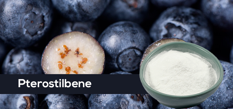
Applications of Pterostilbene in Skincare Products

(I) Overview of Common Product Types

Pterostilbene, with its remarkable skincare and anti-aging effects, has been widely used in various skincare products. A wide variety of skincare products containing pterostilbene are available on the market, catering to different skin types and usage scenarios.

Serums, as highly concentrated skincare products, are one of the common carriers of pterostilbene. These products typically have a light texture and high fluidity, allowing for rapid penetration into the skin and deep nourishment. For example, a well-known brand’s pterostilbene serum has a pterostilbene concentration as high as 0.5%, and also contains moisturizing ingredients such as sodium hyaluronate and various plant extracts. This serum is suitable for dry, normal, and combination skin types. In terms of usage scenarios, it is especially suitable for nighttime skincare. After a day of fatigue and environmental aggressors, the skin needs deep nourishment and repair at night. Using pterostilbene serum at this time can effectively combat free radicals, promote collagen production, help restore skin elasticity, reduce fine lines, and allow the skin to receive ample care overnight, waking up with a healthy glow the next day. The cream has a relatively thick texture and provides excellent moisturizing and nourishing effects. Creams containing pterostilbene form a protective film on the skin’s surface, locking in moisture while continuously releasing the active ingredients of pterostilbene for anti-aging benefits. For example, a cream containing 1% pterostilbene also includes ceramides and squalane to enhance the skin barrier function. This cream is more suitable for dry and sensitive skin. Dry skin already lacks sufficient oil secretion, requiring the cream to provide adequate hydration; while sensitive skin is more fragile, and the various gentle ingredients in the cream can soothe and protect the skin while providing anti-aging benefits. In the dry and cold autumn and winter seasons, skin loses moisture quickly; using this cream effectively combats dryness, maintaining skin’s hydration and elasticity, keeping skin in its best condition.

The lotion has a refreshing texture, is easy to spread and absorb, and is suitable for oily and combination-oily skin types. These skin types have excessive oil secretion and require lightweight skincare products to avoid burdening the skin. Emulsions containing pterostilbene, such as those from a certain brand, also contain oil-controlling ingredients like salicylic acid and various vitamins, in addition to pterostilbene. They not only provide anti-aging benefits but also regulate sebum secretion, keeping skin refreshed. Used in summer or during the day, they effectively control oil, reducing greasiness while protecting against UV rays and environmental pollution, keeping skin refreshed and healthy all day.

(II) Product Selection Guide

With a dazzling array of skincare products containing pterostilbene, choosing the right one is crucial. First, pay attention to the concentration of the ingredient, as this directly affects the product’s efficacy. Generally, skincare products with a pterostilbene concentration between 0.1% and 1% can effectively exert anti-aging effects while ensuring safety. However, higher concentrations are not necessarily better; excessively high concentrations may increase the burden on the skin and even cause discomfort. When choosing, check the product’s ingredient list to understand the specific pterostilbene content and make a judgment based on your own skin condition. If you are using a product containing pterostilbene for the first time, it is recommended to choose a lower concentration to allow your skin to adapt. Then, based on your skin’s tolerance and needs, choose a product with an appropriate concentration.

Brand reputation is also an important factor that cannot be ignored. Choosing a well-known brand means that the product is more reliable in terms of research and development, production, and quality control. Well-known brands usually have professional R&D teams and advanced production equipment, ensuring that the extraction and addition technology of pterostilbene is mature and the product quality is stable. For example, some internationally renowned skincare brands have a good reputation and market recognition worldwide, investing heavily in research and clinical trials to strictly control the safety and efficacy of their products. Skincare products containing pterostilbene from these brands are more trustworthy in terms of ingredient selection, formula rationality, and product stability. At the same time, the brand’s after-sales service provides consumers with more assurance, ensuring timely answers and solutions to any problems encountered during use.


Pterostilbene Compared to Other Anti-Aging Ingredients

(I) Comparison with Classic Anti-Aging Ingredients

Among the vast family of anti-aging skincare ingredients, pterostilbene has unique advantages and characteristics compared to classic anti-aging ingredients such as retinol, peptides, and pro-xylane.

Retinol, as a “star ingredient” in the anti-aging world, can effectively reduce wrinkles and improve skin texture by regulating gene expression and promoting skin cell renewal. However, it also has some limitations. Retinol is somewhat irritating, and some people may experience discomfort such as dry skin, redness, and peeling after use, especially those with sensitive skin who often find it difficult to tolerate. Moreover, retinol is sensitive to light and air, and has poor stability. Special care must be taken to avoid light and seal the product during storage and use, otherwise it is easy to lose its activity.

Pterostilbene, on the other hand, is relatively mild and less irritating, suitable for most skin types, including sensitive skin. It has high stability and can maintain its activity for a long time in skincare products, continuously exerting its anti-aging effects. In terms of efficacy, while retinol excels in promoting cell renewal, pterostilbene’s powerful antioxidant capacity is unique, more effectively scavenging free radicals and preventing skin aging at its source. It is also comparable in stimulating collagen production. Both have their strengths in anti-aging, offering more choices for consumers with different needs.

Peptides are small molecule compounds composed of amino acids. Different types of peptides have different effects. For example, hexapeptide-8 can inhibit nerve conduction and reduce muscle contraction, thereby reducing dynamic wrinkles; pentapeptide-3 can stimulate collagen production and increase skin elasticity. Peptides are advantageous due to their high safety profile, rarely causing allergic reactions, making them suitable for all skin types. However, the effects of peptides are relatively singular, and some peptides have poor transdermal absorption, affecting their efficacy.

Compared to pterostilbene, it not only stimulates collagen production but also comprehensively combats free radicals, regulates skin cell metabolism, and enhances skin barrier function, resulting in a more comprehensive anti-aging effect. In terms of transdermal absorption, pterostilbene, with its strong lipophilicity, can better penetrate the skin barrier and reach deeper layers of the skin to exert its effects, thus exhibiting more powerful efficacy in skincare and anti-aging.

Pro-Xylane, a xylose derivative extracted from beech trees, can stimulate the production of glycosaminoglycans (GAGs), promote the synthesis of collagen and elastin fibers in the extracellular matrix, increase skin firmness and elasticity, and also has a certain moisturizing effect. However, the disadvantage of Pro-Xylane is its high price, resulting in generally higher prices for skincare products containing it, increasing the cost for consumers. Furthermore, its effects are relatively slow, requiring long-term use to see noticeable results.

Pterostilbene shares similarities with Pro-Xylane in its efficacy, both promoting collagen production and improving skin firmness. However, pterostilbene is relatively inexpensive, making skincare products containing it more affordable and allowing more consumers to enjoy the benefits of anti-aging skincare. Moreover, the antioxidant properties of pterostilbene can more quickly improve dullness and roughness of the skin, resulting in a healthier complexion in a shorter period.

(II) The Secret of Synergistic Effect

The combination of pterostilbene and hyaluronic acid creates a synergistic effect in hydration and moisturizing. Hyaluronic acid is a natural moisturizing factor, acting like a “super absorbent sponge,” capable of absorbing and locking in more than a thousand times its own weight in water, replenishing the skin with abundant moisture and keeping it constantly hydrated. However, hyaluronic acid itself has poor stability and is easily degraded by enzymes in the skin, thus shortening its moisturizing time. Pterostilbene, on the other hand, has excellent antioxidant and stability properties. It can inhibit the activity of hyaluronidase, slowing down the degradation rate of hyaluronic acid and prolonging its stay in the skin, allowing hyaluronic acid to better exert its moisturizing effect. At the same time, pterostilbene can also promote skin cell metabolism, enhance the skin’s ability to absorb and utilize water, and work together with hyaluronic acid to build a stronger moisturizing barrier for the skin, making the skin not only hydrated but also able to maintain this hydrated state for a long time, reducing fine lines and roughness caused by dryness.

In terms of whitening and brightening, the combination of pterostilbene and vitamin C is perfect. Vitamin C is a powerful antioxidant that inhibits tyrosinase activity, preventing melanin formation and reducing existing melanin to lighter shades, thus achieving a whitening and fading effect. However, Vitamin C has strong reducing properties and is easily oxidized in air and under light, making it unstable and affecting its whitening efficacy. Pterostilbene, on the other hand, is highly stable and, when combined with Vitamin C, protects it from oxidation, maintaining its good activity. Simultaneously, pterostilbene’s own strong antioxidant capacity works synergistically with Vitamin C to eliminate free radicals in the skin, reducing damage to skin cells and further inhibiting melanin production. The two work together from multiple angles to whiten and brighten the skin while enhancing its antioxidant capacity, preventing dullness and pigmentation caused by UV rays and environmental pollution, resulting in a healthy, radiant complexion from within.


The Correct Way to Use Pterostilbene Essence for Skin Care and Anti-Aging

(I) Usage Methods and Techniques

When using skincare products containing pterostilbene essence, the correct order of application ensures maximum efficacy. Generally, after cleansing and toning, take an appropriate amount of essence in your palm. Gently rub your hands together to distribute the essence evenly, using the warmth of your palms to slightly warm the essence and better release its active ingredients. Then, gently press the essence onto your face, spreading it from the center outwards, massaging gently in an outward and upward direction, avoiding excessive pulling of the skin. For the forehead, gently massage from the center of the eyebrows towards the hairline; for the area around the eyes, gently dab and massage with your ring finger, being very gentle as the skin around the eyes is the most delicate.

When using face cream, pay attention to the amount. Dry skin can use a pea-sized amount, while normal and combination skin can use a mung bean-sized amount. Apply face cream to five areas: forehead, cheeks, nose, and chin. Then, gently spread it with your fingertips, massaging in the same direction as the face: from the center outwards, from the inside out, and from the bottom up. Finally, gently press your hands against your face to promote absorption. The application method for lotion is similar to face cream, but because lotion has a lighter texture, you can apply it slightly faster. Again, ensure even application and massage until fully absorbed.

(II) Frequency and Cycle of Use

The frequency of using skincare products containing pterostilbene varies depending on skin type. Dry skin, due to dehydration and lower sebum secretion, has a relatively slower skin metabolism and requires more nourishment and care. Therefore, skincare products containing pterostilbene can be used every morning and evening to provide continuous nutrition and hydration. Normal skin is more stable, with moderate sebum and moisture secretion. It can be used once daily or every other day, depending on individual needs. Oily skin tends to produce excessive sebum, and overuse may overburden the skin, leading to breakouts. It’s recommended to use it 3-4 times a week, choosing a lightweight product to avoid a greasy feeling. Combination skin tends to have a sebum-rich T-zone and relatively drier areas like the cheeks. Use it 3-4 times a week on the T-zone, and daily or every other day on drier areas like the cheeks, depending on individual needs.

Generally, after using skincare products containing pterostilbene for about 2-4 weeks, you’ll gradually notice subtle changes in your skin, such as increased hydration and radiance. However, to achieve significant anti-aging effects, such as a noticeable reduction in wrinkles and improved skin firmness, you generally need to use it continuously for 2-3 months or more. This is because the skin’s metabolic cycle is approximately 28 days, and collagen synthesis and skin structure improvement require time to accumulate. Consistent use allows pterostilbene to fully exert its effects and gradually improve skin condition. (III) Precautions and Contraindications

When using skincare products containing pterostilbene, avoid mixing them with highly irritating ingredients, such as high concentrations of fruit acids or salicylic acid. Fruit acids and salicylic acid have strong exfoliating properties; mixing them with pterostilbene may over-stimulate the skin, leading to redness, stinging, peeling, and other discomfort. Also, avoid using them with photosensitive ingredients, such as psoralen. Pterostilbene itself has antioxidant properties, and when mixed with photosensitive ingredients, it may trigger allergic reactions under UV radiation, increasing the risk of sunburn and tanning.

If redness, itching, stinging, or other discomfort occurs during use, discontinue use immediately. First, wash your face with water to remove as much of the product as possible, then observe your skin condition. If the symptoms are mild, they will generally subside within 1-2 days after discontinuing use. During this period, you can use some gentle moisturizing products, such as skincare products containing ingredients like ceramides and hyaluronic acid, to help soothe the skin and repair the skin barrier. However, if the symptoms are more severe, such as large areas of erythema, swelling, or even blisters, you should seek medical attention promptly and consult a professional dermatologist for treatment under their guidance to avoid delaying your condition.

The Limitless Potential of Pterostilbene

Pterostilbene has demonstrated multi-dimensional and outstanding advantages in the field of skincare and anti-aging. From its powerful antioxidant capabilities to promoting collagen production, regulating skin cell metabolism, and enhancing skin barrier function, each effect provides strong support for youthful skin. Its excellent performance in clinical trials and its potential for synergistic effects with other ingredients further demonstrate its immense value in skincare.

With continuous technological advancements and in-depth research, pterostilbene is expected to be used to develop more effective and safer skincare products in the future. In terms of extraction technology, scientists will continue to explore and innovate, further improving the extraction rate and purity of pterostilbene, reducing production costs, and enabling more consumers to enjoy its skincare benefits. At the same time, in-depth research into the mechanism of action of pterostilbene will provide a theoretical basis for its rational combination with other ingredients, developing more comprehensive and targeted skincare formulas.

In the future skincare market, pterostilbene is expected to become a core anti-aging ingredient, leading a new trend in skincare and anti-aging. Whether it’s personalized skincare products tailored to different skin types and age groups, or smart skincare products incorporating advanced technology, pterostilbene will play a vital role. It’s believed that in the near future, we will see more high-quality skincare products containing pterostilbene on the market, safeguarding people’s skin health and beauty, and making youthful skin a reality.

Fisetin: The Natural Code for Antioxidant and Anti-aging

Rhus Fisetin, a natural flavonoid compound, shine like a bright pearl, radiating a unique charm. They are widely found in various fruits and vegetables, such as strawberries, apples, onions, cucumbers, and the privet tree (Anacardiaceae). Its chemical name is 3,3′,4′,7-tetrahydroxyflavone, with the molecular formula C₁₅H₁₀O₆. It appears as yellow needle-like crystals, insoluble in water, ether, benzene, chloroform, and petroleum ether, but readily soluble in ethanol, acetone, and acetic acid.

The flavonoid family is vast and diverse, and rhus Fisetin are an important member. Fisetin are widely distributed in plant-based products such as fruits, vegetables, wine, tea, and chocolate, encompassing six major categories: flavonols, flavones, flavanones, flavanols, anthocyanins, and isoflavones. With their inherent antioxidant activity, these substances become powerful allies in the body’s fight against daily toxins. They can regulate cellular activity, combat free radicals, reduce the damage of oxidative stress to the body, and thus lower the risk of chronic diseases. As an outstanding representative of Fisetin, rosin, in addition to possessing the common characteristics of Fisetin, also has more unique antioxidant and anti-aging capabilities, making it highly regarded in the life sciences field.

The “Dark Alliance” of Free Radicals and Aging

In the human body, this exquisite and complex “life factory,” countless chemical reactions occur every moment, and free radicals are one of the byproducts of these reactions. During cellular respiration and metabolism, incomplete electron transfer—a “small episode”—can quietly give rise to free radicals. When we exercise, the body’s energy demand increases significantly, cellular respiration accelerates, and the production of free radicals also rises; during inflammatory responses, immune cells release free radicals as “weapons” to fight pathogens, which also leads to an increase in free radicals in the body. Besides internal physiological activities, the external environment constantly fuels the production of free radicals. Ultraviolet (UV) radiation acts like an invisible killer; when it penetrates the atmosphere and shines unhindered onto our skin, the molecules in skin cells instantly become disorganized, leading to rapid oxidation and a surge of free radicals. Skin exposed to sunlight for extended periods without proper sun protection is relentlessly attacked by free radicals, resulting in sunburn, premature aging, and other problems. Pollutants such as industrial waste gas, vehicle exhaust, and pesticide residues, once inside the body, interfere with normal physiological metabolism, triggering oxidative stress and promoting the massive generation of free radicals. When smoking, harmful substances like nicotine and tar in tobacco trigger a series of oxidation reactions in the body. A small cigarette butt, when burned, can produce trillions of free radicals, which rampage through the body, causing serious damage.

Free radicals are extremely chemically reactive; they act like a group of “restless little devils,” attacking various biomolecules within cells, with DNA, proteins, and lipids bearing the brunt. When free radicals attack DNA, they cause oxidation, alkylation, or deamination of DNA bases, leading to DNA structure damage, base pairing errors, and consequently affecting normal gene expression and replication. Once proteins are targeted by free radicals, their amino acid residues are oxidized and modified, altering their spatial structure and causing functional loss. Lipids, the cell’s “protective membrane,” undergo peroxidation under free radical attack, disrupting cell membrane fluidity and integrity, severely impacting cellular transport and signal transduction.

When these biomolecules are damaged by free radical oxidative stress, cell function gradually declines, accelerating the aging process. In the skin, free radicals damage collagen and elastin fibers, causing loss of elasticity and the appearance of wrinkles, sagging, and other signs of aging. In the immune system, weakened immune cell function reduces the body’s ability to fight pathogens, making it more susceptible to disease. In the cardiovascular system, damaged vascular endothelial cells increase the risk of atherosclerosis, significantly raising the incidence of cardiovascular diseases such as heart disease and stroke. The nervous system is also not spared. Nerve cells are attacked by free radicals, and the synthesis and transmission of neurotransmitters become abnormal, leading to problems such as memory loss and cognitive impairment.

The Antioxidant “Superpower” of Fisetin

A “Sharp Weapon” for Directly Scavenging Free Radicals

Fisetin‘s exceptional antioxidant capacity is closely related to its unique molecular structure, especially the multiple phenolic hydroxyl groups in its molecule. These phenolic hydroxyl groups are its key “weapons” for exerting its antioxidant effect. Superoxide anion radicals, as one of the most common free radicals in the body, are highly reactive and widely produced during cellular metabolism. The phenolic hydroxyl groups in the Fisetin molecule can keenly capture superoxide anion radicals. The hydrogen atoms in the phenolic hydroxyl groups generously provide hydrogen peroxide to the superoxide anion radicals, reducing them to hydrogen peroxide. Hydrogen peroxide has relatively low reactivity, and antioxidant enzymes such as catalase exist in cells, which can quickly decompose hydrogen peroxide into water and oxygen, thus effectively reducing the threat of superoxide anion radicals to cells.

Hydroxy radicals are the “destroyers” among free radicals, with extremely strong oxidizing power and great destructive force on intracellular biomolecules. Fisetin is also fearless in the face of hydroxyl radicals. The hydrogen atom of the phenolic hydroxyl group undergoes an addition reaction with the hydroxyl radical, and the two bind tightly together. The flavonoid itself is then transformed into a relatively stable phenoxy radical. It’s worth noting that the presence of multiple phenolic hydroxyl groups in the flavonoid structure allows the formed phenoxy radical to cleverly stabilize its own structure through resonance and other mechanisms. This is like creating a sturdy “shield” for the phenoxy radical, preventing it from further initiating free radical reactions and successfully stopping the attack of hydroxyl radicals on cells. In lipid peroxidation, peroxide radicals play a crucial “disruptor” role. WhenFisetin encounter peroxide radicals, they react rapidly, like a brave warrior engaging in fierce combat with an enemy. By providing hydrogen atoms, Fisetin transform peroxide radicals into relatively stable products. This process effectively prevents the chain reaction of lipid peroxidation. Just as Fisetin lend a helping hand at the critical moment when a domino is about to fall, blocking the first domino and preventing the subsequent chain reaction from continuing, thus protecting lipids from oxidative damage and maintaining the integrity and normal function of the cell membrane.

The “Conductor” Activating the Antioxidant Enzyme System

In the cell’s antioxidant defense system, antioxidant enzymes such as superoxide dismutase (SOD), glutathione peroxidase (GSH-Px), and catalase (CAT) act as loyal “guardians,” protecting cells from free radical damage. Fuchsia, on the other hand, acts like a wise “conductor,” precisely activating these antioxidant enzymes and enhancing the cell’s antioxidant capacity. SOD, as the first line of defense in antioxidant defense, catalyzes the dismutation reaction of superoxide anion free radicals, converting them into hydrogen peroxide and oxygen. Fuchsia can activate SOD by regulating complex intracellular signaling pathways. For example, it may activate the Nrf2-ARE signaling pathway. Nrf2 is a nuclear transcription factor; when activated by fuchsia, it acts like a “horn” being sounded, rapidly binding to antioxidant response elements (AREs). This combination acts like a cellular antioxidant “switch,” initiating the transcription of antioxidant enzyme genes such as SOD, prompting the cell to synthesize more SOD. More SOD means a stronger ability to scavenge superoxide anion free radicals, thus significantly reducing intracellular superoxide anion free radical levels and alleviating oxidative damage to the cell.

Glutathione peroxidase (GSH-Px) and catalase (CAT) are primarily responsible for removing hydrogen peroxide from cells. Excessive accumulation of hydrogen peroxide can cause severe cellular damage. Urushibrin can promote the function of GSH-Px by regulating the intracellular redox state. It increases the content of glutathione (GSH), an important cofactor for GSH-Px to function. More glutathione provides ample “ammunition” for GSH-Px, enabling it to more effectively remove hydrogen peroxide. For CAT, rutin may subtly modify its enzyme protein structure, much like carefully tuning a precision instrument, or regulate its synthesis and degradation processes to keep CAT in optimal working condition. This allows CAT to more efficiently decompose hydrogen peroxide, converting it into harmless water and oxygen, further enhancing the cell’s antioxidant capacity and enabling it to better resist free radical attacks.


The Anti-Aging “Miracle” of Rhus Fisetin

Calorie Restriction Mimic: Activating Autophagy

In the pursuit of health and longevity, calorie restriction (CR) has been proven to be an effective anti-aging strategy. By reducing calorie intake while ensuring the intake of essential nutrients, calorie restriction can activate a series of complex and sophisticated cellular and molecular mechanisms, thereby slowing down the aging process and reducing the risk of age-related diseases. However, strictly adhering to calorie restriction long-term is not easy; it requires strong willpower and strict control over diet, which deters many people. Fortunately, scientists have discovered calorie restriction mimics (CRMs), and rhus Fisetin is one of the best. As a calorie restriction mimic, rhus Fisetin can cleverly simulate the effects of calorie restriction without the need for people to endure the pain of hunger, activating related signaling pathways. In the complex signaling network of cells, the SIRT1 protein in the Sirtuins family acts as a key “key,” controlling the cellular aging process. Fuchsia can precisely upregulate SIRT1 expression, like injecting powerful energy into this “key,” enabling it to function better.

mTOR (mammalian target of rapamycin) is an important regulator of cell growth and metabolism. When mTOR is overactivated, cells grow and proliferate rapidly, which to some extent accelerates cellular aging. Fuchsia inhibits mTOR activity, acting like a “slow-down button” on cell growth and proliferation, thus slowing down cellular aging. During this process, autophagy is induced. Autophagy is a self-cleaning mechanism within cells, clearing damaged organelles, misfolded proteins, and other metabolic waste, acting like a diligent “cleaner,” constantly maintaining a clean cellular environment.

It is worth mentioning that, compared to other calorie restriction mimics, fuchsia exhibits unique tissue specificity, with a particularly significant effect on promoting autophagy in brain cells. As the body’s “command center,” the brain faces numerous challenges with age, such as an increased risk of neurodegenerative diseases. Autophagy in brain cells is crucial for maintaining normal neuronal function. It clears abnormal proteins accumulated within neurons, protecting them from damage. The promoting effect of Fisetin on autophagy in brain cells offers new hope for the prevention and treatment of neurodegenerative diseases. In animal experiments, feeding animals with a diet rich in Fisetin significantly increased autophagy levels in their brain cells, and also significantly improved memory and cognitive abilities, further confirming the important role of Fisetin in protecting brain health and delaying brain aging.

Senolytics: Clearing Senescent Cells

The continuous accumulation of senescent cells is a key factor in the aging process. These senescent cells are like “time bombs” in the body; although they no longer divide and proliferate normally, they are not “well-behaved.” Senescent cells secrete large amounts of senescence-associated secretory phenotypes (SASPs), which include various inflammatory factors, proteases, and growth factors. These SASPs, like “toxins,” negatively affect surrounding normal cells, interfering with their normal functions and causing them to gradually age as well. As senescent cells accumulate in the body, the functions of tissues and organs gradually decline, leading to various age-related diseases. In the cardiovascular system, the accumulation of senescent cells causes thickening of blood vessel walls and decreased elasticity, increasing the risk of atherosclerosis and cardiovascular disease. In joints, inflammatory factors secreted by senescent cells trigger inflammatory responses, leading to diseases such as arthritis. In the skin, senescent cells disrupt the synthesis of collagen and elastin fibers, causing the skin to lose elasticity, resulting in wrinkles and sagging.

Roseflavin, a powerful senolytic substance, can precisely identify and eliminate these senescent cells. Its mechanism of action is closely related to the upregulated SCAP network (anti-apoptotic pathway) of senescent cells. The reason senescent cells can survive in the body for a long time without undergoing apoptosis is largely due to their upregulated SCAP network. Roseflavin acts like a “precision marksman,” successfully blocking the SCAP network by blocking the PI3k/Akt/mTOR pathway and activating SIRT1. This removes the anti-apoptotic “protective shield” from senescent cells, forcing them to undergo apoptosis and be naturally eliminated by the body.

A wealth of research data has fully demonstrated the remarkable ability of Fisetin to clear senescent cells. In a 2018 study, scientists compared the effects of 10 Fisetin in clearing senescent cells. The results showed that Fisetin stood out, with its effect on clearing senescent cells far exceeding that of other Fisetin such as resveratrol, curcumin, and catechins. In another study on elderly mice infected with COVID-19, the remarkable efficacy of Fisetin was further validated. When these elderly mice were given Fisetin to clear senescent cells, their mortality rate decreased dramatically from nearly 100% to 50%. This astonishing result demonstrates that Fisetin can not only effectively clear senescent cells but also significantly improve the body’s health and reduce disease mortality.

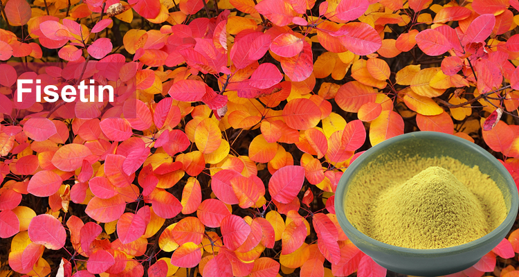
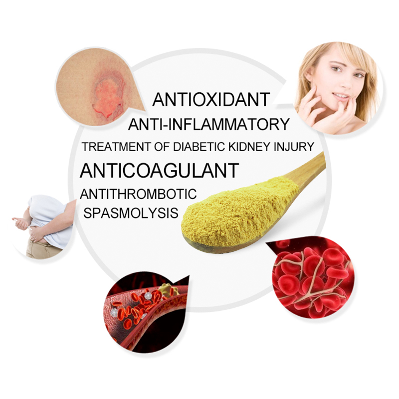
Future Applications of Fisetin

Fisetin, with its excellent antioxidant and anti-aging capabilities, has shown great application potential in multiple fields, bringing new hope for improving human health and quality of life.

In the food industry, Fisetin is expected to become a multifunctional food additive. It can be used as a natural food coloring additive, giving biscuits, bread, pastries, jams, jellies, candies, and other foods a bright yellow or brownish-yellow color, enhancing their appearance and meeting consumer demand for healthy, additive-free foods. Fisetin also has certain antibacterial properties, effectively inhibiting the growth of microorganisms in food, reducing the use of chemical preservatives, improving food safety, and extending shelf life. In jam production, adding an appropriate amount of Fisetin can extend the shelf life of the jam by several days. Fisetin is rich in vitamins and minerals and can be used as a nutritional fortifier in infant food, health products, and other products, providing the body with the necessary nutrients and promoting the growth, development, and health of infants and young children. It also possesses a certain aroma, making it a valuable flavoring agent that enhances the flavor and texture of food, adding a unique taste and improving its quality and market competitiveness.

In the cosmetics field, ursolic acid is an ideal skincare ingredient. It has powerful antioxidant and anti-inflammatory effects, effectively scavenging free radicals in skin cells, reducing oxidative stress damage, and slowing down the skin aging process. Urushibrine can also inhibit inflammatory signaling pathways, reduce the production of inflammatory factors, regulate the function of immune cells in the skin, alleviate skin inflammation, and promote skin healing and recovery. Adding ursolic acid to skincare products such as creams, masks, and serums can protect the skin from environmental damage, improve skin quality, reduce the appearance of fine lines and wrinkles, and maintain skin elasticity and radiance. In a mouse model of photoaging, intervention with ursolic acid significantly reduced the depth and number of wrinkles and increased skin elasticity. Urushibrine can also be used as a natural hair dye to change hair color; its good dyeing effect and minimal damage to hair make it a promising candidate for use in hair dyeing products. It can also provide a vibrant yellow or brownish-yellow hue for lipsticks and lip balms, and a warm brownish-yellow hue for eyeshadows and blushes, increasing the appeal and market competitiveness of these makeup products.

In the pharmaceutical field, Fisetin also have broad application prospects. They possess strong antioxidant capabilities, can scavenge free radicals in the body, and protect cells from oxidative damage. They are widely used in antioxidant drugs, helping to prevent and treat various diseases. Fisetin are expected to play an important role in the treatment of neurodegenerative diseases, cardiovascular diseases, and other diseases closely related to oxidative stress. They also have certain anti-inflammatory effects, relieving inflammatory responses and reducing pain and discomfort, making them a common ingredient in anti-inflammatory drugs and analgesics. Fisetin can promote the metabolism and repair of hepatocytes, reduce the burden on the liver, and protect liver function, and are used in some hepatoprotective drugs. Some studies have shown that Fisetin have certain anti-tumor activity, inhibiting the growth and spread of tumor cells, and have potential application value in the development of anti-tumor drugs.

With ongoing research and technological advancements, the extraction and synthesis techniques for urscin will be continuously optimized, and costs will gradually decrease, providing strong support for its large-scale application. In the future, we can expect to see more foods, cosmetics, and pharmaceuticals containing urscin emerge, safeguarding people’s health and beauty. We also look forward to scientists further exploring the mechanism of action and application effects of urscin, uncovering its greater potential value, and providing more solutions to various health problems facing humanity.

Nervonic Acid: The life code to unlock nerve repair

Nervonic acid, scientifically known as 24-carbon-cis-15-enoic acid, is a long-chain monounsaturated fatty acid. Despite its unfamiliar name, it plays a crucial role in nerve repair. In the intricate and complex “kingdom” of the nervous system, nerve damage is like a disruption of communication lines, hindering information transmission and disrupting various bodily functions. Nervonic acid acts like a remarkable “repairman,” possessing unique abilities to repair these damaged “circuits,” helping the nervous system restore normal function. Its importance in nerve repair is self-evident, attracting numerous researchers to dedicate themselves to its study, striving to uncover more of its mysteries and bring new hope to patients suffering from nerve damage.

What is Nervonic Acid?

Nervonic acid, a substance playing a crucial role in the nervous system, is chemically known as cis-15-tetracosenoic acid. Its chemical structure features a long chain of 24 carbon atoms, with a cis double bond between carbon 15 and carbon 16. This unique structure endows nervonic acid with special chemical and physical properties, making it stand out among many fatty acids as a long-chain monounsaturated fatty acid. It was first discovered by scientists in the brains of sharks, and subsequent research confirmed its widespread presence in the brains and nerve tissues of mammals.

In the complex human body, nervonic acid is mainly distributed in the central nervous system, with relatively high concentrations in the brain and retinal nerve tissue. Taking the brain as an example, as the body’s “command center,” nervonic acid acts as the “core building material” of brain nerve cells and nerve fibers, participating in the formation of nerve cell membranes and playing an indispensable role in maintaining the normal physiological functions of the nervous system. To put it more vividly, if we compare the nervous system to a city’s transportation network, then nervonic acid is like the high-quality road materials, supporting the stable and efficient operation of the entire network. A deficiency in nervonic acid is like a broken road, preventing the smooth flow of information and leading to a series of nervous system problems.

The Grim Reality of Nerve Injury

Nerve injury is not uncommon in life; it’s like a hidden “health killer,” capable of causing immense suffering at any time. Its types are diverse, encompassing damage to both the central and peripheral nervous systems. Common examples include brain injuries and spinal cord injuries, both falling under the category of central nervous system injuries. Among brain injuries, concussions and lacerations are relatively common, often caused by accidents such as traffic accidents and falls from heights. Spinal cord injuries, such as spinal concussions and spinal shock, can be caused by both external trauma and certain spinal lesions. Peripheral nerve injuries are equally prevalent, including mechanical injuries such as cuts from knives or glass lacerations leading to direct rupture of nerve fibers; ischemic injuries, such as prolonged use of tourniquets to block blood supply to nerves; and chemical injuries caused by exposure to toxic chemicals such as heavy metals and organic solvents that interfere with nerve cell metabolism.

Taking stroke, a common type of nerve injury, as an example, it is characterized by high morbidity, high disability rate, and high mortality rate. Data shows that approximately 2 million new stroke patients are diagnosed in my country each year, and the incidence rate is increasing annually. Once a stroke occurs, a patient may instantly fall from a healthy state into hemiplegia, aphasia, and become unable to care for themselves, completely disrupting their normal life rhythm. They need long-term bed rest, and require assistance with eating, drinking, and toileting. Even simple daily activities like dressing and washing become extremely difficult for them.

Cauda equina syndrome is also a serious concern, occurring more frequently in young adults, with a relatively higher proportion of male patients. In today’s social structure, many families rely primarily on the male for economic support. When the family’s “pillar” suffers cauda equina syndrome, the economic pressure becomes immense. Treatment costs are high, and patients lose their source of income during treatment, placing not only a financial burden on the family but also significant pressure on their livelihood, mental well-being, and psychological well-being. Patients with severe conditions, in addition to long-term high treatment costs, require specialized care, making it difficult to live a normal life, undoubtedly a heavy blow to the family. Brachial plexus injury is not uncommon in children, mostly caused by birth trauma, such as malposition of the fetus, incorrect delivery techniques, or forceful traction on the fetal neck and shoulders. This not only affects the child’s normal development but also causes great anxiety for parents, who worry about their child’s future life and growth.

Nerve injury not only brings physical and psychological torment to the individual patient but also places an unbearable burden on the family due to the long-term care and treatment costs, potentially leading to financial collapse. Family relationships can also become tense and strained due to the patient’s condition. From a societal perspective, the large number of patients with nerve injury increases the burden on medical resources and leads to the loss of some labor force, affecting the normal functioning of society and economic development. Therefore, finding effective nerve repair methods is urgent, whether for the sake of the individual patient’s quality of life, family harmony and stability, or the healthy development of society. This is a key reason why nervonic acid is highly anticipated.

Unveiling the Mechanism of Nervonic Acid in Repairing Nerve Damage

Nervonic acid’s ability to repair nerve damage is underpinned by a complex and sophisticated mechanism, much like a precision instrument where each component works collaboratively to achieve the goal of repairing nerve damage. Let’s delve into the specific mechanisms by which nervonic acid repairs nerve damage.

(I) Guardian of the Cell Membrane

In the structure of nerve cells, the cell membrane is a crucial component, acting like the walls of a castle, protecting the cell’s internal “residents”—various organelles and substances necessary for life activities. When nerves are damaged, whether by physical impact, chemical poisoning, ischemia, or hypoxia, the cell membrane is the first to be affected. Once the cell membrane is damaged, it’s like a breach in the wall; intracellular substances leak out, and harmful extracellular substances can easily enter, ultimately leading to the nerve cell’s inability to function properly and even death.

Nervonic acid is precisely the key to solving this problem. It is a key precursor to sphingomyelin, a core component of the nerve cell membrane, much like a high-quality brick in building a castle. When nerve cell membranes are damaged, supplementing with nervonic acid allows for rapid absorption and utilization by nerve cells, accelerating the “lipid remodeling” of the damaged cell membrane. This is analogous to quickly repairing a breach in a city wall with high-quality bricks, restoring the cell membrane’s fluidity and barrier function. Simultaneously, an intact cell membrane maintains the balance of ions inside and outside the cell, such as the sodium-potassium ion (Na⁺/K⁺) balance. Once this ion balance is disrupted, neurons die due to ion disorder. By maintaining ion balance, nervonic acid buys valuable time for nerve cell self-repair, laying a solid foundation for subsequent repair work.

In related animal experiments, researchers constructed an animal model of cerebral ischemia. Supplementing the experimental animals with nervonic acid yielded surprising results. Compared to the control group without nervonic acid supplementation, the neuronal mortality rate in the ischemic area of ​​the animals supplemented with nervonic acid was significantly reduced, and the number of surviving neurons was significantly increased. This directly demonstrates that nervonic acid has outstanding performance in repairing damaged nerve cell membranes and maintaining neuronal survival.

(II) The Engine of Myelin Regeneration

Myelin, the lipid structure surrounding nerve fibers, plays a crucial role in the transmission of nerve signals. It can be imagined as the insulation layer on an electrical wire; if the insulation layer is damaged, the wire is prone to leakage, and signal transmission will be interfered with. In diseases such as multiple sclerosis, diabetic neuropathy, and spinal cord injury, myelin damage, also known as demyelination, often occurs. Once demyelination occurs, nerve signal transmission slows down or even stops, and patients experience a series of symptoms such as numbness in the hands and feet, motor disorders, and cognitive decline.

Nervonic acid plays the role of the “engine” in the process of myelin regeneration. It accounts for more than 20% of the total lipids in myelin and is an “essential raw material” for myelin synthesis. Specifically, nervonic acid can activate the proliferation and differentiation of myelin-forming cells, namely oligodendrocytes in the central nervous system and Schwann cells in the peripheral nervous system. This is like adding more workers to a factory, greatly accelerating the production of myelin. Simultaneously, nervonic acid can directly participate in myelin lipid synthesis, filling structural gaps in demyelinated areas and reconstructing complete “signal transduction pathways.”

Researchers conducted in-depth studies in an experimental autoimmune encephalomyelitis mouse model. This mouse model simulates human multiple sclerosis, and the experimental results after nervonic acid supplementation were encouraging. The demyelinated area of ​​the mice was significantly reduced, the signal transduction velocity of nerve fibers was significantly improved, and motor function was also significantly improved, such as enhanced walking coordination. These experimental data fully demonstrate the powerful role of nervonic acid in promoting nerve myelin regeneration and restoring signal transduction efficiency.

(III) A Facilitator of Synaptic Remodeling

Synapses, as “bridges” for transmitting signals between neurons, play an indispensable role in the realization of neural function. When nerves are damaged, such as in traumatic brain injury or stroke, a large number of synapses are lost or their function weakens. This is like a broken or unstable bridge, naturally leading to impaired neural function. Patients may experience problems in memory, language, motor function, and other areas.

Nervonic acid plays a crucial supporting role in synaptic remodeling. It supports synaptic repair and remodeling by regulating cell membrane properties. Specifically, nervonic acid enhances the release efficiency of neurotransmitters such as acetylcholine and glutamate from the presynaptic membrane. This is analogous to adding more transport vehicles to one end of a bridge, allowing signals to be “sent” out more quickly. Simultaneously, nervonic acid increases the sensitivity of postsynaptic membrane receptors, such as NMDA receptors, optimizing signal “reception,” much like installing a more efficient receiving device at the other end of the bridge. Furthermore, nervonic acid promotes the formation of new synapses, helping damaged neural networks reconnect.

Some observational studies on stroke patients in the recovery phase have shown that patients supplemented with nervonic acid exhibited improved cognitive scores, such as the MMSE scale, and faster recovery of limb motor function. This clearly demonstrates the positive significance of nervonic acid in assisting synaptic remodeling and rebuilding neural functional connections, bringing hope for patient recovery.

(IV) A Purifier of the Microenvironment

Following nerve injury, persistent inflammation and oxidative stress occur locally, creating a post-disaster environment filled with dangers and obstacles. Inflammation leads to excessive activation of microglia, releasing large amounts of inflammatory factors such as TNF-α and IL-1β. These inflammatory factors act like destructive molecules, further damaging nerve cells. Simultaneously, oxidative stress generates excessive free radicals, such as ROS. Free radicals are highly oxidizing, attacking various structures of nerve cells and causing cell damage. These two reactions interact, forming a vicious cycle of “damage-inflammation-re-damage,” severely hindering the nerve repair process.

Nervonic acid acts as a purifier, breaking this vicious cycle. In terms of anti-inflammation, nervonic acid inhibits the release of inflammatory factors and reduces excessive activation of microglia, thereby alleviating inflammatory infiltration in the damaged area. In terms of antioxidation, nervonic acid can scavenge excess free radicals and enhance the activity of intracellular antioxidant enzymes, such as SOD. Through these two methods, nervonic acid creates a friendly microenvironment of “low inflammation and low oxidation” for nerve repair. In such an environment, processes such as cell membrane repair and myelin regeneration can proceed more efficiently, much like rebuilding a home on cleaned-up ruins is much smoother.

How to Obtain Nervonic Acid

After understanding the repairing effect of nervonic acid on nerve damage, people are probably more concerned about how to obtain it. There are two main ways to obtain nervonic acid: through dietary intake and through nutritional supplements.

Regarding food, there are relatively few foods in nature that contain nervonic acid, but there are still some foods that are excellent sources of it. One such source is Acer truncatum seed oil, which is extracted from the seeds of the Acer truncatum tree. Acer truncatum is a tree species unique to China, and its seed oil has a relatively high nervonic acid content, approximately 5%-7%. Besides Acer truncatum seed oil, some deep-sea fish, such as salmon and tuna, also contain a certain amount of nervonic acid. This is because they live in deep-sea environments, and their food sources may be rich in nervonic acid-related precursors, which are then metabolized and accumulated in their bodies. In addition, some nuts and seeds, such as walnuts, almonds, and sesame seeds, also contain nervonic acid. These foods not only replenish nervonic acid but are also rich in protein, unsaturated fatty acids, and other nutrients, which are very beneficial to health. However, obtaining nervonic acid through daily diet often has limitations. On one hand, the nervonic acid content in food is relatively low. For example, even consuming large amounts of nuts containing nervonic acid would not provide enough to meet the high doses required for nerve damage repair. On the other hand, some food sources are not readily available; for instance, Acer truncatum seed oil is not as widely available as common cooking oils. Therefore, in many cases, nutritional supplements become an important way to obtain nervonic acid.

There are many types of nervonic acid supplements on the market, and extra caution is needed when choosing one. First, pay attention to the product’s ingredients and purity. High-quality nervonic acid supplements should have high purity nervonic acid with few impurities. Some products using advanced extraction technologies can effectively improve the purity of nervonic acid. For example, supercritical carbon dioxide extraction technology can extract nervonic acid under relatively mild conditions, reducing damage to its structure and activity, thus obtaining high-purity nervonic acid products. Second, check the product’s certifications and production standards. Legitimate nervonic acid supplements should have passed relevant quality certifications, such as GMP (Good Manufacturing Practice) certification and ISO (International Organization for Standardization) certifications. These certifications indicate that the product strictly adheres to certain quality control standards during the production process, ensuring higher product quality and safety. In addition, you can refer to reviews from other consumers and evaluations from professional organizations. Real-world consumer feedback allows us to understand the actual effects and potential problems of the product more intuitively. Evaluations from professional organizations, based on scientific experiments and analysis, provide us with more objective and accurate product information.

There are also some precautions to take when using nervonic acid supplements. It is essential to strictly follow the product instructions or your doctor’s advice. Different age groups and health conditions have different requirements and tolerance levels for nervonic acid. Arbitrarily increasing or decreasing the dosage may affect the supplementation effect and even cause adverse effects on the body. For example, excessive use of nervonic acid supplements may cause gastrointestinal discomfort, such as nausea, vomiting, and diarrhea; long-term high-dose use may also increase the metabolic burden on the liver and kidneys. Meanwhile, nervonic acid supplements cannot replace normal medical treatment. If you already have a serious neurological injury, such as stroke or spinal cord injury, you should use nervonic acid supplements as an adjunct therapy under the guidance of a doctor, combining them with conventional treatments such as medication and rehabilitation training to promote the recovery of neurological function.

Future Blueprint for Nervonic Acid Research

Nervonic acid has demonstrated enormous potential in the field of nerve repair; however, current research represents only the tip of the iceberg, leaving vast room for future exploration.

In basic research, scientists will further delve into the molecular mechanisms of nervonic acid in nerve repair. While we already know that nervonic acid plays a crucial role in cell membrane repair and myelin regeneration, some specific signaling pathways and molecular targets remain unclear. For example, how does nervonic acid precisely regulate the proliferation and differentiation of oligodendrocytes and Schwann cells? This may involve a series of complex gene expression and protein modification processes. Further research is expected to reveal these molecular mysteries, providing a solid theoretical foundation for developing more efficient nervonic acid treatment strategies.

With the rapid development of gene technology, using gene editing technologies, such as the CRISPR-Cas9 system, to study the effects of nervonic acid on nerve cell gene expression will also become an important research direction. This can help us better understand the interaction between nervonic acid and the nerve cell genome, and may even enhance the efficiency of nerve cell uptake and utilization of nervonic acid through gene editing technology, thereby improving the effectiveness of nerve repair. In applied research, the combined application of nervonic acid with other therapeutic approaches will be a significant future trend. For example, when combined with stem cell therapy, nervonic acid can provide a favorable microenvironment for stem cell survival, differentiation, and function, promoting stem cell differentiation into neural cells and further enhancing nerve repair capabilities. Animal studies have already demonstrated the application of nervonic acid in combination with mesenchymal stem cells for the treatment of spinal cord injury, showing that the combined treatment group exhibited significantly better neurological function recovery compared to using stem cells or nervonic acid alone.

Furthermore, the application of nervonic acid in the treatment of more neurological diseases is promising. Besides the currently well-studied diseases such as stroke and spinal cord injury, nervonic acid may also play an important role in the treatment of neurodegenerative diseases such as Alzheimer’s disease and Parkinson’s disease. These diseases have complex pathogenesis and currently lack effective cures. Through its nerve damage repair, anti-inflammatory, and antioxidant effects, nervonic acid may slow disease progression and improve patients’ quality of life. Some preliminary studies have shown positive effects of nervonic acid in improving cognitive function in Alzheimer’s disease mice, but more large-scale, multi-center clinical trials are needed to validate these findings. From a product development perspective, future efforts will focus on developing more diverse and higher-quality nervonic acid products. In terms of extraction technology, existing extraction processes will be continuously optimized to improve the extraction efficiency and purity of nervonic acid, reduce production costs, and enable nervonic acid products to be more widely used in clinical practice and daily life. Simultaneously, novel nervonic acid dosage forms, such as nano-formulations and targeted formulations, will be developed to improve the bioavailability and targeting of nervonic acid, allowing it to act more precisely on damaged nerve tissue and reduce side effects on other tissues.

The research prospects for nervonic acid in the field of nerve repair are very broad. With the continuous deepening of research and technological advancements, it is believed that in the near future, nervonic acid will bring good news to more patients with nerve damage and make significant contributions to overcoming the challenges of nervous system diseases. Let us look forward to nervonic acid shining even brighter on the future medical stage.

Oleuropein: Unlocking the Natural Code to Antioxidation and Anti-aging

Oleuropein, this mysterious compound, was first discovered by Bourquelot and Vintilesco in 1908. It is mainly found in the leaves and fruits of the olive tree, especially in immature olives and leaves, where the dry matter concentration can reach as high as 140 mg/g in young olives and 60-90 mg/g in leaves. It is precisely this compound that gives immature and unprocessed olives their characteristic bitterness. Chemically, oleuropein is a phenolic schizocyclic ether glycoside composed of hydroxytyrosol, glucose molecules, and oleuropein. This seemingly simple molecular structure contains powerful biological activities, demonstrating remarkable potential in antioxidation, anti-inflammation, neuroprotection, and anti-aging, attracting numerous researchers to continue its in-depth exploration.

Antioxidants: The Natural Nemesis of Free Radicals

The Dangers of Free Radicals

Our bodies are constantly undergoing complex physiological activities, and free radicals, like a group of restless “monsters,” are quietly generated. Under normal circumstances, the body’s metabolism produces free radicals, which are atoms, molecules, or ions containing unpaired electrons and possess extremely strong oxidizing activity. Like a mischievous child, they constantly try to steal electrons from surrounding molecules to achieve their own stable state. When we are exposed to factors such as ultraviolet radiation, environmental pollution, stress, and poor dietary habits, the production of free radicals increases significantly, like opening Pandora’s box, causing a series of damages to our bodies.

Free radicals attack cells in all directions. They relentlessly damage cell membranes, impairing their structure and function, preventing cells from carrying out normal material exchange and signal transmission. Imagine a cell as a house; the cell membrane is the wall and windows. The attack of free radicals leaves the walls riddled with holes, and the doors and windows unable to open and close properly, naturally disrupting all activities within the house. Free radicals also attack proteins and DNA within cells, altering protein structure and causing them to lose their original function. DNA damage can lead to gene mutations, increasing the risk of cancer. This is akin to tampering with the cell’s “blueprint,” disrupting cell growth and division.

Numerous scientific studies have shown that free radicals play a crucial role in the development of many chronic diseases. In cardiovascular disease, free radicals oxidize low-density lipoprotein (LDL), turning it into oxidized LDL. This substance is easily engulfed by macrophages, forming foam cells that gradually accumulate on the blood vessel walls, leading to atherosclerosis. This causes blood vessels to narrow and become blocked, affecting blood supply to the heart and brain—like scale buildup in a pipe reducing water flow or even causing complete blockage. In diabetes, free radicals damage pancreatic islet cells, affecting insulin secretion and action, leading to elevated blood sugar. The body cannot properly utilize sugar, much like a malfunctioning machine in a factory. Free radicals are also closely related to neurological disorders, cancer, inflammation, and many other diseases, becoming a “silent killer” threatening human health.

The Antioxidant Mechanism of Oleuropein

Faced with the onslaught of free radicals, our bodies are not without defenses. Oleuropein is a powerful antioxidant weapon. Its antioxidant capacity stems from its unique molecular structure, especially the ortho-bisphenol group in the molecule. This group acts like a magical “electron scavenger,” acting as a hydrogen donor to react with free radicals, donating its own electrons to stabilize the free radicals and effectively scavenging reactive oxygen species (ROS). Oleuropein can also stabilize oxygen free radicals through intramolecular hydrogen bonding, further enhancing its antioxidant effect, like putting a “restraint” on free radicals, preventing them from causing further damage.

Compared to common antioxidants such as vitamins C and E, oleuropein exhibits unique advantages. Vitamin C is a water-soluble antioxidant that mainly functions in the extracellular fluid, effectively scavenging water-soluble free radicals. Vitamin E, on the other hand, is a fat-soluble antioxidant, mainly found in lipid environments such as cell membranes, protecting cell membranes from free radical attacks. Oleuropein not only possesses strong antioxidant activity, with an antioxidant capacity 5-10 times that of vitamin C, but it also functions effectively in various environments, whether water-soluble or fat-soluble. Like an “all-around warrior,” it combats free radical damage comprehensively. It’s like a superhero with multiple skills, capable of fighting both in water and on land, providing more comprehensive protection for cells.

Scientifically Verified Antioxidant Efficacy

Numerous scientific studies and experimental data strongly support the antioxidant efficacy of oleuropein. In a study of a D-galactose-induced aging mouse model, researchers supplemented aging mice with oleuropein and found a significant reduction in malondialdehyde (MDA), a lipid peroxidation product, in the heart, liver, and brain tissues of the mice. MDA is a product of lipid peroxidation; elevated MDA levels indicate that cells have been attacked by free radicals, resulting in lipid oxidative damage. The effect of oleuropein in reducing MDA levels demonstrates its ability to effectively inhibit lipid peroxidation and reduce free radical damage to cells. Simultaneously, the activities of superoxide dismutase (SOD) and glutathione peroxidase in mice were significantly enhanced. SOD and glutathione peroxidase are important antioxidant enzymes in the human body, catalyzing the decomposition of free radicals and converting them into harmless substances, acting like “cleaners” in the body, specifically cleaning up this “waste” of free radicals. The fact that oleuropein can enhance the activity of these antioxidant enzymes indicates that it can not only directly scavenge free radicals but also activate the body’s own antioxidant defense system, forming a dual protective network to jointly combat free radical damage.

Furthermore, in vitro experiments have shown that oleuropein can effectively scavenge free radicals such as nitric oxide and hypochlorous acid, exhibiting strong antioxidant capabilities. Nitric oxide is a biologically active free radical that participates in various physiological and pathological processes in the body; excessive nitric oxide can lead to oxidative stress and inflammatory responses. Hypochlorous acid is a strong oxidant produced by neutrophil myeloperoxidase at sites of inflammation; it can damage biological macromolecules such as proteins. Oleuropein can scavenge these free radicals, indicating its great potential in preventing and treating diseases related to oxidative stress, thus safeguarding our health.

Anti-aging: The Potential Key to Reversing Time

The Biological Mechanisms of Aging

Aging is a complex and natural physiological process, a slow but inevitable “journey of decline” involving changes at every level of the body. At the cellular level, mitochondria, the organelles hailed as the cell’s “energy factory,” gradually decline in function with age. Increased mitochondrial DNA mutation rates hinder energy production, leading to the accumulation of reactive oxygen species (ROS). Like aging machinery in a factory, this not only reduces efficiency but also generates a large amount of “waste.” These ROS further damage cell structure and function, triggering oxidative stress, destroying important biomolecules such as cell membranes, proteins, and DNA, and accelerating cellular aging.

Inflammation is also a significant characteristic of the aging process, with a chronic, low-grade inflammatory state quietly spreading throughout the body. As we age, the immune system gradually declines, leading to an increase in the secretion of inflammatory cytokines such as interleukin-6 (IL-6) and tumor necrosis factor-α (TNF-α). These cytokines act like a group of uncontrolled “little devils,” triggering inflammatory responses in the body, damaging tissues and organs, and are closely related to the development of many chronic diseases such as cardiovascular disease and neurodegenerative diseases. In tissues such as the skin, collagen loss is also a clear sign of aging. Collagen is an important substance for maintaining skin elasticity and firmness; it acts like the skin’s “support framework.” With age, collagen synthesis decreases, while the activity of enzymes such as matrix metalloproteinases (MMPs) increases. These enzymes act like “scissors,” relentlessly degrading collagen, leading to loose skin, increased wrinkles, and a loss of youthful radiance. Aging is also accompanied by various biological changes, such as abnormal cell cycle regulation, telomere shortening, and altered gene expression. These changes intertwine and jointly drive the aging process, posing various challenges to our bodies.

Multidimensional Anti-aging Effects of Oleuropein

Mitochondrial Activation

Mitochondria play a central role in cellular energy metabolism and aging, and oleuropein acts like a magic key, unlocking the door to mitochondrial vitality. In late 2024, a groundbreaking study published in the top international journal *Cell Metabolism* revealed that scientists from the University of Padua and Nestlé Research Institute in Italy discovered that oleuropein can directly activate the mitochondrial calcium single-channel transporter (MCU). With age, the expression of the regulator MCUR1 declines, leading to a decline in mitochondrial calcium uptake and hindered energy production. The deglycosylated metabolite of oleuropein specifically binds to the MICU1 subunit of MCU, overcoming age-related calcium uptake barriers. In aged animal models, treatment with oleuropein restored mitochondrial calcium uptake to youthful levels, significantly increased ATP production, and substantially improved muscle performance. This discovery makes oleuropein the first natural molecule proven to directly activate mitochondrial calcium channels, opening a new direction in the field of anti-aging. It’s like injecting new vitality into the “energy factory” of aging cells, making it operate efficiently again.

Inflammation Regulation

Chronic low-grade inflammation is one of the important characteristics of aging, and oleuropein acts like an “inflammation brake,” effectively inhibiting inflammatory responses. It has anti-inflammatory properties, working by inhibiting the function of inflammatory cells and reducing oxidative stress caused by various factors. Research results show that oleuropein can reduce the production of nitrite (NO) in cells stimulated by lipopolysaccharide (LPS) by downregulating the expression of inducible nitric oxide synthase gene. NO is an inflammatory mediator; excessive NO can trigger inflammatory responses and damage cells. Oleuropein can also reduce the expression of M1-related pro-inflammatory cytokines and their genes, while increasing the expression of M2-related anti-inflammatory genes and cytokine production, regulating the inflammatory balance. It’s like setting up an “anti-inflammatory defense line” in the body, reducing the damage of inflammation to the body and slowing down the aging process. Collagen Protection

As we age, collagen in our skin is continuously lost, much like the “supporting beams” of a house gradually decaying, leading to sagging skin and wrinkles. Olive bitter glycosides act as a loyal “collagen guardian,” inhibiting the activity of matrix metalloproteinase 1 (MMP-1) by up to 35%. MMP-1 acts like a “destructive sword,” specifically degrading type I collagen. Olive bitter glycosides significantly weaken this “sword,” reducing type I collagen degradation and maintaining skin elasticity and firmness. Simultaneously, excessive accumulation of collagen fibers is a hallmark of many localized fibrotic diseases, such as cardiac fibrosis and keloids. Experiments have shown that olive bitter glycosides inhibit collagen fiber formation. Like a “construction worker,” it maintains existing “building materials” (collagen) while preventing excessive accumulation, maintaining the normal structure and function of body tissues and keeping skin and body tissues youthful.

Regulation of Cellular Longevity

Oleuropein can also activate SIRT1, known as the “longevity protein,” which acts like a “cellular longevity switch,” regulating aging-related proteins such as p53 and FOXO through deacetylation. In cell experiments, oleuropein increased proteasome activity, reduced the accumulation of oxidized proteins, and extended the replication lifespan of human fibroblasts by approximately 15%. Proteins such as p53 and FOXO play crucial roles in cellular senescence, acting like “senescence commanders” within the cell. By regulating these proteins, oleuropein alters the cellular aging process, essentially slowing down the cell’s “aging clock,” allowing cells to maintain vitality and proliferative capacity for a longer period, thus delaying the aging process at the cellular level.

Current Research Status and Challenges

Although oleuropein has shown great potential in the fields of anti-oxidation and anti-aging, current research still faces many challenges. From a bioavailability perspective, the oral absorption rate of natural oleuropein is low, which acts as an obstacle to its efficacy. After oral administration, it may be affected by various factors in the gastrointestinal tract, such as destruction by gastric acid, degradation by intestinal enzymes, and the absorption barrier of the intestinal mucosa, resulting in a significant reduction in the effective dose entering the bloodstream and exerting its effects, making it difficult for it to fully exert its antioxidant and anti-aging capabilities. To overcome this bottleneck, researchers are actively exploring various solutions and developing nanocarrier technology to encapsulate oleuropein in nanoscale carriers such as liposomes and nanoparticles. This improves its stability, reduces degradation in the gastrointestinal tract, and promotes intestinal absorption, essentially giving oleuropein an “invisible protective suit” to help it reach various parts of the body and exert its effects. Modifying the structure of oleuropein through chemical methods alters its molecular structure, enhancing its lipid or water solubility and increasing its affinity for intestinal absorption sites, thereby improving bioavailability. This is like an “upgrade,” allowing it to be better absorbed and utilized by the body.

Regarding dosage optimization, the optimal anti-aging dose of oleuropein in humans has not yet been established. Existing research mostly focuses on cell and animal experiments. Different experimental models, administration methods, and observation indicators lead to variations in research results, making it difficult to directly deduce the effective dose suitable for humans. This is similar to knowing that a drug has a therapeutic effect but not knowing the optimal dosage; too little may not achieve the desired effect, while too much may cause adverse reactions. To solve this problem, extensive clinical trials are needed, recruiting volunteers of different ages, genders, and health conditions, setting up different dosage groups, and conducting long-term follow-up observations. By monitoring the concentration of oleuropein in blood and tissues, oxidative stress indicators, inflammatory factor levels, and aging-related biomarkers, the safety and efficacy of oleuropein at different dosages can be comprehensively evaluated to determine the most suitable anti-aging dose for the human body.

Global olive processing generates over 1 million tons of waste leaves annually, which are rich in oleuropein. However, current extraction processes are not standardized enough, leading to significant resource waste. Different extraction methods and process conditions affect the extraction rate and purity of oleuropein. Some traditional extraction methods may suffer from low extraction efficiency, high energy consumption, and unstable product quality. Establishing standardized extraction processes is urgently needed. Researchers need to conduct in-depth research and optimization of extraction methods, screen suitable solvents, optimize parameters such as extraction temperature, time, and solid-liquid ratio, and develop efficient, environmentally friendly, and low-cost extraction technologies. At the same time, by combining advanced separation and purification technologies, the purity and quality of oleuropein can be improved, enabling the resource utilization of waste leaves and turning these once discarded leaves into valuable resources, thus providing a sufficient source of raw materials for the research and application of oleuropein.

Application Prospects and Future Outlook

With increasing public awareness of health and anti-aging, oleuropein, with its excellent antioxidant and anti-aging properties, has shown broad application prospects in multiple fields.

In the food industry, oleuropein is expected to become a novel natural food additive. It can be added to various beverages, dairy products, and baked goods, not only adding unique flavor but also extending shelf life and preventing spoilage due to oxidation, essentially giving food an “antioxidant armor.” For consumers pursuing a healthy diet, foods containing oleuropein are undoubtedly a superior choice, offering both delicious taste and antioxidant and anti-aging health benefits, meeting people’s dual needs for functional and nutritional food.

In the health supplement industry, oleuropein has enormous development potential. Nutritional supplements with oleuropein as the main ingredient, such as capsules, tablets, and oral liquids, can be developed to provide effective health support for middle-aged and elderly people, those under chronic stress, and consumers focused on anti-aging. These health supplements can help people enhance their body’s antioxidant capacity, reduce free radical damage, slow down the aging process, and improve immunity and resistance, allowing people to maintain a healthier and more energetic state.

In the pharmaceutical field, oleuropein may become a new hope for treating diseases related to oxidative stress and aging. Researchers are conducting in-depth studies on its potential applications in the treatment of cardiovascular diseases, neurodegenerative diseases, diabetes, and other diseases. Perhaps in the near future, new drugs based on oleuropein will emerge, providing more effective solutions for the treatment of these diseases and bringing new vitality and hope to patients.

In the future, research directions for oleuropein will become more diversified. Regarding its mechanism of action, further in-depth exploration of its interactions with various intracellular signaling pathways is needed to fully reveal its antioxidant and anti-aging molecular mechanisms. Like solving a complex puzzle, understanding how it precisely functions in the body will provide a more solid theoretical foundation for its application. In dosage form development, efforts will be focused on researching more efficient and stable formulations to improve bioavailability. For example, nanotechnology will be used to develop nano-level oleuropein preparations, enabling better absorption and utilization by the body to maximize their efficacy. More large-scale, long-term clinical trials are also needed to verify their safety and efficacy in humans, determine the optimal dosage and administration method, and provide reliable data support for their commercial application. With ongoing research and technological advancements, oleuropein is expected to find applications in more fields, making a greater contribution to human health and beauty, and becoming a shining star in safeguarding life and well-being.

Oleuropein, a natural treasure derived from the olive tree, brings new hope to our health with its remarkable antioxidant and anti-aging capabilities. Like a loyal guardian of health, it fiercely battles free radicals in the microscopic world, protecting every corner of the cell. From mitochondrial activation and inflammation regulation to collagen protection and cellular longevity regulation, it slows down the aging process in multiple dimensions, injecting our bodies with a continuous stream of vitality. Although challenges remain in bioavailability, dosage optimization, and extraction processes, researchers are actively exploring solutions and continuously breaking through technological bottlenecks. In the future, with in-depth research and technological advancements, oleuropein is expected to shine brightly in multiple fields such as food, health products, and pharmaceuticals, becoming a powerful assistant in people’s pursuit of health and beauty. Let us look forward to this natural health ingredient shining even brighter in the future, writing a new chapter in human health.

Coenzyme Q10:The dual secrets to cellular energy protection and anti-aging

Coenzyme Q10, chemically known as 2,3-dimethoxy-5-methyl-6-decanopentenyl-1,4-benzoquinone, is a fat-soluble quinone compound. It is widely found in nature, appearing in the cells of everything from microorganisms to higher plants and animals. In the human body, coenzyme Q10 plays an indispensable role, especially in organs with extremely high energy demands, such as the heart, liver, and kidneys, where its content is relatively higher. These organs are constantly engaged in high-intensity physiological activities. For example, the heart needs to continuously and rhythmically contract and relax to maintain blood circulation; the liver undertakes various complex functions such as metabolism and detoxification; and the kidneys are responsible for filtering blood and maintaining water and electrolyte balance. Coenzyme Q10 acts like an “energy manager” behind the scenes, silently ensuring an adequate energy supply and maintaining the normal functioning of these organs. From a molecular structure perspective, the parent nucleus of coenzyme Q10 is p-benzoquinone, linked by a long side chain consisting of 10 isoprene units. This unique structure endows it with lipid solubility, allowing it to easily embed itself in the lipid bilayer of biological membranes. This lipid solubility not only facilitates the function of coenzyme Q10 in lipid-rich cellular environments but is also closely related to its various physiological functions. In biological membranes, coenzyme Q10 not only participates in energy metabolism but also plays a crucial regulatory role in membrane stability and fluidity, acting like a “stabilizer” in a building to ensure the structural integrity and normal function of this vital cellular barrier, providing a stable microenvironment for various intracellular biochemical reactions.

From Energy Factory to Cell Barrier: A Dual Role

The Core Coenzyme for Energy Metabolism: Mitochondria in cells are known as “energy factories,” and coenzyme Q10 is the core “technical backbone” of this factory. In the process of mitochondrial oxidative phosphorylation, coenzyme Q10 plays a crucial role in electron transport, cleverly connecting complexes I/II and III in the respiratory chain. When nutrients are oxidized and broken down within the mitochondria, electrons are released. These electrons act like a “relay baton,” passed sequentially by coenzyme Q10. In this process, coenzyme Q10 efficiently transfers electrons to downstream complex III through its own redox cycle—from oxidized ubiquinone to reduced ubiquinol, and then back to its oxidized form. Each electron transfer is accompanied by protons (H⁺) being pumped from the mitochondrial matrix into the intermembrane space, gradually forming a proton gradient, much like the water level difference on both sides of a dam, containing enormous energy. When protons flow back down their concentration gradient, they drive ATP synthase to work, causing adenosine diphosphate (ADP) to combine with phosphate, generating adenosine triphosphate (ATP), the cell’s “energy currency.” It can be said that the series of processes involving coenzyme Q10 is a crucial link in cellular energy production; its efficiency directly affects the amount of ATP produced, thus determining whether the cell can obtain enough energy to perform its basic functions, such as substance synthesis, signal transduction, and cell division.

Natural Antioxidant Barrier: During normal cellular metabolism, some highly reactive oxygen species are inevitably produced, such as superoxide anions (O₂⁻) and hydroxyl radicals (・OH). These are like “troublemakers” within the cell; if left uncontrolled, they can cause serious damage. Coenzyme Q10 acts as an “antioxidant guardian” within the cell, effectively scavenging these toxic oxygen species with its powerful antioxidant capacity. The benzoquinone structure of coenzyme Q10 gives it excellent electron-donating properties. When it encounters free radicals, it can rapidly donate electrons, reducing the free radicals into stable molecules, thus preventing free radicals from attacking cellular components. For example, superoxide anions are reduced to hydrogen peroxide (H₂O₂) by coenzyme Q10, and then hydrogen peroxide can be further decomposed into water and oxygen by other antioxidant enzymes, avoiding the toxic effects of superoxide anions on cells.

Simultaneously, coenzyme Q10 can also inhibit lipid peroxidation, which is crucial for protecting the integrity of biological membranes. Biological membranes are mainly composed of a phospholipid bilayer, and the unsaturated fatty acids in phospholipids are easily attacked by free radicals, resulting in peroxidation. Once peroxidation occurs, the structure and function of biological membranes are damaged, leading to problems such as intracellular leakage and signal transduction disorders. Coenzyme Q10 acts like a strong “defense line,” preventing free radicals from contacting phospholipids, thereby inhibiting lipid peroxidation and protecting the stability of biological membranes. Furthermore, Coenzyme Q10 can protect the genetic material DNA in cells from damage by free radicals, maintain gene stability, and reduce the risk of gene mutations and cell carcinogenesis caused by DNA damage, thus providing comprehensive protection for cell health.


Multidimensional Mechanisms of Cell Protection

(I) Precise Regulation of Mitochondrial Function

1. Optimization of the Electron Transport Chain

In the core production line of the mitochondrial “energy factory”—the electron transport chain—coenzyme Q10 plays a crucial “coordinator” role. The electron transport chain consists of a series of protein complexes (complexes I-IV), coenzyme Q10, cytochrome c, etc., which work together to gradually transfer electrons released from the oxidation of nutrients, ultimately combining with oxygen to form water and generating ATP in the process. Coenzyme Q10 acts as a “bridge” connecting these key links, especially at complex III, where it plays a vital role in electron transport.

Coenzyme Q10 significantly enhances the activity of complex III by stabilizing the coenzyme Q10-cytochrome c redox cycle. When electrons are transferred to coenzyme Q10, it can rapidly transfer electrons to cytochrome c, ensuring the smooth progress of electron transport. This efficient process not only guarantees the normal generation of ATP but also reduces the risk of electron leakage. Electron leakage is a highly dangerous process. When electrons cannot be transported along the normal pathway, they leak out and react with oxygen molecules, generating a large number of free radicals. These free radicals act like “time bombs” within cells, causing severe oxidative damage to various cellular components such as DNA, proteins, and lipids. Coenzyme Q10 effectively reduces the generation of free radicals by optimizing the electron transport chain, thus reducing oxidative stress damage to cells at its source.

Numerous experimental studies have also fully demonstrated the positive impact of coenzyme Q10 on mitochondrial function. In cell culture experiments, researchers found that supplementing cells with an appropriate amount of coenzyme Q10 significantly increased mitochondrial ATP production, reaching 15%-20%. This means that cells can obtain more energy, thereby better maintaining their normal physiological functions, such as cell growth, division, and substance transport. Simultaneously, coenzyme Q10 can also delay the aging-related degradation of cristae structures in mitochondria. Creistae are structures formed by the inward folding of the inner mitochondrial membrane, which greatly increases the surface area of ​​the inner membrane, providing more attachment sites for enzymes and proteins related to the electron transport chain, and are an important structural basis for efficient energy metabolism in mitochondria. As cells age, the cristae structure gradually degenerates, leading to decreased mitochondrial function. Coenzyme Q10, by optimizing the electron transport chain, maintains the integrity and stability of the cristae structure, thereby slowing mitochondrial aging and enabling mitochondria to maintain efficient energy production over time, providing continuous support for cellular health and vitality.

2. Maintaining Mitochondrial Homeostasis

Mitochondria are not static organelles; they are constantly undergoing dynamic changes, fusing and fissioning. This dynamic balance is crucial for maintaining normal mitochondrial function. Mitochondrial fusion connects multiple mitochondria, allowing them to share materials and information, repair damaged mitochondria, and enhance mitochondrial function. Mitochondrial fission, on the other hand, facilitates mitochondrial proliferation and distribution to meet the needs of cells in various physiological states. However, this dynamic balance can be disrupted when mitochondria are exposed to external stimuli or experience internal metabolic abnormalities. Excessive mitochondrial fission, in particular, can trigger a series of problems and even activate apoptosis, leading to cell death.

Coenzyme Q10 plays a key role in maintaining mitochondrial homeostasis by inhibiting the activation of apoptosis caused by excessive mitochondrial fission. Research has shown that Coenzyme Q10 can maintain normal mitochondrial morphology and function by regulating the expression and activity of proteins involved in mitochondrial fission and fusion. For example, dynamin-related protein 1 (DRP1) is a key protein mediating mitochondrial fission. Overactivation of DRP1 leads to excessive mitochondrial fission. Coenzyme Q10 inhibits DRP1 phosphorylation, reducing its activity and thereby reducing excessive mitochondrial fission. Furthermore, Coenzyme Q10 promotes the expression and activity of mitochondrial fusion proteins, such as mitofusin 1 (MFN1) and mitofusin 2 (MFN2). These proteins promote mitochondrial fusion, repair damaged mitochondria, and maintain normal mitochondrial morphology and function.

The role of Coenzyme Q10 in maintaining mitochondrial homeostasis has been well-established in a cardiomyocyte ischemia-reperfusion model. When cardiomyocytes undergo ischemia-reperfusion injury, mitochondria are often severely damaged, resulting in excessive fission, decreased membrane potential, and decreased ATP synthesis, which can lead to cell apoptosis and myocardial damage. After intervention with coenzyme Q10, researchers found that mitochondrial fragmentation was significantly reduced, by about 30%. This indicates that coenzyme Q10 effectively inhibited excessive mitochondrial division, promoted mitochondrial fusion and repair, and enabled mitochondria to maintain a relatively normal morphology and structure. Simultaneously, mitochondrial function was significantly improved, membrane potential stabilized, ATP synthesis was restored, and energy metabolism returned to homeostasis. This not only helps reduce cardiomyocyte damage but also improves cardiomyocyte survival, playing a crucial role in protecting cardiac function. By maintaining mitochondrial homeostasis, coenzyme Q10 provides cells with a stable energy supply environment, ensuring normal cellular physiological function under various stress conditions, and is a vital guardian of mitochondrial health within cells.

(II) A Three-Dimensional Defense Network Against Oxidative Stress

1. A “Double Blow” in Free Radical Scavenging

Direct Neutralization: The generation of free radicals is unavoidable in cellular redox reactions. Coenzyme Q10, with its unique molecular structure, has become a powerful weapon against free radicals. Its phenolic hydroxyl structure acts like a “free radical trap,” rapidly capturing highly reactive free radicals such as hydroxyl radicals (・OH) and superoxide anions (O₂⁻). When a free radical comes into contact with coenzyme Q10, the hydrogen atom on the phenolic hydroxyl group is captured by the free radical, thus giving the free radical an electron and reducing it to a relatively stable molecule, blocking the chain oxidation reaction initiated by the free radical. This ability to directly neutralize free radicals makes coenzyme Q10 play an important first-line defense role in the cellular antioxidant defense system.

In vitro experiments have quantitatively evaluated the free radical scavenging ability of coenzyme Q10. The results show that its scavenging rate of hydroxyl radicals is as high as 65%, which is significantly better than vitamin E under the same concentration conditions. Vitamin E is also a common antioxidant that plays an important role in cells, but Coenzyme Q10 is superior at scavenging hydroxyl radicals. This advantage makes Coenzyme Q10 more effective in protecting cells from oxidative damage when faced with high concentrations of free radicals. For example, during inflammation or when cells are exposed to radiation, large amounts of hydroxyl radicals are generated within cells. Coenzyme Q10 can rapidly respond and neutralize these free radicals, reducing their oxidative damage to intracellular biomolecules such as proteins, nucleic acids, and lipids, thereby maintaining a stable intracellular environment.

Indirectly enhancing defense: In addition to directly scavenging free radicals, Coenzyme Q10 also has a more profound antioxidant defense strategy by upregulating the expression of endogenous antioxidant enzymes, creating a more powerful synergistic antioxidant effect. Important antioxidant enzymes within cells, such as superoxide dismutase (SOD) and catalase (CAT), work synergistically to convert reactive oxygen species such as superoxide anions and hydrogen peroxide produced during cellular metabolism into harmless water and oxygen, thereby protecting cells from oxidative damage. Coenzyme Q10 can promote the transcription and expression of antioxidant enzyme genes, such as SOD and CAT, by activating relevant signaling pathways within cells. Specifically, Coenzyme Q10 may interact with certain transcription factors within the cell, modulating the activity of the promoter regions of these antioxidant enzyme genes, facilitating their transcription into mRNA and subsequent translation into the corresponding antioxidant enzyme proteins. When the levels of antioxidant enzymes such as SOD and CAT increase within cells, they form a tightly coordinated antioxidant network with Coenzyme Q10. Coenzyme Q10 first directly neutralizes free radicals, reducing their concentration and lowering oxidative stress. Antioxidant enzymes such as SOD and CAT then completely convert intermediate products, such as hydrogen peroxide, produced by the action of Coenzyme Q10 into harmless substances, preventing their accumulation within cells and causing secondary damage. This Coenzyme Q10-dependent antioxidant synergistic effect significantly enhances cells’ antioxidant defenses, enabling them to better cope with various oxidative stress challenges. Whether it originates from free radicals generated by normal cellular metabolism or oxidative stress induced by external environmental factors such as ultraviolet light and chemicals, cells can effectively resist free radical attacks through this synergistic defense system, maintaining cellular health.

2. Molecular Mechanisms of Biomembrane Protection

Biomembranes are a crucial barrier separating cells from the external environment and are also crucial for many cellular physiological activities, including material transport and signaling. However, the phospholipid bilayers within biomembranes are rich in unsaturated fatty acids. The carbon-carbon double bonds in these unsaturated fatty acids are susceptible to free radical attack, leading to lipid peroxidation and damage to the structure and function of the biomembrane. As a fat-soluble substance, Coenzyme Q10 can cleverly embed itself into the phospholipid bilayers of cell and organelle membranes, acting like a protective shield for the biomembrane and playing a vital role in protecting it. When biological membranes are damaged by free radicals and undergo oxidative damage, Coenzyme Q10 can act promptly to repair damaged membrane proteins. Membrane proteins play a critical role in the function of biological membranes. For example, ion channel proteins are responsible for maintaining the balance of ions inside and outside the cell. Free radical attack can alter the structure of these ion channel proteins, leading to dysfunction. Coenzyme Q10 can neutralize the oxidative effects of free radicals on membrane proteins by donating electrons, restoring the damaged membrane protein structure and restoring normal function. For example, Na⁺/K⁺-ATPase is a key membrane protein responsible for maintaining a high-potassium, low-sodium ion environment within the cell, crucial for normal cellular function. Under conditions of oxidative stress, the activity of Na⁺/K⁺-ATPase is easily inhibited. Coenzyme Q10 can protect the enzyme’s structure, maintaining its activity and ensuring stable intracellular ion balance.

In a liver cell model, researchers have observed a significant protective effect of Coenzyme Q10 on biological membranes. When hepatocytes are damaged by oxidative stress, levels of malondialdehyde (MDA), a product of membrane lipid peroxidation, increase significantly, a key marker of oxidative damage to the biomembrane. However, treatment with Coenzyme Q10 reduced MDA levels in hepatocytes by approximately 40%. This indicates that Coenzyme Q10 effectively inhibits membrane lipid peroxidation, reducing free radical oxidative damage to lipids in the biomembrane, thereby maintaining the integrity and stability of the biomembrane. Furthermore, Coenzyme Q10 regulates the fluidity of the biomembrane, maintaining an optimal state, which facilitates the normal function of membrane proteins and the smooth flow of substances across the membrane. By providing comprehensive protection for the biomembrane, Coenzyme Q10 ensures the normal and orderly conduct of various physiological activities within the cell, providing a solid foundation for cellular health and acting as a key guardian of biomembrane homeostasis.


Empirical Evidence on Cell Protection in Multiple Organ Systems

(I) Cardiovascular System: Targeted Protection by High-Energy Cells

1. Energy Sustain and Damage Repair of Cardiomyocytes

In the cardiovascular system, the health of cardiomyocytes is directly related to the normal function of the heart, and coenzyme Q10 plays a crucial role in this process. Taking myocardial damage caused by COVID-19 infection as an example, the attack of the novel coronavirus on cardiomyocytes is multi-dimensional. It not only directly invades cardiomyocytes but also triggers a series of immune responses, leading to myocardial energy metabolism disorders and seriously threatening heart health.

Coenzyme Q10 provides sufficient energy support to cardiomyocytes by increasing their ATP reserves, with an increase of approximately 25%. ATP, as the “energy currency” of cells, is crucial for cardiomyocytes to maintain normal contraction and relaxation functions. When cardiomyocytes are attacked by the novel coronavirus, energy metabolism is hindered, ATP production decreases, myocardial contractility weakens, and the heart’s pumping function declines. Coenzyme Q10 can effectively improve myocardial contractility by optimizing the mitochondrial electron transport chain, promoting oxidative phosphorylation, and increasing ATP synthesis, thus enabling the heart to maintain normal pumping function and ensuring systemic blood supply. Simultaneously, coenzyme Q10 also possesses strong anti-apoptotic capabilities, inhibiting mitochondrial cristae damage induced by viral spike proteins and reducing cardiomyocyte apoptosis rate by up to 35%. Mitochondrial cristae are crucial structures for mitochondrial energy metabolism; the SARS-CoV-2 spike protein damages the structure of mitochondrial cristae, leading to impaired mitochondrial function and subsequently triggering apoptosis. Coenzyme Q10 stabilizes mitochondrial membrane potential, regulates intracellular apoptosis signaling pathways, reduces apoptosis, and protects the number and function of cardiomyocytes, playing a vital role in maintaining normal cardiac rhythm and function.

2. Barrier Strengthening of Vascular Endothelial Cells

Vascular endothelial cells are a single-cell barrier on the inner wall of blood vessels. They not only maintain vascular integrity but also participate in important physiological processes such as vasodilation, vasoconstriction, and substance exchange. In the development of cardiovascular diseases, damage to vascular endothelial cells is often a key initiating step. Oxidized low-density lipoprotein (ox-LDL) is a major factor leading to endothelial cell damage, inducing apoptosis, disrupting the integrity of the vascular endothelium, and subsequently triggering a series of cardiovascular diseases.

Coenzyme Q10 can reduce ox-LDL-induced endothelial cell apoptosis, mainly due to its strong antioxidant capacity. ox-LDL generates a large number of free radicals in the body, which attack vascular endothelial cells, leading to lipid peroxidation of the cell membrane, damaging the normal structure and function of cells, and ultimately inducing apoptosis. Coenzyme Q10 can effectively scavenge these free radicals, inhibit lipid peroxidation, protect the cell membrane integrity of vascular endothelial cells, and reduce the occurrence of apoptosis. Furthermore, coenzyme Q10 can also promote nitric oxide (NO) synthesis and improve vasodilatory function. NO is an important vasodilator that relaxes vascular smooth muscle, increases blood vessel diameter, lowers blood pressure, and improves blood circulation. Coenzyme Q10 activates relevant intracellular signaling pathways, promoting the synthesis and release of NO, thus enhancing vasodilatory function and allowing blood to flow more smoothly in the blood vessels. Clinical studies have shown that coenzyme Q10 can lower systolic blood pressure by 8-12 mmHg in hypertensive patients, demonstrating its significant effects in regulating blood pressure and improving vascular function. By reducing endothelial cell apoptosis and promoting NO synthesis, coenzyme Q10 effectively delays the formation of atherosclerotic plaques, reduces the risk of cardiovascular disease, and provides a solid guarantee for the health of the cardiovascular system.

(II) Protective Synergy Between the Liver and Immune Cells

1. Support for Hepatocyte Metabolic Detoxification

The liver, as the largest metabolic and detoxification organ in the human body, bears the important responsibility of maintaining normal metabolism and homeostasis. Mitochondria play a crucial role in liver metabolism, participating in important physiological processes such as fatty acid β-oxidation, energy production, and the metabolism of harmful substances. When the liver is infected by viruses, such as in viral hepatitis models, mitochondrial function of hepatocytes is severely affected, leading to disordered energy metabolism and the accumulation of toxic metabolites, thereby impairing normal hepatocyte function.

Coenzyme Q10 enhances mitochondrial β-oxidation function, accelerating the clearance of toxic metabolites and providing strong support for hepatocyte metabolic detoxification. Under normal circumstances, fatty acids are broken down into acetyl-CoA in mitochondria through the β-oxidation pathway, which then participates in the tricarboxylic acid cycle to generate energy. However, under pathological conditions such as viral infection, mitochondrial β-oxidation function is impaired, fatty acid metabolism is hindered, and toxic metabolites such as lipid peroxides accumulate in large quantities in hepatocytes, causing oxidative damage. Coenzyme Q10 can activate mitochondrial β-oxidation-related enzymes, promote fatty acid metabolism, and accelerate the clearance of toxic metabolites, thereby reducing the burden on hepatocytes and protecting their normal function. Simultaneously, coenzyme Q10 can also inhibit the NF-κB pathway, reducing intrahepatic inflammatory infiltration. NF-κB is an important transcription factor that plays a crucial regulatory role in inflammatory responses. When the liver is infected by a virus, the NF-κB pathway is activated, leading to the release of a large number of inflammatory factors, such as tumor necrosis factor-α (TNF-α) and interleukin-6 (IL-6). These inflammatory factors attract inflammatory cells to infiltrate liver tissue, further aggravating hepatocyte damage. Coenzyme Q10 can reduce the release of inflammatory factors by inhibiting the activation of the NF-κB pathway, thereby alleviating intrahepatic inflammation and protecting hepatocytes from inflammatory damage. Clinical studies have shown that coenzyme Q10 can reduce ALT and AST levels by 20%-30%. These two indicators are important markers reflecting the degree of hepatocellular damage. The decrease in their levels fully demonstrates the protective effect of coenzyme Q10 on hepatocellular cells, effectively improving liver function and promoting liver repair and regeneration.

2. Enhancement of Immune Cell Function

Immune cells are an important component of the human immune system. They are responsible for recognizing and eliminating pathogens, senescent cells, and tumor cells in the body, maintaining the body’s health. Mitochondria also play an indispensable role in the functioning of immune cells, providing energy support for their activation, proliferation, and cytokine secretion. Coenzyme Q10 can increase the mitochondrial membrane potential of T cells, promote the secretion of cytokines (such as IFN-γ), and enhance the immune activity of T cells. T cells are important immune cells that play a key role in cell-mediated immunity, capable of recognizing and attacking cells infected by pathogens and tumor cells. Mitochondrial membrane potential is a crucial indicator for maintaining normal mitochondrial function. Coenzyme Q10 enhances mitochondrial function by increasing the mitochondrial membrane potential of T cells, providing sufficient energy for T cell activation and proliferation, and simultaneously promoting the secretion of the cytokine IFN-γ. IFN-γ is an important immunomodulatory factor that can activate macrophages, enhance NK cell activity, and promote antibody production by B cells, thereby strengthening the body’s immune function.

Furthermore, coenzyme Q10 can enhance the ability of NK cells to recognize and eliminate senescent cells. In vitro experiments show that it can increase the activity of immune cells by 18%-25%. NK cells, short for natural killer cells, are one of the important lines of defense in the human immune system. They can directly kill senescent cells, tumor cells, and virus-infected cells without prior contact with antigens, exhibiting rapid response characteristics. During the aging process, senescent cells gradually accumulate. These senescent cells secrete a series of inflammatory factors and proteases, causing damage to surrounding tissues and affecting the body’s normal functions. Coenzyme Q10 enhances the ability of NK cells to recognize and kill senescent cells, promptly clearing senescent cells from the body, maintaining homeostasis, and slowing down the aging process. By improving the function of immune cells such as T cells and NK cells, coenzyme Q10 effectively enhances the body’s immunity, enabling the body to better resist the invasion of pathogens, prevent the occurrence of diseases, and also help maintain normal physiological functions, promoting health and longevity.

(III) Intervention Potential for Neurological and Age-Related Diseases

In the field of neuroscience, Parkinson’s disease is a common neurodegenerative disease. Its main pathological feature is the progressive degeneration and death of dopaminergic neurons in the substantia nigra of the midbrain, leading to a significant decrease in striatal dopamine levels, thereby causing a series of clinical symptoms such as motor disorders, tremors, and rigidity. Studies have shown that mitochondrial dysfunction plays a key role in the pathogenesis of Parkinson’s disease, especially the reduction in mitochondrial complex I activity, which leads to energy metabolism disorders, increased oxidative stress, and abnormal aggregation of α-synuclein, further accelerating neuronal degeneration. Coenzyme Q10 has shown potential therapeutic value for Parkinson’s disease by protecting the activity of mitochondrial complex I in dopaminergic neurons. It maintains normal mitochondrial function, ensures a stable energy supply, and reduces neuronal damage caused by energy deficiency. Simultaneously, the antioxidant properties of coenzyme Q10 play a crucial role, effectively scavenging excess free radicals within cells, reducing oxidative stress levels, and inhibiting the aggregation of α-synuclein, thereby slowing the degenerative process of dopaminergic neurons and providing new hope for neuroprotection in Parkinson’s disease patients.

In aging-related research, cellular senescence is considered the foundation of overall aging, and telomere shortening is one of the important markers of cellular senescence. Telomeres are repetitive DNA sequences at the ends of chromosomes, acting like “caps” to protect the integrity of chromosomes. With continuous cell division, telomeres gradually shorten; when telomeres shorten to a certain extent, the cell enters a senescent state. Studies on skin fibroblasts have shown that coenzyme Q10 can reduce the rate of telomere shortening by 15%, meaning that coenzyme Q10 can effectively delay the process of cellular aging. It stabilizes telomere length and delays the appearance of cellular senescent phenotypes by regulating the intracellular redox state, reducing free radical damage to telomeres, and maintaining telomerase activity. This intervention in cellular aging is not only observed in skin fibroblasts but also provides important clues for studying the anti-aging effects of coenzyme Q10 in other tissues and organs, opening up new research directions for delaying aging and preventing age-related diseases.

Future Research and Industrial Transformation Directions

(I) Cutting-Edge Breakthroughs in Mechanism Research

In future research, elucidating the regulatory role of coenzyme Q10 on the gut microbiota-mitochondrial axis will be an important cutting-edge direction. Increasing research shows that the gut microbiota is closely related to human health; it not only participates in food digestion and nutrient absorption but also influences host metabolism, immunity, and neural function through multiple pathways. Mitochondria, as the cell’s energy factories, play a crucial role in cellular health and survival. Coenzyme Q10 may indirectly affect mitochondrial function by regulating the composition and metabolic activity of the gut microbiota, thereby protecting distant organs.

Studies have found that certain gut microbiota can produce metabolites such as short-chain fatty acids. These metabolites can enter other tissues and organs through blood circulation, regulating cellular metabolism and function. Coenzyme Q10 may regulate mitochondrial energy metabolism and oxidative stress levels by influencing the production of gut microbiota metabolites. When the gut microbiota is imbalanced, it may lead to a decrease in beneficial metabolites such as short-chain fatty acids and an increase in the production of harmful metabolites, thereby affecting mitochondrial function and triggering inflammatory responses and oxidative stress damage. Coenzyme Q10 may indirectly protect distant organs from damage by improving intestinal mucosal barrier function, regulating the composition and metabolism of the gut microbiota, increasing the production of beneficial metabolites, and reducing the accumulation of harmful metabolites.

The rapid development of single-cell sequencing technology has provided a powerful tool for revealing the epigenetic regulatory mechanisms of coenzyme Q10 on stem cell differentiation. Stem cells have the ability to self-renew and differentiate into various cell types, playing a crucial role in tissue repair and regeneration. Coenzyme Q10 may influence stem cell differentiation fate by regulating epigenetic modifications within stem cells, such as DNA methylation and histone modifications. Using single-cell sequencing technology, comprehensive gene expression analysis and epigenetic modification detection can be performed on individual stem cells, thereby providing a deeper understanding of the specific regulatory mechanisms of coenzyme Q10 on stem cell differentiation. Researchers can compare the gene expression profiles and epigenetic modification maps of stem cells before and after adding coenzyme Q10 at the single-cell level, identifying key genes and signaling pathways regulated by coenzyme Q10 and revealing its molecular mechanisms in stem cell differentiation. This will help develop coenzyme Q10-based stem cell therapy strategies, providing new ideas and methods for tissue repair and regenerative medicine.

(II) Expansion into Precision Medicine Scenarios

In the context of precision medicine, developing disease-specific dosage regimens is an important direction for the future application of coenzyme Q10. Different diseases have different pathogenesis and pathophysiological processes, and patients’ needs and responses to coenzyme Q10 may also differ. For patients with heart failure, studies have shown that a recommended dose of 200 mg twice daily may have a good therapeutic effect. Heart failure is a serious cardiovascular disease in which patients’ cardiomyocytes have abnormal energy metabolism and impaired mitochondrial function. Coenzyme Q10 can improve the energy supply of cardiomyocytes, enhance myocardial contractility, and improve cardiac function by optimizing mitochondrial function. A dose of 200mg twice daily can, to a certain extent, meet the Coenzyme Q10 needs of heart failure patients and maximize its therapeutic benefits. However, for other conditions, such as diabetes and neurological diseases, different dosage regimens may be necessary based on the characteristics of the disease and the individual patient’s condition. In diabetic patients, Coenzyme Q10 primarily regulates blood sugar levels by improving insulin sensitivity. However, the required Coenzyme Q10 dosage may vary among patients, depending on the degree of insulin resistance and blood sugar control. Therefore, further large-scale clinical studies are needed to explore the optimal Coenzyme Q10 dosage for patients with different diseases to improve therapeutic efficacy and reduce the occurrence of adverse reactions.

Combining genetic testing to assess individual differences in response to SNPs related to the COQ10 biosynthesis pathway is also a key component of precision medicine. Some genes in the COQ10 biosynthesis pathway, such as the COQ2 gene, contain single nucleotide polymorphisms (SNPs). Variations in these SNPs may affect the synthesis and metabolism of Coenzyme Q10, leading to individual differences in Coenzyme Q10 requirements and responses. Genetic testing can identify the genotype of SNP sites related to the COQ10 synthesis pathway in patients, thereby predicting their response to coenzyme Q10 supplementation. If a patient carries certain SNP mutations, it may lead to insufficient coenzyme Q10 synthesis, making them more sensitive to coenzyme Q10 supplementation and requiring an appropriate increase in the supplementation dose. Conversely, patients with other genotypes may have a relatively lower need for coenzyme Q10, allowing for a corresponding reduction in the supplementation dose. Thus, guiding coenzyme Q10 supplementation through genetic testing enables personalized treatment, improving the precision and effectiveness of treatment and providing patients with higher-quality medical services.

A comprehensive guardian from energy metabolism to cellular homeostasis

Coenzyme Q10’s cellular protective effects are essentially achieved through a three-dimensional mechanism: “energy supply – oxidative defense – membrane maintenance,” building a resilient buffer system for cells to withstand both internal and external insults. From the energy production workshops of mitochondria to the cell membrane’s frontline defense against free radical invasion, Coenzyme Q10 plays an indispensable role in various key cellular components. In the cardiovascular system, it protects the continuous beating of cardiomyocytes and strengthens the healthy endothelial barrier. In the liver and immune system, it aids hepatocyte metabolism and detoxification, enhancing the defense function of immune cells. In research on neurological and aging-related diseases, it has also demonstrated potential to intervene in disease progression and delay cellular aging.

With the advancement of delivery technologies and precision medicine, this naturally occurring “cell guardian” is evolving from an adjunct therapeutic ingredient to a core component of multi-disease intervention and holistic anti-aging strategies. Whether optimizing dosage forms to enhance bioavailability or using combination therapies to enhance therapeutic efficacy, the value of Coenzyme Q10 in clinical applications is continuously being explored and expanded. In the future, in-depth research into its mechanism of action will unlock more health secrets, providing new scientific pathways for delaying organ aging and improving the prognosis of chronic diseases, and continuing to write its own chapter in humanity’s pursuit of health and longevity.

Quercetin Dihydrate:From cell repair to barrier renewal

When we look in the mirror, we might notice, in a fleeting moment, that the fine lines around our eyes have deepened, and our skin is no longer as firm and radiant as it was in our youth—these are all visible signs of skin aging. At the microscopic level, however, the process of skin aging is far more complex than what we see with the naked eye. It is essentially the result of the long-term accumulation of endogenous and exogenous damage, a process involving changes at multiple levels. Among exogenous factors, ultraviolet (UV)-induced photoaging is a major culprit in skin aging. Ultraviolet rays in sunlight, especially long-wave ultraviolet (UVA, wavelength 320–400 nm), have strong penetrating power and can reach deep into the skin. UVA can damage important structures in the dermis, such as collagen and elastin fibers, acting like a “molecular scissor,” cutting through the fibers that were originally arranged in an orderly manner and gave the skin firmness and elasticity. This gradually makes the skin rough, wrinkles creep onto the face, and may also cause aging signs such as telangiectasia and abnormal pigmentation. Long-term cumulative exposure to low doses of long-wave ultraviolet (UVA) radiation is sufficient to initiate the photoaging process of the skin. This low-dose radiation is ubiquitous in daily life; for example, small amounts of UVA are present in indoor fluorescent lights. Endogenous factors are equally significant, with SASP (Self-Aging Activated Skin Acids) caused by fibroblast aging being a key factor. Fibroblasts are the main cell type in the dermis and play a crucial role in maintaining skin structure and function. With age, fibroblasts gradually age, entering a special physiological state known as SASP. SASP contains various pro-inflammatory factors (such as IL-6 and IL-8), extracellular matrix components (such as TGF-β1 and IGF-binding proteins), and proteases. The continuous release of these substances is like igniting a “chronic inflammation fire” within the skin, disrupting the original homeostasis of the skin microenvironment. Pro-inflammatory factors trigger chronic inflammatory responses, stimulating the aggregation of more inflammatory cells and further damaging skin tissue; abnormalities in extracellular matrix components interfere with normal skin metabolism and repair processes; and excessive activity of proteases accelerates the degradation of the skin matrix, causing the skin to lose support and become loose and sagging. In this aging process, chronic inflammation plays a crucial role in accelerating the process. It interacts with collagen fiber breakage and SASP release, creating a vicious cycle. Inflammation accelerates matrix degradation, and the destruction of the matrix further exacerbates the inflammatory response, thus accelerating the skin aging process. A 2024 study from Keio University in Japan provided a new perspective for understanding this complex aging mechanism and brought Quercetin Dihydrate Dihydrate, a natural compound, into the public eye, offering hope for breaking this vicious cycle. The study confirmed that Quercetin Dihydrate, through multi-pathway intervention, can effectively delay the skin aging process, demonstrating powerful anti-aging potential.

Triple Anti-Aging Mechanism: Quercetin Dihydrate Rewrites the Aging Trajectory

(I) Targeted Elimination of Senescent Cells: Precisely

Dismantling “Zombie Cells” Senescent cells, figuratively called “zombie cells,” no longer divide and proliferate, but stubbornly “occupy” skin tissue, continuously releasing SASP, like a “time bomb” hidden deep in the skin, constantly eroding the skin’s healthy microenvironment. The “golden duo” of Quercetin Dihydrate and dasatinib—the D+Q combination—acts like a precise “surgical knife,” specifically identifying and eliminating these senescent cells. In the laboratory, researchers constructed a model of senescent dermal fibroblasts. When treated with the D+Q combination, a remarkable change occurred. By activating intracellular pro-apoptotic pathways, Quercetin Dihydrate played a crucial role, acting like an “apoptosis switch,” downregulating the expression of Bcl-2 protein. Bcl-2 protein, normally a “brake” on apoptosis, is downregulated, allowing pro-apoptotic signals to flow freely, inducing up to 80% of senescent cells to undergo apoptosis. Like autumn leaves swept away by the wind, these “zombie cells” are cleared from the skin tissue. To further verify this effect, researchers established a human-mouse chimeric model, transplanting human skin tissue into mice and then treating them with the D+Q combination. The results showed a significant reduction in SA-β-gal positive cells in the treatment group, with a reduction of up to 60%. SA-β-gal is an important marker of senescent cells, and its significant decrease clearly indicates the effective removal of senescent cells. Simultaneously, p21 protein expression also decreased by 45%. p21 protein is closely related to cellular senescence, and its reduction further confirms the targeted removal effect of Quercetin Dihydrate on senescent cells. This purifies the skin microenvironment at its source, laying the foundation for healthy skin regeneration, like replacing polluted soil with fertile new soil, allowing skin cells to thrive in a healthy environment.

(II) Dual-Effect Antioxidant and Anti-inflammatory Defense

Free Radical Scavenging: Free radicals are the “number one enemy” of skin aging. They are like a group of active “small bombs,” rampaging through skin cells, attacking important biomolecules such as cell membranes, proteins, and DNA, leading to oxidative damage and accelerating the skin aging process. Quercetin Dihydrate, as a natural flavonol, possesses a unique polyphenolic hydroxyl structure, making it a “nemesis” of free radicals.
When free radicals rampage within skin cells, Quercetin Dihydrate can quickly step in, using its polyphenolic hydroxyl structure to react with free radicals by providing hydrogen atoms, capturing them and converting them into relatively stable substances, thereby preventing further damage to cells. In in vitro cell experiments, researchers exposed skin cells to a free radical environment and then treated them with Quercetin Dihydrate Surprisingly, the intracellular ROS (reactive oxygen species, a type of free radical) level was significantly reduced, by 40%-60%. Compared to vitamin C at the same concentration, Quercetin Dihydrate exhibits superior free radical scavenging capabilities, demonstrating powerful antioxidant properties. Like a valiant guardian, it constantly protects skin cells from free radical damage. Inflammation cascade blocking: Chronic inflammation is another significant contributor to skin aging. Like a “slow fire,” it continuously burns within the skin, damaging its normal structure and function. Quercetin Dihydrate possesses strong anti-inflammatory capabilities, capable of blocking the inflammatory cascade reaction at multiple stages, building a robust “anti-inflammatory defense line” for the skin. The occurrence of inflammatory responses is often accompanied by the activation of a series of signaling pathways, among which the NF-κB pathway is one of the key pathways for inflammatory signal transduction. When the skin is stimulated by external stimuli or inflammatory factors released by aging cells, the NF-κB pathway is activated, leading to the massive secretion of inflammatory factors such as IL-6 and TNF-α, triggering an inflammatory response. Quercetin Dihydrate can precisely target the NF-κB pathway, inhibiting its activity, much like installing a “brake” on this inflammatory signaling pathway, thereby reducing the secretion of inflammatory factors such as IL-6 and TNF-α, and curbing inflammation at its source. Simultaneously, Quercetin Dihydrate can downregulate the activity of COX-2 enzymes. COX-2 enzymes participate in the synthesis of prostaglandins during inflammation, and downregulating their activity can effectively alleviate skin redness and inflammatory symptoms. In a UV-induced dermatitis model, treatment with Quercetin Dihydrate significantly repaired skin inflammation symptoms, with a marked reduction in erythema and edema, fully demonstrating the remarkable anti-inflammatory efficacy of Quercetin Dihydrate.

(III) Collagen Ecosystem Remodeling: A Complete Chain of Protection from Synthesis to Restoration

Collagen is a key substance for maintaining skin firmness and elasticity. It acts like the “steel bars” of the skin, supporting its structure and keeping it smooth and firm. As we age and are affected by external factors such as ultraviolet radiation, collagen synthesis in the skin decreases while degradation accelerates, leading to loss of skin elasticity, deepening wrinkles, and sagging. Quercetin Dihydrate plays a crucial role in collagen remodeling, protecting collagen in the skin from synthesis to protection. In promoting collagen synthesis, Quercetin Dihydrate can enhance the expression of the HES1 gene in fibroblasts. The HES1 gene acts like a “collagen synthesis commander,” and increased expression effectively boosts the synthesis of type I collagen. Studies have shown that Quercetin Dihydrate-treated fibroblasts can increase type I collagen synthesis by more than 30%, providing the skin with ample “reinforcing steel” and restoring firmness and elasticity. Simultaneously, Quercetin Dihydrate can inhibit the activity of matrix metalloproteinase-1 (MMP-1). MMP-1 acts like a “collagen scissors,” specifically responsible for degrading collagen; increased MMP-1 activity accelerates collagen degradation, leading to skin laxity. Quercetin Dihydrate reduces collagen degradation by up to 25% by inhibiting the activity of MMP-1, acting like a lock on the “collagen scissors” to protect skin collagen from excessive damage. Quercetin Dihydrate derivatives in rhododendron extract exhibit powerful collagen-remodeling capabilities, increasing the density of dermal elastic fibers by 18%, further enhancing skin elasticity, effectively delaying the deepening of wrinkles and skin sagging, and restoring youthful radiance to the skin.

Application Scenarios: Unlocking the Anti-Aging Potential of Quercetin Dihydrate in Multiple Dimensions

(I) Functional Skincare Products: Precise Anti-Aging Solutions Delivered Transdermally

In the pursuit of youthful skin, functional skincare products have become a powerful assistant in daily anti-aging, and Quercetin Dihydrate, with its outstanding anti-aging effects, occupies an important position in the field of functional skincare products. From the perspective of core formulas, some high-end skincare products cleverly utilize rhododendron extract, which is rich in 14 flavonoid components, of which Quercetin Dihydrate accounts for as much as 25%, making it a well-deserved “anti-aging champion.” Its combination with dihydro Quercetin Dihydrate produces a remarkable synergistic effect, significantly increasing transdermal absorption by 3 times. This is like finding a “transdermal key” for Quercetin Dihydrate, allowing it to more easily penetrate the skin barrier and reach deep into the skin’s layers to exert its anti-aging effects. Well-known products such as Proya’s Double Anti-Aging Essence and SK-II’s R.N.A. Power Cream employ similar formulation strategies, incorporating highly effective anti-aging ingredients like Quercetin Dihydrate to provide comprehensive anti-aging care for the skin, making them popular among consumers. Quercetin Dihydrate also exhibits a powerful synergistic effect in functional skincare products. When used in combination with niacinamide, it works synergistically, increasing NAD+ levels by 20%. NAD+ is an important coenzyme within cells, participating in numerous cellular metabolic processes. Increased NAD+ levels help accelerate DNA repair, allowing damaged cellular DNA to be repaired promptly, maintaining normal cell function, and thus delaying skin aging. When combined with Pro-Xylane, Quercetin Dihydrate promotes glycosaminoglycan synthesis. Glycosaminoglycans are an important component of the extracellular matrix in the dermis; they act like the skin’s “springs,” strengthening the supporting structure of the dermis, making the skin firmer and more elastic, reducing wrinkles, and restoring youthful radiance.

(II) Dietary Supplementation: Building the Foundation for Endogenous Anti-Aging

Besides topical skincare products, dietary supplementation with Quercetin Dihydrate is also an effective way to achieve endogenous anti-aging, laying a solid foundation for healthy skin. Studies have shown that daily intake of 50-100mg of Quercetin Dihydrate, roughly equivalent to the Quercetin Dihydrate content in 500g of onions or 200g of blueberries, can increase plasma antioxidant capacity by 30%. This means that the body’s antioxidant defense system is strengthened, better able to resist free radical damage, reduce oxidative stress damage to body cells, and slow down the aging process from within. Simultaneously, serum IL-6 concentration decreases by 15%. IL-6 is an important inflammatory factor; its decrease indicates that the level of chronic inflammation in the body is effectively controlled, further reducing the adverse effects of inflammation on the skin and helping to maintain healthy skin. To better leverage the anti-aging effects of Quercetin Dihydrate, clinical recommendations suggest combining Quercetin Dihydrate supplementation with vitamin C and pterostilbene. Vitamin C acts as an “absorption booster” for Quercetin Dihydrate, promoting its absorption and allowing the body to better utilize its nutrients. Pterostilbene, on the other hand, forms a powerful “combination punch” with Quercetin Dihydrate in terms of anti-inflammatory effects. Its excellent anti-inflammatory capabilities, working synergistically with Quercetin Dihydrate, enhance the anti-inflammatory effect and jointly create a healthy internal environment for the body. This “external protection and internal repair” anti-aging combination works from both internal and external levels to comprehensively protect skin health, making it an ideal choice for delaying skin aging.

(III) Pharmaceutical Applications: From Problem Skin to Photodamage Repair

In the pharmaceutical field, Quercetin Dihydrate has shown even greater application potential, especially in addressing photoaging-related diseases, bringing new hope to patients. For photoaging-related diseases such as seborrheic keratosis and photodermatitis, the Quercetin Dihydrate nanoliposome targeted delivery system has become an effective treatment method. This targeted delivery system acts like a precise “missile,” accurately delivering Quercetin Dihydrate to the lesion site. Upon reaching its target area, it reduces inflammatory infiltration, acting like a “firefighting operation” to quickly alleviate symptoms. Simultaneously, it accelerates keratinocyte renewal and promotes skin cell metabolism, allowing damaged skin cells to be replaced by new cells more quickly, thus repairing the skin. In clinical trials, this treatment has achieved significant results, with an effectiveness rate of up to 78% and a 50% reduction in recurrence rate. This means that most patients experience effective control of their condition after treatment, with a significantly reduced likelihood of recurrence. This opens a new avenue for the treatment of photoaging-related diseases, allowing patients to escape the torment of skin diseases and regain healthy skin.

Future Outlook: Advanced Directions for Quercetin Dihydrate’s Anti-Aging Effects

With ongoing research, Quercetin Dihydrate has shown a broader prospect for development in the field of delaying skin aging. Currently, improving the water solubility and bioavailability of Quercetin Dihydrate is one of the key research directions. Due to its poor water solubility, Quercetin Dihydrate’s absorption and utilization in the body are limited to some extent, affecting the full realization of its anti-aging effects. To solve this problem, scientists are actively exploring new technologies and methods. Nanocrystal preparation technology has become a key means to overcome this bottleneck. Through nanocrystal preparation technology, the solubility of Quercetin Dihydrate has been significantly improved, by as much as 50 times. This allows Quercetin Dihydrate to dissolve better in solution, laying a good foundation for its subsequent absorption and utilization. At the same time, the transdermal absorption rate has also significantly increased from the original 5% to 22%, meaning that more Quercetin Dihydrate can penetrate the skin barrier and reach the deeper layers of the skin to exert its anti-aging effects. This discovery opens a door to the deeper layers of the skin for Quercetin Dihydrate, allowing it to act more effectively on skin cells and slow down the aging process. The development of single-cell sequencing technology has also provided a powerful tool for further elucidating the mechanism of action of Quercetin Dihydrate. Through single-cell sequencing, scientists have discovered that Quercetin Dihydrate can upregulate the KLF6 transcription factor. KLF6 plays a crucial regulatory role in the growth, differentiation, and aging of skin cells, and Quercetin Dihydrate’s upregulation of it further reveals the potential mechanism by which Quercetin Dihydrate slows down skin aging. This discovery provides a more solid theoretical foundation for the application of Quercetin Dihydrate and points the way for the development of novel anti-aging products based on Quercetin Dihydrate. Based on these research findings, Quercetin Dihydrate is expected to evolve from a single anti-aging ingredient into a core component of multi-target anti-aging solutions. In the future, scientists may rationally combine Quercetin Dihydrate with other anti-aging ingredients to create a synergistic effect, achieving multi-target, comprehensive intervention in skin aging. Just like forming a powerful anti-aging “joint force”, the various ingredients work together to fight skin aging, bringing people more significant anti-aging effects, allowing the skin to regain its youthful glow, and opening up a new path to delaying skin aging.

Redefining the Logic of Anti-Aging—From “Repairing Damage” to “Active Anti-Aging”

The anti-aging value of Quercetin Dihydrate lies not only in removing senescent cells or inhibiting wrinkles, but also in building a three-dimensional anti-aging system of “prevention-repair-regeneration.” Whether delivered transdermally in skincare products or through dietary supplementation to regulate the internal environment, its fundamental role is to activate the skin’s ability to rejuvenate itself. With breakthroughs in delivery technology and mechanism research, this natural flavonoid is leading the anti-aging field in a paradigm shift from “symptomatic treatment” to “precision prevention.”

In the future, with continued in-depth research and technological innovation, Quercetin Dihydrate will shine even brighter in the field of delaying skin aging, bringing more surprises about youth and beauty to people, allowing skin to truly “reverse aging” and ushering in a new chapter in proactive anti-aging.